3. Pathology Flashcards

1
Q

Dementia (defined)

A

Decreased cognitive ability, memory, fxn, with intact consciousness.

2
Q

Alzheimer’s dz: Epidemiology? Groups at increased risk?

A

Most common cause of dementia in the elderly. Familial form (10%) associated with early onset (APP, presenilin-1, presenilin-2) and late onset (ApoE4). Down syndrome pts are at increased risk of developing AD.

3
Q

Alzheimer’s dz: genetics?

A
Familial form (10%) assocaited w/ genes on chromosomes 1, 14, 19 (APOE4 allele; autosomal dominant), and 21 (p-App ) gene.
 ApoE2 (19) is protective.
4
Q

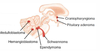
Alzheimer’s dz: pathogenesis?

A

Widespread cortical atrophy with decreased ACh. Senile plaques (extracellular beta-A core, may cause amyloid angiopathy -> intracranial hemorrhage; Ab-amyloid synthesized by cleaving APP. Neurofibrillary tangles: intracellular, abnormal phosphorylated tau proteins=insoluble cytoskeletal elements; tangles correlate with degree of dementia

5
Q

Pick’s dz (frontotemporal dementia). Presentation, Etiology, Histologic/gross findiings

A

Dementia, aphasia, parkinsonian aspects; Associated w/ Pick bodies (intracellular, aggregated tau protein), frontotemporal lobe atrophy. Spares parietal lobe and posterior 2/3rds of superior temporal gyrus.

6
Q

Lewy body dementia. Presentation, Histologic/gross findings

A

Parkinsonism with dementia and hallucinations.Caused by alpha-synuclein defect.

7
Q

Creutzfeldt-Jakob dz (CJD). Presentation, histologic/gross finding, Etiology

A

Rapidly progressive (6 wks-12mo) dementia w/ myoclonus, spongiform cortex; associated w/ prions (PrPc-> PrPsc sheet, resistant to proteases).

8
Q

What are sourses of dementia other than Alzheimer’s, Pick’s, Lewy body dementia and CJD?

A

Multi-infarct (2nd most common cause of dementia in the elderly), Syphilis, HIV, Vitamin B12 deficiency, Wilson’s dz, normal pressure hydrocephalus

9
Q

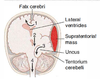
Multiple sclerosis: What is it?

A

Autoimmune inflammation and demyelination of CNS (brain and spinal cord).

10
Q

Multiple sclerosis: How do pts present? What is the course of the dz?

A

Optic neuritis (sudden loss of vision) MLF syndrome (internuclear ophthalmoplegia) Hemiparesis Hemisensory Sx’s Bowel/bladder incontinence. Relapsing and remitting course.

11
Q

Multiple sclerosis: Who is affected?

A

Most often affects women in their 20s and 30s; more common in whites.

12
Q

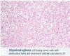
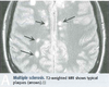
Findings in multiple sclerosis

A

Elevated protein (IgG) in CSF. Periventricular plaques (areas of oligodendrocyte loss and reactive gliosis) w/ preservation of axons.

13
Q

Charcot’s triad of MS

A

Charcot’s traid of MS is a SIN : S canning speech I ntention tremor (+I ncontinence, I nternuclear ophthalmoplegia) N ystagmus

14
Q

Tx for multiple sclerosis

A

Beta-interferon or immunosuppressant therapy. Symptomatic Tx for neurogenic bladder (catheterization, muscarinic agonists), spasticity (baclofen, GABA receptor agonist), and pain (opiods)

15
Q

Guillan-Barre syndrome (acute inflammatory demyelinating polyradiculopathy) What is it/what are the main Sx?

A

Inflammation and demyelination of peripheral nerves and motor fibers of ventral roots (sensory effect less severe than motor), causing symmetric ascending muscle weakness beginning in distal lower extremities. Facial paralysis in 50% of cases. Autonomic fxn may be severely affected (e.g., cardiac irregularities, HTN, or hypotension).

16
Q

Guillan-Barre syndrome (acute inflammatory demyelinating polyradiculopathy) What is the prognosis?

A

Almost all pts survive; the majority recover completely after wks to months.

17
Q

Guillan-Barre syndrome (acute inflammatory demyelinating polyradiculopathy): Findings?

A

Elevated CSF protein w/ normal cell count (albuminocytologic dissociation). Elevated protein -> papilledema.

18
Q

Guillan-Barre syndrome (acute inflammatory demyelinating polyradiculopathy): is associated with…?

A

Associated with infections -> autoimmune attack of peripheral myelin due to molecular mimicry (e.g., Campylobacter jejuni or herpesvirus infxn), inoculations, and stress, but no definitive link to pathogens.

19
Q

Guillan-Barre syndrome (acute inflammatory demyelinating polyradiculopathy): Management/Tx?

A

Respiratory support is critical until recovery. Additional Tx: plasmapheresis, IV immune globulins (1st choice).

20
Q

Progressive multifocal leukoencephalopathy (PML). Presentation, Etiology, Risk Group and Progession

A

Demyelination of CNS due to destruction of oligodendrocytes. Associated w/ JC virus and seen in 2-4% of AIDS pts (reactivation of latent viral infxn). Rapidly progressive, usually fatal.

21
Q

Acute disseminated (postinfectious) encephalomyelitis. Etiology

A

Multifocal perivenular inflammation and demyelination after infxn (e.g., chickenpox, measles) or certain vaccinations (e.g., rabies, smallpox)

22
Q

Metachromatic leukodystrophy. Genetics, Etiology and Presentation

A

Autosomal-recessive lysosomal storage dz fromd eficient arylsulfatase A deficiency. Build up of cerebral sulfatides leads to impaired production of myelin sheath.
Central and peripheral demyelination with ataxia and dementia

23
Q

Charcot-Marie Tooth disease (aka hereditary motor and sensory neuropathy, HMSN) Etiology and Presentation

A

Group of progressive hereditary nerve d/o’s related to defective production of proteins involved in the structure and fxn of peripheral nerves or the myelin sheath.
Numbness in feet, slapping gait

24
Q

What are seizures?

A

Characterized by synchronized, high-frequency neuronal firing. Variety of forms.

25
Q

What are partial seizures? Where do they commonly originate from and progresss to?

A

1 area of brain. Most commonly originates in the mesial temporal lobe. Often preceded by seizure aura; can secondarily generalize.

26
Q

What are the 2 types of partial seizures?

A

1.) Simple partial (consciousness intact): motor, sensory, autonomic, psychic 2.) Complex partial (impaired consciousness)

27
Q

What are generalized seizures?

A

Diffuse seizures

28
Q

What are the 5 Types of generalized seizures?

A

1.) Absence (petit mal, 3Hz, no postictal confusion) - blank stare 2.) Myoclonic - quick, repetitive jerks 3.) Tonic-clonic (grand mal) - alternating stiffening and movement 4.) Tonic - stiffening 5.) Atonic - “drop” seizures (falls to floor); commonly mistaken for fainting

29
Q

What is epilepsy?

A

A d/o of recurrent seizures (febrile seizures are not epilsepsy)

30
Q

Causes of seizures by age: Children?

A

Genetic, Infxn (febrile), Trauma, Congenital, Metabolic

31
Q

Causes of seizures by age: Adults?

A

Tumors, Trauma, Stroke, Infxn

32
Q

Causes of seizures by age: Elderly?

A

Stroke, Tumor, Trauma, Metabolic, Infxn

33
Q

What are headaches?

A

Pain due to irritation of structures such as dura, cranial nerves, or extracranial structures, not brain parenchyma itself.

34
Q

What is Migraine headache? Duration? Presentation? Etiology?

A

4-72 hours of unilateral pulsating pain w/ nausea, photophobia, or phonophobia. +/- “aura” of neurologic Sx before HA, including visual sensory, speech disturbances. Due to irritation of CN V and release of substance P, CGRP, vasoactive peptides.

35
Q

What are treatments for acute migraines?

A

Propranolol, NSAIDs, sumatriptan

36
Q

Tension headache. Presentation

A

>30 minutes of bilateral steady pain. Not aggravated by light or noise; no aura.

37
Q

Cluster headache. Presentation and Treatment

A

Unilateral, repetitive brief headaches. Periorbital pain associated with ipsilateral lacrimation, rhinorrhea. Horner’s syndrome. > in males. (15 min-3 hr)
Treatment: inhaled O2, sumatriptan

38
Q

Other causes of headache (besides migraine, tension and cluster)?

A

Subarachnoid hemorrhage (“worst headache of my life”), Meningitis, Hydrocephalus, Neoplasm, Arteritis

39
Q

Vertigo (definition)

A

Illusion of movement, not to be confused w/ dizziness or lightheadedness.

40
Q

Peripheral vertigo. Etiology

A

More common type. Inner ear etiology (e.g., semicircular canal debris, vestibular nerve infxn, Meniere’s dz). Positional testing -> delayed horizontal nystagmus.

41
Q

Central vertigo. Etiology.

A

Brain stem or cerebellar lesion (e.g., vestibular nuclei, posterior fossa tumor). Positional testing –> immediate nystagmus in any direction; may change directions.

42
Q

Sturge-Weber syndrome. Presentation, Etiology. What can it cause?

A

Neurocutaneous disorders. Congenital with port-wine stains (nevus flammeus). typically in V1 opthalmic distribution; ipsilateral leptomeningeal angiomas, pheochromocytomas. Can cause glaucoma, seizures, hemiparesis, and mental retardation. Occurs sporadically.

43
Q

Tuberous sclerosis. Presentation

A

HAMARTOMAS
Hamartomas in CNS/skin. Adenoma sebaceum (cutaneous angiofibromas). M. Ash-leaf spots (hypopigmented). cardiac Rhabdomyoma (Tuberous sclerosis). autOsomal dominant. Mental retardation. renal Angiomyolipoma. Seizures

44
Q

Neurofibromatosis type I (von Recklinghausen’s disease). Genetics, Presentation.

A

Neurocuteanous disorder. Autosomal dominant, 100% penetrant, variable expression. Mutated NF-1 gene on chromosome 17.
Café-au-lait spots, Lisch nodules (pigmented iris hamartomas), Neurofibromas in skin, optic gliomas, pheochromocytoma

45
Q

von Hippel-Lindau dz. Genetics and Presentation

A

Autosomal dominant. Mutated tumor suppressor VHL on chromosome 3. Cavernous hemangiomas in skin, mucosa, organs; benign renal cell carcinoma hemangioblastoa in retina, brain stem, cerebellum; pheochromocytomas

46
Q

Primary brain tumors. Presentations? Progression? Location?

A

Clinical presentation due to mass effects (e.g., seizures, demential, focal lesions); Primary tumors rarely undergo metastasis. The majority of adult primary tumors are supratentorial, while the majority of childhood primary tumors are infratentorial. Note: half of adult brain tumors are metastases (well circumscribed; usually present at the gray-white junction).

47
Q

Glioblastoma multiforme (grade IV astrocytoma). Incidence, Prognosis, Location, Classic Presentations

A

Most common primary brain tumor. Prognosis grave. <1yr life expectancy. Found in cerebral hemispheres. Can cross corpus callosum (“butterfly glioma” [below]) Stain astrocytes for GFAP. “Pseudopalisading” pleomorphic tumor cells – border central areas of necrosis and hemorrhage

48
Q

Most common primary brain tumor. Poor prognosis (

A

Glioblastoma multiforme (grade IV astrocytoma)

49
Q

Meningioma. Incidence, Classic Presentations

A

2nd most common primary brain tumor. Most often occurs in convexities of hemispheres and parasagittal region. Arises from arachnoid cells external to brain. Resectable.
Spindle cells concentrically arranged in a whorled pattern; psammoma bodies (laminated calcifications).

50
Q

2nd most common primary brain tumor. Most often in convexities of hemispheres and parasagittal region; from arachnoid cells external to brain. Spindle cells in whorled patterns and psammoma bodies (laminated calcifications)

A

Meningioma

51
Q

Schwannoma. Incidence, Presentation, Clinical Findings

A

3rd most common primary brain tumor. Schwann cell origin; often localized to CN VIII -> acoustic schwannoma. Resectable. Usually at cerebellopontine angle. Bilateral schwannoma found in neurofibromatosis type II.

52
Q

Oligodendroma. Progression, Location, Clinical finding

A

Relatively rare, slow growing. Most often in frontal lobes. Chicken-wire capillary pattern. ‘ Oligodendrocytes = “fried egg” cells – round nuclei w/ clear cytoplasm. Often calcified in oligodendroglioma.

53
Q

Pituitary adenoma. Presentation

A

Most commonly prolactinoma. Bitemporal hemianopia (due to pressure on optic chiasm) and hyper- or hypopituitarism are sequelae. Rathke’s pouch.

54
Q

Pilocytic (low-grade) astocytoma. Location, Prognosis, Clinical findings

A

Usually well circumscribed. In children, most often found in posterior fossa. May be supratentorial. GFAP positive. Benign; good prognosis. Rosenthal fibers – eosinophilic, corkscrew fibers. cystic + solid (gross)

55
Q

Usually well circumscribed. In children, most often found in posterior fossa. May be supratentorial. GFAP positive. Benign; good prognosis. Rosenthal fibers – eosinophilic, corkscrew fibers. What tumor does this describe?

A

Pilocytic (low-grade) astocytoma

56
Q

Medullo-blastoma. Originates, Presentation, Clinical Findings

A

Childhood peak incidence tumors. Highly malignant cerebellar tumor. A form of primitive neuroectodermal tumor (PNET). Can compress 4th ventricle, causing hydrocephalus. Rosettes or perivascular pseudorosette pattern cells. Radiosensitive.

57
Q

Highly malignant cerebellar tumor. A form of primitive neuroectodermal tumor (PNET). Can compress 4th ventricle, causing hydrocephalus. Rosettes or perivascular pseudorosette pattern cells. Radiosensitive. What tumor does this describe?

A

Medullo-blastoma

58
Q

Ependymoma. Location, clinical presentation, findings.

A

Childhood peak incidence tumors. Ependymal cell tumors most commonly found in 4th ventricle. Can cause hydrocephalus. Poor prognosis. Characteristic perivascular pseudorosettes. Rod-shaped blepharoplasts (basal ciliary bodies) found near nucleus.

59
Q

Ependymal cell tumors most commonly found in 4th ventricle. Can cause hydrocephalus. Poor prognosis. Characteristic perivascular pseudorosettes. Rod-shaped blepharoplasts (basal ciliary bodies) found near nucleus. What tumor does this describe?

A

Ependymoma

60
Q

Hemangioblastoma. Location, clinical findings

A

Most often cerebellar; associated w/ von Hippel-Lindau syndrome when found w/ retinal angiomas. Can produce EPO –> secondary polycythemia. Foamy cells and high vascularity are characteristic.

61
Q

Most often cerebellar; associated w/ von Hippel-Lindau syndrome when found w/ retinal angiomas. Can produce EPO –

A

Hemangioblastoma

62
Q

Craniopharyngioma. Location, clinical findings

A

Childhood peak incidence tumors. Benign childood tumor, confused w/ pituitary adenoma (can also cause bitemporal hemianopia). Most common childhood supratentorial tumor. Derived from remnants of Rathke’s pouch. Calcification is common (tooth enamel-like).

63
Q

Benign childood tumor, confused w/ pituitary adenoma (can also cause bitemporal hemianopia). Most common childhood supratentorial tumor. Derived from remnants of Rathke’s pouch. Calcification is common (tooth enamel-like). What tumor does this describe?

A

Craniopharyngioma

64
Q

Herniation syndromes: Cingulate (subfalcine) herniation under falx cerebri

A

Can compress anterior cerebral artery. #1 below:

65
Q

Can compress anterior cerebral artery. #1 below: What herniation syndrome is this?

A

Cingulate (subfalcine) herniation under falx cerebri

66
Q

Herniation syndromes: Downward transtentorial (central) herniation

A

2 below

67
Q

Herniation syndromes: Uncal herniation

A

(hint: Uncus = medial temporal lobe) #3 below:

68
Q

Herniation syndromes: Cerebellar tonsillar herniation into the foramen magnum

A

4 below:

69
Q

Hernation syndromes: what is the biggest danger?

A

Coma and death result when these hernations compress the brain stem.

70
Q

Clinical signs of uncal herniation: Ipsilateral dilated pupil/pstosis. Cause?

A

Stretching of CN III (innervates levator palpebrae)

71
Q

Clinical signs of uncal herniation: Contralateral homonymous hemianopia. Cause?

A

Compression of ipsilateral posterior cerebral artery

72
Q

Clinical signs of uncal herniation: Ipsilateral paresis

A

Compression of contralateral crus cerebri (Kernohan’s notch)

73
Q

Clinical signs of uncal herniation: Duret hemorrhages – paramedian artery rupture. Cause?

A

Caudal displacement of brain stem.

74
Q

What are some diagnoses of ring-enhancing lesions?

A

Metastases (lung>breast>melanoma>kidney>GI); abscesses; toxoplasmosis; primary CNS lymphoma (assoc. AIDS, EBV)

75
Q

What are diagnoses of uniformly enhancing brain lesions?

A

Metastatic lymphoma (often B cell non-Hodgkin’s), meningioma, metastases (usually ring enhancing)

76
Q

What are the diagnoses of heterogenously enhancing brain lesions?

A

Glioblastoma multiforme