TBL 15 Flashcards

1
Q

What is the abdominopelvic cavity?

A

It consists of the large abdominal cavity and small pelvic cavity and extends between the thoracic and pelvic diaphragms

2
Q

Where can the cavity ascend to?

A

Even superiorly to the 4th ICS so more superiorly positioned abdominal organs (liver, stomach, spleen) are partially protected by the thoracic cage

3
Q

Name the quadrants of the abdominal cavity and which planes divide the body into the four quadrants?

A

Right Upper Quadrant Right Lower Quadrant Left Upper Quadrant Left Lower Quadrant transumblical and medial

4
Q

Which planes demarcate the regions of the abdomen?

A

midclavicular subcostal transtubercular

5
Q

What are the regions of the body? What do they do?

A

epigastric, umbilical, and hypogastric (pubic) They localize visceral pain referred from the abdominal organs onto the anterior abdominal wall.

6
Q

What is the anterolateral abdominal wall mainly formed by?

A

A musculocutaneous sheet consisting of three laterally positioned muscle layers (external and internal obliques and transverse abdominis) with their fused aponeuroses forming the anterior aspect of the sheet.

7
Q

What causes prune belly syndrome and why does the abdomen become distended?

A

Partial or complete absence of abdominal musculature causes the abdominal wall to become so thin that organs are visible/ easily palpated. It is associated with malformations of the urinary tract/ bladder so there is an accumulation of fluid so this distends the abdomen and there is atrophy of the abdominal muscles.

8
Q

Where is the sheet forming the anterolateral abdominal wall derived from?

A

myoblasts and fibroblasts of the parietal mesoderm

9
Q

What forms the superficial muscle layer of the wall and where does its fibers run?

A

External oblique forms the superficial muscle layer and its fibers run inferomedially from the lateral surfaces of the 5th-12th ribs to the illiac crest.

10
Q

Which muscle forms the intermediate muscle layer and where do its fibers run?

A

Internal obliques an they run superomedially from the illiac crest to the inferior borders of the 10th-12th ribs.

11
Q

What do the external oblique and contralateral internal oblique form? What does it do?

A

They form a two bellied muscle sharing a common aponeurosis and synergistic actions of the muscle bellies cause flexion and rotation for torsional movement of the trunk.

12
Q

Which muscle forms the innermost muscle layer? Describe this muscle. What is it ideal for?

A

Transverse abdominis and the transverse circumferential orientation of its fibers (from the internal surfaces of the 7th-12th ribs to the linea alba) is ideal from compressing abdominal contents and increasing intraabdominal pressure.

13
Q

What does the fusion of the aponeuroses of the three muscle layers form?

A

It forms the rectus sheath that encloses the paired rectus abdominis muscles.

14
Q

What forms the linea alba? What is it used for?

A

The midline fusion of the bilateral rectus sheaths form the vertical linea alba that seperates the rectus abdominis muscles. It is used surgically for rapid midline incisions that are relatively bloodless and avoid major nerves.

15
Q

What do the rectus abdominus muscles do?

A

The extend vertically from the pubic symphysis to the 5th-7th costal cartilages and the muscles powerfully flex the vertebral column, especially the lumbar region.

16
Q

Why does lack of anterolateral wall muscle tone contribute to visceroptosis (sinking of abdominal organs) and excessive lordosis?

A

When muscles are atrophic, they provide insufficient tonus to resist the increased weight of a protuberant abdomen on the anterior pelvis. The six common causes of abdominal protrusion: food, fluid, fat, feces, flatus and fetus. The pelvis tilts anteriorly at the hip joints when standing (the pubis descends and the sacram ascends) producing excessive lordosis.

17
Q

Why do palpation-induced spasms of anterolateral wall muscles provide a clinical sign of acute abdomen?

A

Cold hands during palpation can cause involuntary muscle spasms known as intense guarding, board-like reflexive muscular rigidity cannot be willfully suppressed, occurs during palpation when an organ (such as the appendix) is inflamed and constitutes a clinically significant sign of acute abdomen. The involuntary muscular spasms attempt to protect the viscera from pressure, which is painful when an abdominal infection occurs. The common nerve supply of the skin and muscles of the wall explain why these spasms occur.

18
Q

Why can transverse incisions of the rectus abdominis be made without permanent damage to the muscle?

A

This muscle may be divided transversely without serious damage because a new transverse bands forms when the muscle segments are rejoined.

19
Q

Artery stuff?

A

See paper! Remember the abdominal aorta branches at L4 to give rise to: 1. bilateral external iliac –> femoral and inferior epigastric 2. internal iliac which supplies the pelvis

20
Q

Describe the superficial fascia below the umblicus.

A

The superficial fascia is divided into the superficial fatty layer (Camper’s fascia) and the deep fibrous layer (Scarpa’s fascia). It is reinforced by elastic and collagen fibers.

21
Q

What innervates the anterolateral abdominal wall? What provide somatic sensory fibers to the epigastric, umbilical and pubic regions?

A

Anterolateral abdominal wall: 1. Intercostal Nerves (T5-T11) 2. Subcostal Nerve (T12) 3. Iliohypogastric (L1) 4. Iliolinguinal (L1) Peripheral fibers from the DRG at: T5-T9 (epigastric) T10-T11 (umbilical) T12-L1 (pubic)

22
Q

Where do lymphatic vessels reside? Which nodes receive lymph formed above and below the transumbilical plane?

A

They reside the Camper’s fascia. Lymph from below the transumbilical plane drain into the SUPERFICIAL INGUINAL lymph nodes. Lymph from above the plane drains into AXILLARY lymph nodes.

23
Q

What is endoabdominal fascia?

A

It lines the internal surface of the musculotendinous sheet and is called transversalis fascia where it lines the internal surface of the transverse abdominal muscle.

24
Q

What is the tissue layer that separates the parietal peritoneum from the transversalis fascia?

A

Extraperitoneal fat

25
Q

Why is the endoabdominal fascia of special importance to abdominal surgery?

A

It provides a plane that can be opened, enabling the surgeon to approach structures on or in the anterior aspect of the posterior abdominal wall, such as the kidneys or bodies of lumbar vertebrae, without entering the membranous peritoneal sac containing abdominal viscera. This way the risk of contamination is minimized.

26
Q

What forms the median, medial and lateral umbilical folds and what do they cover?

A

They are formed by longitudinal folds of the parietal peritoneum from the internal surface of the anterior abdominal wall and cover embryonic remnants and blood vessels. Specifically: Median umbilical fold (apex of urinary bladder to umbilicus): median umbilical ligament Medial umbilical folds (lateral to median umbilical fold): medial umbilical ligaments Lateral umbilical folds (lateral to both): inferior epigastric vessels

27
Q

What is the falciform ligament? What are the patent and obliterated veins in its inferior edge?

A

It is a reflection of parietal peritoneum off the superoanterior abdominal wall that extends into the liver. It encloses the round ligament of the liver (fibrous remnant of the umbilical vein) and the para-umbilical veins.

28
Q

What forms the inguinal ligament?

A

dense inferiormost part of the external oblique aponeurosis forms the inguinal ligament that extends from the ASIS to the pubic tubercle

29
Q

Describe the location of the inguinal canal and rings

A

Inguinal canal: superior and parallel to the medial half of the inguinal ligament Superficial inguinal ring: superolateral to the pubic tubercle Deep inguinal ring: superior to the midpoint of the inguinal ligament and lateral to the inferior epigastric artery

30
Q

What is the main occupant of the inguinal canal in males? In females?

A

Males: spermatic cord (spermatic cord and testis traverse the deep inguinal ring to enter the inguinal canal and exit via the superficial inguinal ring) Females: round ligament of the uterus

31
Q

Why does palpation of an impulse at the superficial inguinal ring and a mass at the deep inguinal ring define an indirect inguinal hernia? How is a direct inguinal hernia detected by palpation?

A

Direct inguinal: palpate inguinal triangle and superficial ring indirect: impulse at superficial ring and mass at deep inguinal ring because processus vaginalis persists.

32
Q

Where does an undescended testis commonly lie and what is its clinical risk? How can an undescended testis be distinguished from an inguinal hernia in infants?

A

The undescended testis commonly lies somewhere along the normal path of its prenatal descent, commonly in the inguinal canal. Increased risk for developing malignancy in the undescended testis. feel the scrotum. If nothing is there then it is an undescended testis.

33
Q

Where does an undescended testis commonly lie and what is its clinical risk? How can an undescended testis be distinguished from an inguinal hernia in infants?

A

The undescended testis commonly lies somewhere along the normal path of its prenatal descent, commonly in the inguinal canal. It is not palpable!

34
Q

What is the processus vaginalis?

A

Diverticulum beginning in the deep inguinal ring of the parietal peritoneum created by the testis pushes muscular and fascial layers of the anterolateral wall ahead of it into the inguinal canal Eventually… stalk part collapses and distal part forms the tunica vaginalis which adheres to the testis and epididymis

35
Q

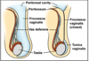
How is a persistent processus vaginalis related to a hydrocele of the testis and how does a hydrocele of the spermatic cord differ from a hydrocele of the testis?

A

A hydrocele is the prescence of excess fluid in a persistent processus vaginalis. A hydrocele of the testis is confined to the scrotum and distends the tunica vaginalis. A hydrocele of the cord is confined to the spermatic cord and distends the persistent part of the stalk of the processus vaginalis.

36
Q

What provides somatic motor fibers to the cremaster muscle?

A

genital branch of the genitofemoral nerve

37
Q

What provides somatic sensory fibers to the skin of the superomedial thigh?

A

ilioinguinal nerve

38
Q

Which nerves constitute the cremasteric reflex?

A

genital branch of the genitofemoral nerve (somatic motor) ilioinguinal nerve (somatic sensory)

39
Q

How is the cremasteric reflex tested?

A

Contraction of the cremaster muscle is elicited by lightly stroking the skin on the medial aspect of the superior part of the thigh with an applicator stick or tongue depressor. The ilioinguinal nerve supplies this area of the skin. The rapid elevation of the testis on the same side is the cremasteric reflex.

40
Q

What does the spermatic cord contain?

A

Ductus (vas) deferens, testicular artery, and pampiniform venous plexus

41
Q

What does the pampiniform plexus do? What does it converge superiorly to form?

A

It cools blood in the testicular artery to maintain the testis a few degrees below body temperature thereby insuring normal spermatozoa production. It converges superiorly as the testicular vein.

42
Q

What does the smooth muscle of the vas deferens do?

A

The mucosa of the vas deferens is surrounded by circular bundles of smooth muscle and the sympathetic mediated contraction of the muscle propels spermatozoa along the ductus deferens during ejaculation.

43
Q

What is a hematocele of the testis?

A

A hematocele of the testis is a collection of blood in the tunica vaginalis that results from (i.e.) rupture of branches of the testicular arty by trauma to the testis.

44
Q

Why is torsion of the spermatic cord a surgical emergency?

A

Necrosis of the testis may occur. Torsion (or twisting) obstructs the venous drainage, with resultant edema and hemorrhage and subsequent arterial obstruction.

45
Q

Why does palpable varicocele seem to disappear when the patient lies down?

A

The vine like pampiniform plexus of veins may become dilated (varicose) and tortuous, producing a varicocele, which is usually visible only when the man is standing or straining. The enlargement disappears when lying down, particularly if the scrotum is elevated while supine, allowing gravity to empty the veins.

46
Q

Where does the round ligament of the uterus extend from?

A

It extends from the lateral uterine wall through the inguinal canal into subcutaneous tissue of the labia majora. The processus vaginalis and round ligament degenerate. The inguinal canal is less relevant in females.

47
Q

How is the round ligament related to the metastasis of uterine cancer?

A

Metastatic uterine cancer cells (especially from tumors adjacent to the proximal attachment of the round ligament) can spread from the uterus to the labium majus and from there to the superficial inguinal nodes which leaves lymph from the skin of the perineum (including the labia)

48
Q

What are the layers of the developing testis? and what are they made from?

A

Processus Vaginalis Internal spermatic fascia (transversalis fascia) Cremaster muscle and fascia (Internal oblique muscle and aponeurosis) External spermatic fascia (aponeurosis of external oblique)