Chapter 13 - Blood vessels Flashcards

1
Q

Review:

What is the intima?

What is the media?

What is the adventitia?

How are they supplied?

A
  • Intima
    • Single layer of endothelial cells with little subendothelial CT
    • Supplied by the lumen
    • Separated from media by the internal elastic lamina
  • Media
    • Smooth muscle
    • Inner layers: lumen
    • Outer layers: small arterioles (vasa vasorum)
  • Adventitia
    • CT, Nerves, and Vasa Vasorum
2
Q

Review

Difference between large, medium, and small arteries?

A
  • Large (elastic arteries)
    • Media = rich in elastic fibers that alternate with layers of smooth muscle
  • Medium (muscular arteries)
    • Media = mostly smooth muscle
  • Small (less than 2mm)
    • Structurally similar to medium arteries
    • Found within tissue and organs
3
Q

Review still:

Arterioles:

Composition?

Importance?

Capillaries:

Composition?

Importance?

A
  • Arterioles
    • Small amount of smooth muscle
    • NO elastic lamina
    • *Principle sites of BF resistance*
  • Capillaries
    • Endotheilal lining but NO media
    • BF slows dramatically here obbbbbviously for nutrient exchange
4
Q

Review:

Where does blood flow after capillaries? What is important about these?

Veins in comparison to arteries?

Composition of lymphatics?

A
  • Postcapillary venules (after cap)
    • Site of inflammation induced leakage
  • Veins
    • Large lumens and walls (less organized than art.)
    • Contains 2/3 of all blood
  • Lymphatics
    • Thin-walled, endotheium lined channels
    • Drain intersitial fluid and inflammatory cells
5
Q

How are endothelial cells identified immunohistochemically?

A

Endothelial cells identified by:

  • PECAM-1 (CD31)
  • CD34
  • vWF
6
Q

Functions of endothelial cells?

A

Endothelial cell function:

  • Maintenance of permeability barrier
  • Elaboration of compounds that control coagulation
  • Produce ECM proteins for healing/angiogenesis
  • Production of vasoconstrictors (endothelin/angiotensin)
  • Production of vasodilators (NO/prostacyclin)
  • Important mediators of inflammation
    • Produce IL-1/6/8
    • Express VCAM-1, ICAMS, E/P selectin
  • Produce growth factors
7
Q

What is endothelial dysfunction associated with?

A

Endothelial dysfunction:

  • Abnormal thrombus formation
  • Atherosclerosis
  • Vascular lesions of hypertension
8
Q

Functions of vascular smooth muscle cells?

A

Vascular smooth muscle cells:

  • Vasodilation/constriction in response to mediators
  • Synthesis extracellular basement membrane proteins
    • Collagen, elastin, and proteoglycan
  • They can also proliferate and be migratory
    • In response to cytokines (IL-1, IFN)
    • In response to GF (PDGF, endothelin-1, and FGF)
9
Q

What layer of the artery thickens in response to vessel injury?

A

The intima

10
Q

What is the pathology of intimal thickening in response to vessel injury?

A

Pathology

  • Endothelial cells lose the ability to contract but they proliferate and produce ECM proteins, creating what is known as the neointima*
  • If chronic - associated with atherosclerosis
11
Q

Arteriosclerosis:

Generic term for?

What are the 3 variations?

A

Arteriosclerosis:

  • Arterial wall thickening and the loss of wall elasticity

3 variations:

  • Monckeberg medial calcific sclerosis
  • Arteriolosclerosis
  • Atherosclerosis
12
Q

What is monckeberg medial calcific sclerosis?

A

Characterized by excessive Ca++ deposits in muscular arteries (50+ yo) that are usually benign because the deposits do not cause obstruction

13
Q

What is arteriolosclerosis?

A

Involves arterioles and small arteries

Hyaline and hyperplastic forms occur and are associated with thickening of the vessel walls and obstruction of the lumen

14
Q

What is atherosclerosis characterized by?

A

Characterized by intimal lesions known as atheromas or atheromatous fibrofatty plaques

(they may obstruct the lumen and weaken the media)

15
Q

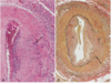
What is this an example of?

A

This is Monckeberg medial calcific sclerosis with the collection of calcium deposits (arrows) in the media of small muscular arteries

16
Q

Where do atherosclerotic lesions primarily develop?

Atherosclerotic symptomatic disease most often involves the arteries supplying what organs?

A
  • Lesions develop in:
    • Elastic arteries (aorta, carotid, and iliac)
    • Large/medium muscular arteries (coronary/popliteal)
  • Symptomatic disease involves arteries supplying:
    • Heart
    • Brain
    • Kidneys
    • Lower extremities
17
Q

Major outcomes of atherosclerosis?

A

Atherosclerosis major outcomes:

  • MI
  • Stroke
  • Aortic aneurysms
  • Peripheral vascular disease
18
Q

Earliest lesion of atherosclerosis

A

Earliest lesion:

  • Fatty streaks in the intima
    • Lipid filled macrophages (foam cells)
    • Not raised so no disturbance in BF
19
Q

Atherosclerotic plaques typically have what 3 principal components?

A
  • Cells
    • Smooth muscle, macrophages, and lymphocytes
  • ECM proteins
    • Collagen, elastic fibers, and proteoglycan
  • Intra/Extracellular Lipid
    • Cholesterol clefts are common
20
Q

Top risk factors for CVD?

A
  • Hyperlipidemia
  • Hypertension
  • Cig smoking (oops)
  • Diabetes
  • Advanced age
21
Q

Response to injury hypothesis implies?

A

Implies that atherosclerosis is a chronic inflammatory response of the artery wall initiated by injury to the endothelium

22
Q

Lesion progression of atherosclerosis is sustained by?

A

Sustained by interactions between

  • lipoproteins
  • Macrophages
  • T cells
  • Smooth muscle cells
23
Q

Steps in lesion development?

A
  • Chronic injury
  • Accumulation of lipoproteins (mostly LDL)
  • Increased adhesion of monocytes/leukocytes to endo.
  • Adhesion of platelets
  • Release of factors that induce smooth muscle migration
  • Smooth muscle proliferation
  • Elaboration of extracellular matrix proteins
24
Q

What is C-reactive protein?

A

CRP = one of the cheapest and most sensitive predictors of the risk of MI, stroke, peripheral arterial disease, and sudden cardiac death

25
Q

How can the endothelium become damaged to create plaques?

A

Endothelial damage:

  • May be associated with infection
  • Initiated by normal hemodynamic disturbances
    • Eg: at branch points
  • Hypercholesterolemia
26
Q

Inflammatory role associated with plaque formation?

A
  • Increases expression of adhesion molecules by endothelial cells
    • Allows for infiltration of inflammatory cells to intima
  • This is initially protective until T cells become activated
    • Secrete proinflammatory cytokines that induce smooth muscle migration/proliferation
27
Q

What are the major lipids in atheromatous plaques?

Correlation between the severity of atherosclerosis and?

A
  • Major lipids
    • Plasma derived cholesterol and cholesterol esters
  • Correlation of severity with:
    • High levels of plasma cholesterol or LDL
28
Q

Smooth muscle cells convert the fatty streak into what?

Catastrophic evens are associated with what aggregation?

A
  • Smooth muscle cells convert fatty streak to
    • Fibrofatty atheroma
  • Catastrophic events are associated with platelet aggregation over the plaque
    • This is followed by eventual disruption of the fibrous cap
29
Q

When does atherosclerotic coronary artery disease begin?

A

Childhood (modification should begin then)

30
Q

Hyptertensive vascular disease BP:

Normal?

Prehypertensive?

Stage 1 hypertension?

Stage 2 hypertension?

A
  • Normal
    • <120/80
  • Prehypertension
    • 120-129/80-89
  • Stage 1
    • 140-159/90-99
  • Stage 2
    • >160/100
31
Q

Types of hypertension?

A
  • Essential hypertension
    • idiopathic and some develop potentially fatal BP
  • Secondary
    • Underlying renal or adrenal disease
32
Q

What relationship is altered in arterial hypertension?

A

The relationship between cardiac output and TPR

( BP = CO x TPR )

33
Q

What is cardiac output dependent upon?

Where is TPR determined?

A
  • CO
    • Blood Vol influenced by Na+ homeostasis
  • TPR
    • Determined at the arterioles
34
Q

Role of the kidney in BP regulation?

A
  • Renin converts A to AI which is then converted to AII bye ACE
  • AII increases BP by increasing peripheral resistance and increasing the blood volume by increasing Na+ resorption
  • Kidney also produces antihypertensive substances (PG/NO)
    • Counterbalances AII
  • Natriuretic Factors inhibit Na+ resorption and induce vasodilation
35
Q

What three pathological dysfunctions alter Renin secretion?

A
  • ( - ) Fibromuscular dysplasia
    • Genetic disorder
  • ( + ) Renal artery stenosis
    • Acquired Disorder
  • ( + ) Renin-secreting tumor
    • Acquired Disorder
36
Q

What two things alter angiotensin (A) secretion?

A
  • ( + ) Oral contraceptives
    • Acquired
  • ( - ) Angiotensinogen Variants
    • Genetic disorder
37
Q

What three pathological dysfunctions alter Na+ reabsorption?

A
  • ALL ( - ) Genetic Disorders
    • Liddle Syndrome
    • Pseudohypoaldosteronism
    • Gitelman syndrome
38
Q

Hyptertension is associated with what two forms of arteriolosclerosis?

A

Hyaline and Hyperplastic

39
Q

What is hyaline arteriolosclerosis?

A
  • Lesion consists of a homogeneous hyaline thickening of the wall with eventual narrowing of the lumen
    • These reflect the leakage of plasma components across the endothelium and excessive ECM production by smooth muscle secondary to the hemodynamic stress associated with hypertension
  • Contribute to hypertension related renal failure
40
Q

What is hyperplastic ateriolosclerosis?

A
  • Associated with severe hypertension
  • Concentric, laminated thickening of the arterioles composed of smooth muscle and basement membrane components
  • May become necrotic and infiltrated with fibrin
  • Also common in the kidney
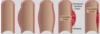
41
Q

What is this?

A

Hyperplastic arteriolosclerosis

(Concentric - looks like an onion )

42
Q

What is this?

A

Hyaline Arteriolosclerosis

43
Q

What is a TIA?

Characterization of a TIA?

A
  • TIA
    • Mini stroke that lasts less than 24 hours
    • Characterized by focal neurological defects that resolve within 24 hours
44
Q

Difference between an aneurysm and a false aneurysm?

What is a Dissection?

A
  • Aneurysm
    • Abnormal dilation of a vascular wall
  • False aneurysm
    • Breach in the vascular wall leading to an extravascular hematoma
  • Dissection
    • Blood enters the artery wall and travels for a distance

Note* aortic aneurysm/dissections are clinically the most important

45
Q

Despite that the picture kindddda tells you.. what are these from left to right (1-4)?

A
  1. True aneurysm (saccular)
  2. True aneurysm (fusiform)
  3. False aneurysm
  4. Dissection
46
Q

Saccular versus fusiform aneurysms?

A
  • Saccular
    • Spherical and involve only a portion of the vessel wall
    • Usually contain a thrombus
    • 5-20 cm in diameter
  • Fusiform
    • Vary in diameter/length
    • May involve the entire segment
47
Q

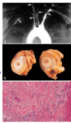
Major cause and location of abdominal aortic aneurysms?

A

Atherosclerosis usually right below the renal arteries and above the bifurcation

48
Q

What predisposes to aneurysms and dissections?

A
  • Genetic defects in CT structure and/or assembly
    • Example: Marfan syndrome
  • Overactive metalloproteinases (w/ chronic inflammation) may degrade CT and weaken the vessel wall
49
Q

Clinical course of the abdominal aortic aneurysm?

A
  • May rupture with massive hemorrhage, obstruction of lumen, obstruction of neighboring vessels/organs, and/or emolism
  • May present as a mass simulating a tumor
  • Most expand until they rupture
50
Q

What occurs in syphilitic aneurysms?

A
  • This occurs in tertiary syphilis as an aortic obliterative endarteritis
    • Inflammation and fibrosis weaken the media
51
Q

What is aortic dissection characterized by?

A
  • Dissection of blood between and along the laminar planes of the media
  • Often rupture outward causing a massive hemorrhage

(90% in men btwn 40-60yo with HT, others due to CT abnormalities or as a result of arterial cannulation)

52
Q

Most common pathologic abnormality of aortic dissection

A
  • Cystic medial degeneration
    • Characterized by fragmentation of the elastic tissue and separation of the elastic/fibromuscular element by clefts
    • Frequently occurs in Marfan syndrome
53
Q

Major risk factor for dissection? Pathology?

A

Hypertension

Path = varying degrees of elastic tissue fragmentation

54
Q

Classic symptoms of aortic dissection?

A
  • Sudden onset of pain
    • Begins in chest
    • Radiates to the back
    • Moves downward as the dissection progresses
55
Q

Vasculitis (inflammation) typically affects what vessels?

Two most common causes?

A
  • Typically affects arterioles, venules, and capillaries
    • Small vessel Vasculitis
  • May have systemic symptoms - Systemic necrotizing vasculitides
  • Causes
    • Infection
    • Immune mediated reactions
56
Q

3 most common pathologies of non-infectious vasculitis?

A
  1. Immune complexes
  2. Antineutrophil cytoplasmic antibodies
  3. Anti-endothelial antibodies
57
Q

How do Immune complexes create vasculitis?

A
  • ICs and Complement components deposit in vessel walls
    • ICs induce neutrophil accumulation/activation/degran.
      • Results in tissue necrosis
  • IC formation may be drug-related, associated with infection or autoimmune disease
58
Q

Role of Antineurophil cytoplasmic antibodies (ANCA) in vasculitis?

A

Abs form to mostly granule enzymes in neutrophils (also monocytes and endothelial cells)

59
Q

Anti-endothelial antibodies are seen in what 2 diseases?

A

SLE and Kawasaki disease

60
Q

Most common form of systemic vasculitis in adults?

What does it involve?

A

Giant cell (temporal) arteritis

  • Ganulomatous inflammation principally affecting the arteries of the head (esp temporal arteries)
  • May also involve vertebral/ophthalmic arteries and the aorta (giant cell aortitis)
61
Q

Morphology of Giant cell arteritis?

Where does granulomatous inflammation occur?

A

Nodular intimal thickening (varying lumen occlusion)

Inflammation occurs in the media with a mononuclear infiltrate (T cells/macrophages) with both foreign body and Langhans giant cells common

Heals with fibrosis

62
Q

Clinical manifestations of giant cell arteritis?

A

Either:

  • Vague and systemic
    • Fever/fatigue/wt. loss
  • Or Localized
    • facial pain/headache
  • Most serious when ophthalmic artery involved (blindness)
63
Q

Diagnosis?

A

ESR is nearly always elevated

Biopsy

64
Q

What is Takayasu arteritis?

A

Ganulomatous vasculitis of medium and large arteries

Predominantly the aortic arch and its branches with irregular thickening of the intima

65
Q

Inflammatory infiltrate of Takayasu arteritis?

Clinical Manifestation?

A

Similar to giant cell arteritis and is composed of mononuclear cells and giant cells

Clinical:

  • Weak pulses in upper extremities
  • May also involve eyes/CNS
66
Q

How to diagnose the difference between Takayasu and Giant cell arteritis in the aorta?

A

Same infiltrate thus AGE:

Takayasu: under 50

Giant cell: over 50

67
Q

What is polyarteritis nodosa (PAN)?

A

Systemic vasculitis involving small/medium muscular arteries

Often involve the renal/visceral vessels but SPARE the pulmonary circulation

68
Q

What is PAN characteristically associated with?

A

Necrotizing inflammation with a mixed infiltrate (neutro/eosinophils and mononuclear cells)

69
Q

Hallmark of PAN?

A

All stages of inflammation coexist in different or the same vessels

70
Q

Clinical manifestations of PAN?

A
  • Fever and weight loss
  • Abdominal pain (mesenteric arteritis)
  • Peripheral neuritis
  • Myalgia and muscle weakness
  • Renal involvement (doesnt EVERYTHING involve the kidneys???!)
71
Q

What is polymyalgia rheumatica (PMR)?

A

Systemic inflammatory disease that occurs in 50% of patients with Giant cell arteritis (GCA)

72
Q

Differences between GCA (giant cell ateritis) and PMR (polymyalgia rheumatica) symptoms?

A

GCA: systemic inflammation accompanies the vascular manifestations

PMR:same systemic symptoms but the vascular lesions are subclinical

73
Q

Similarities between GCA (giant cell) and PMR (polymyalgia rheumatica)?

A
  • Both have an increased ESR
  • Both present with anorexia/wt. loss, fever, depression, and maliase
74
Q

Additional presentation of PMR?

A
  • Normochromic normocytic anemia
  • Pain/stiffness in the shoulders, upper arms, hips, thighs, and/or neck (associated with synovitis of the affected joints)
75
Q

What is the most common form of systemic vasculitis in kids?

A

Henoch-Schonlein purpura

76
Q

Classical manifestation of Henoch-Schonlein purpura?

A
  • Palpable purpura
  • Arthralgia/arthritis
  • Abdominal pain
  • Renal disease: mild porteinuria to insufficiency
77
Q

Pathological finding in Henoch-Schonlein purpura?

A

IgA immune complex deposition

78
Q

What is this?

A

Giant cell arteritis:

Granulomas in the media (black arrows)

Lumen is almost entirely occluded (yellow arrow)

79
Q

What is this?

What is the black arrow pointing at in figure A? Figure B?

A

Giant cell arteritis

A: degenerated internal elastic lamina in active arteritis

B: focal destruction of internal elastic lamina and intimal thickening of long-standing/healed arteritis

80
Q

What is this?

A

PAN: Polyarteritis Nodosa

Shows inflammation, necrosis, and fibrosis (and karyorrhexis as a bonus if you remember nuclear fragmentation)

81
Q

What disease is this?

A

Takayasu arteritis

A: narrowing of aortic arch branches

C: destruction of the media w/ mononuclear infiltrates, giant cells, and fibrosis

82
Q

What disease are you likely to find this in?

(THIS ONES A TOUGHY!)

A

Henoch-Schonlein purpura:

IgA deposits limited to the mesangium and thus the capillary walls are not outlined

83
Q

Leading cause of acquired heart disease in children?

A

Kawasaki disease

84
Q

Kawasaki disease:

AKA?

Acute symptoms?

A

Kawasaki AKA mucocutaneous lymph node syndrome:

  • Fever
  • cervical adenopathy
  • buccal erythema
  • strawberry tongue
  • erythematous rash involving the palms and soles

(resembles TS/Scarlet fever)

85
Q

Associated with what mediators?

A

T cell and macrophage activation

Anti-endothial/smooth muscle Antibodies

86
Q

The vasculitis of Kawasaki resembles?

What can this progress to?

A

Vasculitis resemples that of PAN

Can progress to vasculitis of the coronary vessels (can cause aneurysms/thrombosis/MI)

87
Q

Diagnosis of Kawasaki?

A

Fever at least 5 days + 4 of the 5:

  • Conjunctival injection
  • Oral mucous membrane changes
  • Peripheral extremity changes (erythema of palms/soles)
  • Polymorphous rash
  • Cervical lymphadenopathy
88
Q

What two diseases demonstrate large globular mesangial IgA-IC deposits?

A

IgA nephropathy

Henoch-Schonlein purpura (just makin sure youre learning something over derr)

89
Q

What is Wegener’s granulomatosis?

A

Necrotizing vasculitis characterized by:

  1. Acute necrotizing granulomas of the U/L Respiratory tract
  2. Necrotizing or granulomatous vasculitis of small/med vessels
  3. Focal necrotizing glomerulonephritis

(pathogenesis resembles PAN)

90
Q

Wegener’s URT lesions? Cells?

A

URT lesions range from inflammatory sinusitis to ulcerations

Macrophages, giant cells, lymphocytes, and plasma cells

91
Q

What could happen to the Wegener’s lesions in the lungs?

A

The lesions may coalesce and undergo cavitation

92
Q

Early vs. Late kidney lesions in wegeners?

A

Early

  • Focal necrotizing glomerulonephritis w/ acute focal proliferation, necrosis, and thrombosis (isolated)

Late

  • Diffuse necrosis, proliferation, and crescent formation
93
Q

Wegener’s granulomatosis presentation?

A
  • Persistent pneumonitis
  • Chronic sinusitis
  • Mucosal ulceration
  • Palpable purpura
  • Myalgia/arthralgia
  • Evidence of renal disease
94
Q

What is microscopic polyangitis?

A

Necrotizing vasculitis of arterioles, capillaries, and venules

95
Q

How do lesions in microscopic polyangiitis compare to those in PAN?

Where do they occur?

A

They all are the same age and commonly occur in the skin, lungs, and kidney

96
Q

Characteristic infiltrate of microscopic polyangiitis?

A

Mononuclear and neutrophil infiltrate w/ fibrinoid necrosis

-neutrophils migrate into the wall and disintegrate releasing cytoplasmic components (leukocytoclastic vasculitis)

97
Q

Differences in the morphology between microscopic polyangiitis and Wegeners?

A

MP has NO granulomas in the lesions and wegeners does

98
Q

Differences between P-ANCA and C-ANCA?

(ANCA = antineutrophil cytoplasmic antibodies)

A
  • P-ANCA
    • Stain is limited to perinuclear region and cyto is nonreactive
    • Abs directed at myeloperoxidase
  • C-ANCA
    • Heavy stain in cyto w/ nonreactive nuclei
    • Abs directed against proteinase 3
99
Q

Which is P-ANCA? C-ANCA?

What Diseases are each associated with?

A

P-ANCA: on the left; associated with Microscopic Polyangiitis

C-ANCA: center and right; associated with Wegener’s

100
Q

What is Thromboangiitis obliterans?

A

Buerger’s Disease:

Occurs in conjunction with cig. smoking and affects the distal medium sized A/V of the extremities

101
Q

Characteristics of Thromboangiitis obliterans?

A

Acute/Chronic inflammation with thrombosis (organization/recanalization)

The thrombi often contain abscesses

102
Q

What happens in thromboangiitis obliterans without treatment? What is the treatment?

A

Without treatment it spreads into other vessels/nerves and all structures become fibrotic (looks gangrenous and will require amputation)

Treatment = quit smoking

103
Q

Review (will have more ?? per slide)

What is Raynaud’s phenomena?

Primary vs secondary?

A

Paroxysmal pallor of the digits of hands/feet (white), progressing to cyanosis with pain/numbness (blue), followed by hyperemia (red)

Primary: exaggeration of vasomotor responses to cold/stress

Secondary: associated with CT diseases (slceroderma/SLE/RA), occlusive diseases, drugs, neurologic disorders, and trauma. Arterial insufficiency may result in ulceration or gangrene

104
Q

Most common manifestation of chronic venous disease?

A

Varicose veins

105
Q

Varicose veins are most common where?

How do they occur?

What do they look like?

A

Most common in superficial veins of legs often due to long periods of standing (pressure increases leading to stasis/edema) and they are dilated, tortorous veins.

106
Q

Morphology of varicose veins?

What patients may have esophageal varicosities?

A

Veins with varicosities have thing walls, imcompetent valves, with thus stasis, congestion, pain, and edema

Patients with cirrhosis and portal hypertension may have esophageal varicosities

107
Q

What is thrombophlebitis/phlebothrombosis?

Predisposing conditions?

A

DVT that can result in life threatening venous thromboembolism

Predisposing conditions:

  • HF
  • Neoplasia
  • Pregnancy
  • Obesity/prolonged immobilization
  • Surgery/Previous DVT
108
Q

Clinical manifestations of DVT?

A

Leg pain, tenderness, swelling, dilation of the superficial veins, and cyanosis

109
Q

What is lymphangitis?

A

Bacterial infections that spread through lymphatics causing inflammation

110
Q

What is a Hemangioma?

A

Localized superficial cutaneous lesion of childhood

111
Q

Capillary vs. Cavernous hemangioma?

A
  • Capillary hemangioma
    • Skin, subcutaneous tissue, and oral/lip mucous memb.
    • Covered with an intact epithelium
    • Unencapsulated aggregates of thin capillaries filled with blood and separated by scant CT
  • Cavernous
    • Larger than capillary, well defined, unencapsulated
    • Thrombosis and dystrophic calcification is common
112
Q

What is a pyogenic granuloma?

What is this during pregnancy?

A

Rapidly growin exophytic red nodule attached to the skin or oral mucosa that bleeds easily and often ulcerates?

Pregnancy = granuloma gravidarum

113
Q

Histological presentation of a pyogenic granuloma?

A

Proliferating capillaries

Edema with neutrophils

114
Q

What is a Lymphangioma?

Difference between capillary and cavernous lymphangiomas?

A

Lymphangioma = benign lymphatic analog of hemangioma

  • Capillary:
    • Lesion = small channels beneath the epidermis
    • Resemble hemangioma but w/o blood cells
  • Cavernous
    • Occur mostly in children in the neck/axilla
    • Resemble cavernous hemangioma but w/o blood cells
115
Q

What is a glomus tumor?

A

A painful benign tumor that arises from the modified SMC of the glomus body (specialized arteriovenous anastomosis involved in thermoregulation/BF)

116
Q

Histology and common locations of glomus tumors?

A
  • Histology
    • vascular channels separated by CT w/ masses of glomus cells
  • Commonly occurs:
    • Distal digits (esp under fingernails)
117
Q

What is Vascular ectasias? 3 major examples?

A

Localized dilation of pre-existing channels

Nevus Flammeus

Spider Telangeictasia

Hereditary hemorrhagic telangiectasia

118
Q

What is nevus flammeus?

A

“Birthmark” on head or neck that ranges from pink-deep purple with dilation of vessels in the dermis.

Most regress, others (like port wine stains) may grow proportionately with the child

119
Q

What is spider telangiectasia and where is it likely to occur?

A

Form an array of dilated subcutaneous arteries/arterioles that blanch with pressure

Common on face, neck, or chest in pregnancy/pts w/ cirrhosis

120
Q

What is Hereditary hemorrhagic telangiectasia?

A

The most common genetic (AD) cause of vascular bleeding.

The most frequent symptom is epistaxis

Capillaries are dilated with a loss of subendothelial structures

121
Q

What is Bacillary angiomatosis?

A

Opportunistic infection caused by Bartonella Henselae

Associated with cat bites/scratches

122
Q

Two main histologic forms of bacillary angiomatosis?

A

Bacillary angiomatosis

Cat-scratch disease (granulomatous)

123
Q

Pathology of bacillary angiomatosis?

Characteristics of the Lesions?

A

Proliferation of capillaries, acute inflammatory infiltrate, and bacteria

Lesions = cutaneous or mucocutaneous and tender –> may ulcerate and crust (disseminated in AIDS)

124
Q

Pathology of Cat-scratch disease?

A

Granulomas in the lymph nodes

(treat with azithromycin)

125
Q

Virus of Kaposi sarcoma?

Clinical and histological manifestations?

A

Kaposi sarcoma = HHV-8

  • Clinical
    • _​_cutaneous lesions that are red-purple macules that become raised plaques
  • Histological
    • Spindle cells and inflammatory infiltrate that line vascular channels
126
Q

Different forms of Kaposi sarcoma?

A
  • Chronic/Classic/European
    • Older men
    • Indolent and confined to lower extremities
  • Lymphadenopathic/African/Endemic
  • Immunosuppressive-associated KS
    • Transplants and AIDS
127
Q

What is angiosarcoma?

A

Malignant endothelial neoplasms

Occurs in older adults

Initially involve the skin, heart, breast, or liver

128
Q

Angiosarcoma is the most common sarcoma arising in what two organs?

Morphology and clinical course?

A

Most common arising in the liver and heart

All degrees of endotheial differentiation occurs

These neoplasms are locally invasive, prone to metastases, and yield a generally poor prognosis

129
Q

What is percutaneous transluminal coronary angioplasty?

A

Placement of a balloon, catheter, or stent to open a stenotic vessel.

Use of this + thrombolytic therapy is best for an MI

High success rate (lower with complete occlusion)

130
Q

Restenosis issues with PTCA? How to make better?

A

Elastic recoil: occurs w/o placement of a stent and is a mechanical renarrowing caused by adventitial constriction

Neointimal hyperplasia: proliferation of SMC and production of ECM proteins in response to the injury caused by the balloon and/or stent

Use a stent that elutes antiproliferative agents (sirolimus/paclitaxel) however, may increase risk for stent-associated thrombosis