3.10 - 3.12 Cell to cell communication and signalling Flashcards

(69 cards)

1
Q

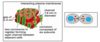
What is the difference between contact dependent or paracrine signalling?

A
  • This is for short distance
  • Contact dependent is where cells are in close contact, membrane to membrane
  • paracrine is where there is an extracellular release of signal that acts only locally on neighbouring cells
2
Q

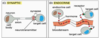
What are the long distance types of cell signalling?

A
  • Synaptic where neurons have an electrical signal along the axon (long distance) resulting in release of neurotransmitter across the synapse (short distance)
  • Endocrine where there is a release of hormone into the bloodstream which acts widely throughout the body
3
Q

What type of signalling occurs at a very short distance?

A
  • Autocrine where cells can stimulate themselves if they have receptor for the ligand
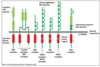
  • Groups of identical signalling cells (community) can reinforce signals, for example in development it ensures cells follow the same differentiation pathway
  • Cancer cells use autocrine signals to stimulate their own survival and proliferation
4
Q

How can signalling occur in a gap junction?

A
  • Allows direct communication between cytoplasm of adjacent cells by small intracellular signalling molecules such as Ca2+ or cAMP
  • Allows neighbouring cells to coordinate responses to signal such as noradrenaline response in liver cells Cx32
5
Q

What are the two types of receptors?

A
  • Cell surface receptors for a hydrophilic ligand that cannot cross the membrane. For example peptide growth factors bind cell surface receptor
  • Intracellular for a hydrophobic or lipophillic ligand which can cross the membrane, for example steroids or small molecules diffuse across the membrane, bind intracellular/nuclear receptors
6
Q

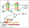
What is the basic cell signalling pathway?

A
7
Q

How does the same signal (acetylcholine) cause different responses in different cells?

A
  • Due to different receptor types
  • Muscarinic (G protein) vs Nicotinic (ion channel) receptors
  • Different intracellular mediators
8
Q

What are the characteristics of steroid hormones?

A
  • They are transported in blood by carrier proteins (steroid-hydrophobic)
  • Cross plasma membrane
  • Bind intracellular receptors that have DNA binding domains (receptors are homo or heterodimers)
9
Q

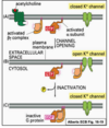
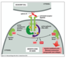
How are intracellular receptors activated?

A
  • Receptors kept inactive as they have inhibitory proteins bound to them
  • When ligand goes into cytosol it kicks off inhibitory protein, binds to the pocket and induces conformational changes
10
Q
A
11
Q

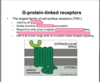
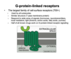
What are the 3 major classes of cell surface receptors?

A
  • Ion channel couple receptors
  • G-protein couple receptors (indirectly linked to enzymes)
  • Enzyme coupled receptors
12
Q

What are the two important classes of enzyme - coupled receptors?

A
  • Receptor tyrosine kinases – have kinase activity and phosphorylate ‘Tyr’ on intracellular signal proteins
  • Receptor serine/threonine kinases – have kinase activity and phosphorylate ‘Ser’ and ‘Thre’ on target proteins
13
Q

What are the two classes of ligands for receptor tyrosine kinases?

A

Secreted growth factors and hormone

  • Epidermal growth factor (EGF)
  • Fibroblast growth factor (FGF)
  • Platelet-derived growth factors (PDGF)
  • Hepatocyte growth factor (HGF)
  • Insulin, Insulin-like growth factor (IGF)
  • Vascular endothelial growth factor (VEGF)
  • Macrophage colony stimulating factor (M-CSF) • Neurotrophin (eg. nerve growth factor NGF)

Membrane-bound ligands

• Ephrins

14
Q

What are the domains like for receptor tyrosine kinases?

A
  • Single transmembrane domain
  • Highly variable extracellular domains
  • Similar intracellular domains (tyrosine kinase domains)
15
Q

How do ephrins/eph receptors function together?

A
  • very large family
  • Can function in bidirectional signalling
  • Ligand can signal back to the cell, main signal is via phosphorylation of Eph receptors, but ephrins often linked to the cytoskeleton so the signalling cell can receive a response as well
  • Often functions in cell migration and axon guidance (attraction or repulsion cues)
16
Q

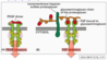
What is the signalling pathway for eph?

A
  1. Ephrins causes clustering of the eph receptors. Receptor on the migrating cell engages with ephrins and dimerises due to cross phosphorylation on a specific tyrosine
  2. A kinase binds resulting in phosphorylation of ephexin (GEF)
  3. Ephexin activates RhoA
  4. RhoA results in myosin-actin interactions and growth cone collapse
  5. No gene transcription, very rapid response
17
Q

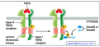
What conformational change do receptor tyrosine kinases undergo when a ligand binds?

A
  • Ligand (dimer or multimer) binding causes receptors to dimerise (unlike G protein coupled receptors)
  • Dimerisation causes cross phosphorylation of each receptor (autophosphorylation)
18
Q

What does the phosphorylated receptor tyrosine kinase bind?

A
  • The phosphorylated (activated) receptor binds other intracellular proteins via phospho-tyrosines
  • Enzymes such as phospholipase Cgamma, phosphatidylinositol-3’-kinase, Src
  • Docking proteins such as Grb2 which act as intermediary for enzyme to bind
19
Q

What are Src Homology domains?

A
  • Binding proteins have homologous phsopho-tyrosin binding domains
  • SH2 binds activated phospho-tyrosines on receptor
  • SH3 binds domains in other intracellular proteins
  • Where that protein sits depends on its folding but once folded the SH3 domain interact with phosphotyrosine
20
Q

What is the SH2 domain of the binding protein for receptor tyrosine kinase?

A
  • it has two binding pockets, one for phosphotyrosine and one for amino acid side chain which is usually adjacent to phosphorylated tyrosine
  • Phosphotyrosine same shape no matter what
  • Plug and socket
21
Q

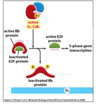
How is Ras activated from receptor tyrosine kinase signalling?

A
  1. Activated RTK binds SH2 domain of Grb-2
  2. Grb-2 = docking protein (via SH3 domain) for guanine nucleotide exchange factors (GEFs) – eg Sos.
  3. SOS (GEF) activates Ras by exchange of GDP for GTP
  4. Ras now binds GTP and activates molecules downstream
22
Q

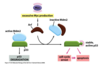
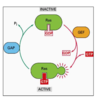
How does Ras function as an ON/OFF switch?

A
  • Superfamily of monomeric GTPases such as Ran, Rab
  • In the inactive state it binds GDP
  • Activated by guanine exchange factors (GEFs) such as sos, where it exchanges GDP for GTP
  • GTPase activating proteins (GAPs) result in increased hydrolysis of GTP
  • Hyperactive Ras mutants are resistant to GAP which leads to cancer
23
Q

Is the receptor tyrosine kinase and Ras active indefinitely?

A
  • No receptor tyrosine kinase and Ras are active only for very short periods
  • Action of phosphatases on receptors and GAPs (GTPase activating proteins) on Ras
  • Therefore need signalling pathway to rapidly propagate these signals for proliferation and differentiation
24
Q

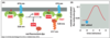
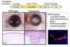
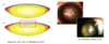
What is happening in this experiment?

A
  • This experiment shows how short the activation of Ras is
  • Ras is manipulated genetically and expressed in cells
  • Linked GTP to a red fluorescent dye. When red comes in close contact with yellow there is resonance energy transfer
  • When Ras binds GTP two fluoro molecules come close and you can measure the fluorescence coming off the red GTP
  • Ras only active for 4-5 minutes
25
What cascade does Ras activate?
* Ras activates downstream Ser/Thr kinase cascade (MAP kinase) * Serine/threonine-PO4 longer lived than tyrosine-PO4 * MAPK (Erk) has both Thr and Tyr which ensure activation is specific, only by MAPKK (Mek) * Acive Erk enters the nucleus and activates multiple gene regulatory proteins (eg G1 cyclins) * MAPKK (Mek) is activated by MAPKKK (Raf)
26
How does the MAPK pathway stimulate proliferation?
* Ras activates the MAPK cascade * MAPK (Erk) leads to expression of immediate early response genes such as Myc, Fos, Jun * Myc activates the cell cycle through various mechanisms * Immediate early response genes are transcribed very quickly and in the absence of protein synthesis
27
What does Myc do from the MAPK pathway?
* Myc activates expression of delayed response genes including cyclin proteins that act in G1 of cell cycles * D cyclins bind and activate cdk proteins (G1 phase cdk)
28
What does active G1-cdk go on to do in the MAPK pathway?
* Actuve cyclin/cdk complex phosphorylates and inactivated Rb protein * Rb normally binds and keeps E2F protein inactive * Phospho-RB dissociates from E2F * Active E2F activates transcription of cell cycle genes (S phase cyclins) * Rb tumour suppressor gene
29
What are the feedback loops in the MAPK pathway?
* S phase cyclins cause DNA synthesis and entry into S phase * feedback loops in place so that Rb remains phosphorylated * E2F positive feedback onto itself
30
What happens during cell cycle arrest to the G1/S-Cdk?
* Active p53 activates transcription of a CDKI (p21) * p21 binds and inactivates the G1 Cdk/cyclin complex so there is no progression into S phase * Failure to correct DNA damage results in accumulation of mutations (cancer)
31
What happens if cell cycle is overactivated?
* Hyperactivated Ras may cause excessive Myc * Myc duplications cause excessive Myc * Cells often display cell cycle arrest or death * Excess myc activity leads to increased arf expression * Arf inactivates the mdm/p53 complex leading to cell cycle arrest or cell death
32
What is the superfamily for receptor serine/threonine kinases?
33
Are the receptor serine/threonine kinases always active?
* Ligands are often inactive/latent, pro-peptide, binding proteins, ECM * Activated by acidic conditions, inflammation, enzymes (MMPs), integrins
34
What are the roles of receptor serine/threonine kinases in the TGFB superfamily?
* Important regulators of cell processes in development and adult tissues * Pattern formation in the embryo * Tissue specification * Extracellular matrix production * Wound healing, fibrosis * Cell death, anti-proliferative/ proliferative (tissue type dependent) * Epithelial-mesenchymal transition (EMT).
35
What are the two major types of receptor serine/threonine kinases?
* Type I receptors (ALK1-8) * Type II receptors - specific for each type of ligand (Act RIIA, ActRIIB, TBRIIs, BMPRII) but can be promiscuous with type I receptors * Ligand induces tetramerisation where type II phosphorylates Type 1 * Smad proteins recruited to phosphorylated type I receptor
36
What are the 4 different types of smads?
* Smads are transcription factors that illicit the responses * **Receptor smads** include smads 2,3 which mediate activin/TGFB signals and smads 1,5,8 which mediate BMP singlas * **Common smad** includes smads 4 which binds Receptor-smads and activates transcription - SBE * **Inhibitory smad** included smads 6,7 which inhibits Receptor-smads from being phosphorylated or the trimeric complex from going in to the nucleus
37
What happens when TGFbeta receptors are activated?
* Activated TGFB receptors are endocytosed via clathrin coated pits (receptor mediated endocytosis) * Most of the signalling occurs in early endosomes (SARA protein facilitates Smad docking and phosphorylation)
38
How does TGFB induce EMT?
* Represses epithelial genes (Eg. Adherens junction proteins) * Activates mesenchymal genes (Eg. ECM proteins, Intermediate filaments)
39
What happens to cells in epithelial mesenchymal transition?
40
What is shown in this experiment?
* Constituitionally active human TGFB1 cDNA does not have the propeotide so it is always active * Trans gene inserted into early embryo of mice * The pink line is the basal membrane * In trans gene lots of ECM produced, huge proliferation and change in phenotype * Change phenotype of epithelial cells by regulating TGFB signalling
41
How is EMT a feature of human cataracts?
* Anterior subcapsular cataract is a plaque of transformed epithelial cells which have excessive abnormal extracellular matrix (collagen I/III, fibronectin) * Myofibroblasts have loss of epithelial characteristics * Mesenchymal markers have alpha-smooth muscle actin * Associated with inflammatory diseases of the eye and physical damage with high incidence in korea
42
What are ion linked channel receptors?
* Ionotropic * Where ligand binds directly to ion channel receptor, no second messengers
43
44
What is the structure of G proteins?
* They have three subunits alpha beta gamma, of which alpha and gamma are membrane tethered * Various types of G protein specific for groups of GPCR * Inactive state has alpha subunit GDP bound and active state alpha subunit GTP bound
45
How does the G protein conformation change when the ligand binds?
* Ligand binding gives conformational change in GPCR so G protein can bind GPCR * GPCR acts as a guanine exchange factor (GEF) * Active GPCR causes release of GDP and binding of GTP * GTP causes conformational change in G protein and activates alpha subunit and the beta,gamma subunit
46
What happen in G protein signalling when the alpha subunit is activated?
* Once activated the alpha subunit (+GTP) binds to a target protein and activates it * The alpha subunit is alpha GTPase which causes hydrolysis of GTP to GDP * GTPase activity of alpha subunit is enhanced by binding to target or RGS proteins (Regulator of G protein signalling or GAP protein for Ras inactivation?
47
How does the G protein get switch off?
* GTP-GDP hydrolysis causes dissociation of alpha subunit from the target protein - switch OFF * Hydrolysis of GTP to GDP inactivates the alpha subunit and it reforms inactive G protein with beta,gamma subunits
48
How does G-protein-receptor signalling target cAMP?
* Common target for G proteins is adenylyl cyclase which catalyses ATP to cAMP * Cyclic AMP has many targets and affects many processes * cAMP binds PKA regulatory subunits * PKA dissociates from regulatory subunits and becomes activated
49
What is an example of a toxin targetting cAMP production?
Cholera toxin overactives G protein which affects the Cl- channel and causes diarrhoea
50
In G protein receptor signalling what does active PKA go on to do?
Active PKA enters nucleus and activates gene transcription by phosphorylating a transcription factor (CREB), which binds associated binding protein (CBP)
51
52
How does the beta,gamma subunit of the G protein cause signaling?
* Activated beta,gamma subunits can also bind to a target protein and activate them * In the heart muscle acetylcholine can bind G-protein linked receptor. Activated beta,gamma subunit binds to a K+ channel and opens it * Loss of K+ ions out of cell decreases contraction and decreases the heart rate
53
What sort of rapid signalling pathways are G-protein linked receptors involved in?
* Smell receptors activated by food and stimulation of saliva * Adrenaline stimulation of heart rate * Fastest is the response of photoreceptors to light (~20 ms) * Achieved by rhodopsin (G- protein coupled light receptor) – affects cGMP-gated Na+ channel
54
What is the pathway for phototransduction in rods?
1. Rhodopsin (G coupled light receptor) is linked to cis-retinal 2. Light-induced isomerisation of cis-retinal (cis to trans) causes conformational change in rhodopsin 3. Transducin (G-protein) alpha subunit activates cGMP phosphodiesterase 4. Drop in cGMP closes cGMP gated Na+ channels causing hyperpolarisation 5. Hyperpolarisation causes calcium channels to close and low calcium reduces glutamate release
55
How is the light signal switched off in phototransduction?
1. RGS (GAP) protein binds to transducin (G protein) which hydrolyses GTP to GDP 2. Rhodopsin kinase phosphorylates rhodopsin which inhibits rhodopsin activation 3. Arrestin binds phospho-rhodopsin and further inhibits activity 4. Low calcium stimulates Gulanylate cyclase which stimulates cGMP production
56
How are bipolar cells in the retina excited or inactivated?
* In the dark photoreceptors release glutamate to inhibit ON bipolar cells and excite OFF bipolar cells * In the light, photoreceptor hyperpolarisation stops inhibition of ON and inactivates OFF bipolar cells * Activated bipolar cells synapse and transmit signals to ganglion cells and then to the brain.
57
58
Where does the Wnt ligand come from?
* 19 different Wnt ligands identified in mammals * Highly glycosylated * Originally identified in Drosophila (Wingless, Wg). * Int gene found in mice – common integration site for mammary tumour virus (MMV). * **Wg + Int = Wnt**
59
What are the 3 different pathways that Wnt proteins can activate?
**Canonical:** * Wnt/B-catenin – relies on regulating degradation of B-catenin protein **Non-canonical:** * Ca2+ pathway * Planar cell polarity (PCP) pathway – Rho GTPases, PCP proteins
60
Where is B-catenin released to at adherens junctions?
* Adherens Junctions in epithelial cells are dynamic * AJ reassembly results in B- catenin release in cytoplasm * Cell needs to recycle OR get rid of cytoplasmic B-catenin (Proteolysis)
61
What does the B-catenin destruction complex comprise of?
* Complex of cytoplasmic complex of proteins target B- catenin for ubiquitylation and degradation by proteasomal enzymes * Includes Axin, glycogen synthase Kinase 3B (GSK3B), Casein Kinase 1 (CK1), Adenomatous polyposis coli (APC) * CK1 and GSK3B phosphorylate B-catenin on phospho Ser/Thr residues, which are targets for E3 ubiquitin ligase complex
62
What is the Wnt/B-catenin signalling pathway with and without Wnt signal?
63
How does B catenin control stem cell differentiation in hair follicles?
* Knockout of B-Catenin inactivates Wnt pathway. * Inactivation of Wnt pathway in the hair follicle cells of skin inhibits stem cell adopting a follicle fate but adopt an epidermal fate instead. * No hair follicles form (loss of stem cell proliferation)
64
How does this show that Wnt signal controls lens epithelial cell fate?
* When you knockout B catenin those stem cells cannot replciate and do not produce more epithelial cells * If you knockout APC10 there is no longer a funcitonal complex that can degrade B catenin so B catenin levels rise, increasing expression of myc and producing lots of proliferating cells * CatX10 is where B-catenin is mutated so that it cannot be phosphorylated * Remove one exon which contained all the phosphorylation sites, still produces functional b catenin that functions in adhesion junctions but when it gets into destruction complex it can’t be phosphorylated * Protein of B catenin can’t be phosphorylated or degraded
65
66
How does over active Wnt cause proliferation?
* APC is a tumour suppressor which controls cell cycle by controlling beta-catenin * Mutations in APc or Catnnb (B-catenin) cause activation of Wnt pathway * Wnt signals regualte Myc and cyclin D expression causing G1/S phase transition * Wnt pathways mutations cause over proliferation
67
How does overactive Wnt lead to cell death?
* In lenses with mutations of the wnt pathway there is increased cell death * Activation of p53 means cellc cannot arrest the cell cycle because of myc * p53 causes apoptosis
68
How are mutations in the Wnt-pathway (APC) associated with colon polyps?
* LOH of Apc or oncogenic B-catenin causes increased proliferation (stem cells) * Failure to differentiate (fate switch) to form polyps * At this stage still not malignate cancer need the other hallmarks of cancer
69