Thoracic Wall and Lungs Picture Cards Flashcards
Name the landmarks on this thoracic vertebrae.

- Vertebral foramen
- Body
- Superior costal facet
- Pedicle
- Transverse costal facet
- Lamina
- Spinous process
- Superior articular facet
- Superior vertebral notch

Name the landmarks on this thoracic vertebrae.

- Body
- Superior costal facet
- Superior articular process and facet
- Pedicle
- Transverse costal facet
- Transverse process
- Inferior articular process
- Spinous process
- Inferior vertebral notch
- Inferior costal facet


Name which structures are auscultated at each point and also describe in words the position of each.

- Aortic valve - this is the second intercostal space just right of the sternum.
- Pulmonic valve - this is the second intercostal space just left of the sternum.
- Tricuspid valve - this is the 5th intercostal space just left of the sternum.
- Mitral valve - 5th intercostal space at the midclavicular line.

What are these two anatomical lines called (in red) and what is the part of the ribs called marked by the arrows?

Scapular line on the left
Paravertebral line on the right
Angle of the ribs
What is depicted by the red dotted line?

The diaphragm.
Name these prominences/landmarks on the rib and what they do.

- Shaft
- Angle - where the rib angulates anteriorly
- Neck
- Head
- Superior facet - articulates with inferior costal facet on the body at a superior level.
- Crest
- Inferior facet - articulates with superior costal facet on the body of vertebrae at the same level.
- Articular facet for transverse process of vertebrae.
- Tubercle - forms joint with the transverse process of vertebrae.
- Costal groove

Which rib is this? Name the landmarks.

It is the 1st rib.
- Anterior scalene muscle attachment
- Groove for subclavian vein
- Groove for subclavian artery
- Middle scalene muscle attachment

What muscles originate/insert at these locations?

- Serratus anterior 2nd digitation (origin)
- Posterior scalene muscle insertion

Name these ligaments

- Costovertebral ligaments
- Costotransverse ligaments
Name these conditions.

Left: Pectus excavatum (funnel chest)
Middle: Pectus carinatum (pigeon chest)
Right: Kyphoscoliosis (a hump with lateral deviation of the thoracic vertebrae)
Name the muscle, innervation, and action. In what direction do their fibers point?
External intercostal muscles. Innervated by the intercostal nerves. Action is to aid in INSPIRATION by increasing the CIRCUMFERENCE of the thoracic cavity (the ribs are raised). The fibers travel anteroinferiorly from the upper rib to the rib below.
Name the muscle, innervation, and action.

Internal intercostal muscles. Innervated by intercostal nerves. Action is to aid in EXPIRATION by decreasing intrathoracic volume.

- Scalenes
- Sternocleidomastoid muscle
- External obliques
- Internal obliques
- Transversus abdominis muscle
- Rectus abdominis muscle
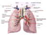
Name the lymphatic elements

- Paratracheal nodes
- Superior tracheobronchial nodes
- Inferior tracheobronchial nodes
- Thoracic duct
- Bronchomediastinal (hilar) nodes
- Pulmonary nodes
Name the nerves.

- Right vagus nerve
- Left vagus nerve
- Right sympathetic chain
- Left sympathetic chain
- Right phrenic nerve
- Left phrenic nerve

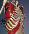
Name the things as indicated by the yellow and black arrows.

Yellow: Internal thoracic artery
Black: Transverse thoracis muscles

What is the significance of the area circled in green?

The green circled area is the “bare area”, where a pericardiocentesis can be safely performed. This is the spot where the pulmonary pleura has been pushed out of the way, and will not be affected by a puncture.

What are the two areas indicated by blue numbers?

- Costomediastinal recess
- Costodiaphramatic recess


Azygos vein

Brachiocephalic vein

ACCESSORY hemiazygos vein (not the same as hemiazygous vein - that is the inferior part)

Hemiazygos vein

Posterior intercostal veins
Name this and describe what it is.

The lingula of the left lung. Considered to be equivalent to the middle lobe of the right lung, the lingula is occasionally separated from the rest of the superior lobe by a fissure.


Fill in the blanks and what not.


- T2
- T1 (medial forearm)
What happened?

Pleural effusion - fluid accumulation in the costodiaphragmatic recess with blunting seen on CXR
What is that called?

Carina