Skin Flashcards

1
Q

which organ is the heaviest comprising 16% of body weight?

A

skin

2
Q

what are the three layers of skin?

A

epidermis, dermis and subcutaneous tissues

3
Q

what are the functions of skin?

A
  1. homeostasis
  2. provides boundaries for body fluids, protecting underlying tissues from microorganisms, harmful substances, and radiation
  3. Modulates body temperature and synthesizes vitamin D.
4
Q

Thin and devoid of blood vessels and divided into outer horny layer and inner cellular layer. Depends on dermis for nutrition.

A

epidermis

5
Q

Supplied with blood; Contains connective tissue, sebaceous glands, sweat glands, and hair follicles

A

dermis

6
Q

Fat layer

A

Hypodermis/Subcutaneous tissues

7
Q

name the four pigments of skin

A

melanin, carotene, oxy and deoxyhemoglobin

8
Q

the brownish pigment of the skin, genetically determined, increased by sunlight.

A

melanin

9
Q

a golden yellow pigment that exists in subcutaneous fat and in heavily keratinized areas like palms and soles.

A

carotene

10
Q

a bright red pigment, predominates in arteries and capillaries

A

oxyhemoglobin

11
Q

darker somewhat bluer pigment from oxyhemoglobin losing its oxygen to the tissues. Increase causes cyanosis.

A

deoxyhemoglobin

12
Q

name the two types of hair

A

vellus and terminal

13
Q

short fine, inconspicuous, and relatively unpigmented. (arms)

A

vellus

14
Q

coarser, thicker, more conspicuous, and usually pigmented (ex. Scalp hair and eyebrows).

A

terminal

15
Q

what is the function of nails?

A

Protects the distal ends of the fingers and toes.

16
Q

where do fingernails get their pinkish color?

A

underlying vascular nail bed

17
Q

be able to identify the following: Lunula (whitish moon), proximal nail fold, cuticle, lateral nail fold.

A
18
Q

at what rate do fingernails grow?

A

Fingernails grow 0.1 mm daily. Toenails grow more slowly.

19
Q

what are the two basic types of glands?

A

sebaceous and sweat

20
Q

what is the purpose of sebaceous glands?

A

produce a fatty substance that is secreted onto the skin surface through the hair follicles

21
Q

where are sebaceous glands located?

A

Present on all skin surfaces except palms and soles

22
Q

name the two types of sweat glands

A

eccrine and apocrine

23
Q

______ glands: widely distributed and open directly onto the skin surface. Control body temperature.

A

eccrine

24
Q

________ glands: found chiefly in axillary and genital regions, open onto hair follicles, and stimulated by emotional stress.

A

apocrine

25
Q

what are the ABCDE of screening moles for melanoma?

A

A for Asymmetry

B for irregular borders, especially ragged, notched, or blurred

C for variation or change in color, especially blue or black

D for diameter > or equal to 6 mm or different from others, especially if changing, itching, or bleeding

E for elevation or enlargement

26
Q

what is another term for a mole?

A

nevus

27
Q

is this likely benign or malignant?

A

malignant

28
Q

what are the 6 things you look for in a skin exam?

A
  1. color
  2. moisture
  3. temperature
  4. texture
  5. lesions
  6. mobility/turgor
29
Q

what is a Café-Au-Lait Spot?

A

-A slightly but uniformly pigmented macule or patch with a somewhat irregular border -Usually 0.5-1.5 cm in diameter

30
Q

6 or more cafe-au-lait spots each with a diameter > 1.5 cm suggests …….

A

neurofibromatosis

31
Q

what is this and example of?

A

Café-Au-Lait Spot

32
Q

what is vitiligo?

A

Depigmented macules appear on the face, hands, feet, extensor surfaces.

33
Q

what is cyanosis?

A

bluish discoloration of the skin as a result of poor perfusion

34
Q

what is jaundice and where can it be seen?

A

yellowing skin -Seen most easily and reliably in the sclera. -May also be seen in mucous membranes.

35
Q

what can cause jaundice?

A

Causes include: liver disease and hemolysis of red blood cells.

36
Q

what is Erythema and what is another name for it?

A

Red hue, increased blood flow, seen here as the “slapped cheeks” of “Fifth Disease”

37
Q

what is psoriasis?

A

An immune-mediated disease that affects the skin. It occurs when the immune system mistakes the skin cells as a pathogen, and sends out faulty signals that speed up the growth cycle of skin cells.

38
Q

how and where does psoriasis present?

A

Presents as silvery scaly lesions. Mainly on extensor surfaces. (Associated with strokes.)

39
Q

what is eczema and what are the symptoms?

A

An allergic disease associated with asthma. -Include dryness and recurring skin rashes that are characterized by redness, skin edema (swelling), itching and dryness, crusting, flaking, blistering, cracking, oozing, or bleeding.

40
Q

what are the characteristics of a Lupus-Malar Rash?

A

-red or purplish and mildly scaly rash in the shape of a butterfly across the face -Spares the nasolabial folds of the face -Macular with sharp edges and not itchy. present in approximately 46–65% of lupus sufferers

41
Q

what are plaques?

A

A patch of closely grouped papules more than 2/5 in (1 cm) across is called a plaque.

42
Q

what are papules?

A

A solid, raised lesion less than 2/5 in (1 cm) across.

43
Q

what are some examples of papules?

A

Seen in warts, syphilis, psoriasis, seborrheic and actinic keratoses, lichen planus, and skin cancer.

44
Q

what is a nodule?

A

A solid lesion (> 0.5 cm) that has distinct edges and that is usually more deeply rooted than a papule.

45
Q

what are some examples of nodules?

A

Nodules are associated with: keratinous cysts, lipomas, fibromas, and some types of lymphomas

46
Q

what is a cyst?

A

Nodule filled with expressible material, either liquid or semisolid.

47
Q

what is a wheal?

A

A skin elevation caused by swelling that can be itchy and usually disappears soon after erupting.

48
Q

what is angioedema?

A

A vascular reaction involving the deep dermis or subcutaneous or submucosal tissues, representing localized edema caused by dilatation and increased permeability of the capillaries, and characterized by the development of giant wheals.

49
Q

what are vesicles?

A

A raised lesion less than 1/5 in (5 mm) across and filled with a clear fluid

50
Q

what is a bulla?

A

Vesicles that are more than 1/5 in (5 mm) across are called bullae or blisters.

51
Q

what are are two forms of a life-threatening skin conditions, in which cell death causes the epidermis to separate from the dermis?

A

Stevens–Johnson syndrome (SJS) and Toxic epidermal necrolysis (TEN)

52
Q

what is Erythema Multiforme

A

Thought to be a hypersensitivity complex that affects the skin and the mucous membranes. The majority of cases are idiopathic or medications, followed by infections and, rarely, cancers. (SJS and TEN)

53
Q

what are pustules?

A

A raised lesion filled with pus. A pustule is usually the result of an infection, acne, imptigo, or boils (abscesses).

54
Q

what are Burrows-Scabies?

A

A minute, slightly raised tunnel in the epidermis, commonly found on finger webs and on the sides of fingers. Appears as short (5-15 mm) linear or curved lines ending in a tiny vessicle

55
Q

A dry, horny build-up of dead skin cells that often flakes off the surface of the skin.

A

scale

56
Q

Diseases that promote scale include ….

A

fungal infections, psoriasis, and seborrheic dermatitis.

57
Q

what is crust? (i’m not referring to pies)

A

A dried collection of blood, serum, or pus. Also called a scab, a crust is often part of the normal healing process of many infectious lesions.

58
Q

Rough, thick epidermis with exaggerated skin lines. This is often a characteristic of scratch dermatitis and atopic dermatitis.

A

lichenification

59
Q

Discolored, fibrous tissue that permanently replaces normal skin after destruction of the dermis.

A

scars

60
Q

Hypertrophic scarring that extends beyond the borders of the initiating injury.

A

keloid

61
Q

what is erosion? (not talking about geography)

A

Nonscarring loss of superficial epidermis. Surface is moist but does not bleed Ex. Chickenpox (after rupture of a vesicle).

62
Q

Linear or punctate erosions caused by scratching or picking at a primary lesion.

A

excoriation

63
Q

what is a fissure?

A

A Linear crack in the skin, often resulting from excessive dryness. Ex. Athlete’s Foot

64
Q

what is an ulcer?

A

A deeper loss of epidermis and dermis; may bleed and scar.

65
Q

Stage 1 of Pressure Ulcers

A

Stage 1: Skin is not broken but is red or discolored.

66
Q

Stage 2 of Pressure Ulcers

A

Stage 2: The epidermis is broken, creating a shallow open sore.

67
Q

Stage 3 of Pressure Ulcers

A

The break in the skin extends through the dermis into the subcutaneous and fat tissue.

68
Q

Stage 4 of Pressure Ulcers

A

The breakdown extends into the muscle and can extend to the bone. Usually lots of dead tissue and drainage are present

69
Q

what is cellulitis?

A

Localized or diffuse inflammation of connective tissue with severe inflammation of dermal and subcutaneous layers of the skin.

70
Q

what are some causes of cellulitis?

A

Cellulitis can be caused by normal skin flora or by exogenous bacteria, and often occurs where the skin has previously been broken. Ex. Cracks, blister, insect bites, and surgical wounds. Common in diabetics and immunocompromised.

71
Q

what are three types of vascular lesions?

A

cherry angioma, spider angioma, spider veins

72
Q

what is a spider angioma?

A

Fiery red, up to 2 cm Central body surrounded by erythema and radiating legs Found on face, neck, arms, and upper trunk associated with liver disease, pregnancy, and vitamin B def.

73
Q

what are spider veins?

A

Bluish, variable size resemble a spider or can be linear, irregular, or cascading found on legs near veins or anterior chest accompanies increased pressure in the superficial veins, as in vericose veins.

74
Q

what is a cherry angioma?

A

Bright or ruby red 1-3 mm Round, flat, sometimes raised Found on trunk or extremities increase size and numbers with aging.

75
Q

what is Petechia/Purpura?

A

Deep red or redish purple Petchia (1-3 mm) Purpura larger Rounded, sometimes irregular; flat Distributed variably suggests blood outide the vessels; may suggest a bleeding disorder or if petechia, emboli to skin; palpable purpura in vasculitis

76
Q

what is echmosis?

A

purple or purplish blue, fades to green, yellow, and brown with time. Variable size, larger than petechia > 3mm Rounded, oval, or irregular; may have central subcutaneous flat nodule (a hematoma) Variable distribution Suggests blood outside the vessels; secondary to bruising or trauma, also seen in bleeding disorders.

77
Q

A small linear subungual hemorrhage which is red when fresh and brown when aged, located at the distal 1⁄3 of the nail bed, classically associated with mitral stenosis

A

splinter hemorrages

78
Q

what is clubbing? (not like night clubs)

A

Build-up of tissue in the fingers, causing the end of the fingers to become enlarged and the nails to curve downward. Results in the fingers appearing bulb-like, or like an upside down spoon.

79
Q

clubbing is a comorbidity of what?

A

Seen most commonly with diseases of the heart and lungs that result in less oxygen in the blood.

80
Q

what is a basal cell carcinoma?

A

Grows slowly and seldom metastasizes Most common in fair-skinned adults over age 40 and usually appears on the face. Initial translucent noduledepressed center and firm elevated border

81
Q

what is a squamous cell carcinoma?

A

usually appears on sun exposed skin of fair-skinned adults > 60 yrs age. Grows more quickly than a BCC, is firmer, and looks redder. Face and back of hands often affected.

82
Q

characteristics of AIDS-Kaposi’s Sarcoma

A

KS lesions are nodules or blotches that may be red, purple, brown, or black, and are usually papular. They are typically found on the skin, but spread elsewhere is common, especially the mouth, gastrointestinal tract and respiratory tract. Growth can range from very slow to explosively fast, and is associated with significant mortality and morbidity.

83
Q

The least serious burns are those in which only the outer layer of skin is burned, but not all the way through.

A

1st degree burn

84
Q

When the first layer of skin has been burned through and the second layer of skin (dermis) also is burned, the injury is called a

A

2nd degree burn

85
Q

The most serious burns involve all layers of the skin and cause permanent tissue damage. Fat, muscle and even bone may be affected. Areas may be charred black or appear dry and white. Difficulty inhaling and exhaling, carbon monoxide poisoning, or other toxic effects may occur if smoke inhalation accompanies the burn.

A

3rd degree burn

86
Q

what is commonly known as flesh-eating disease or flesh-eating bacteria syndrome.

A

necrotizing fasciitis

87
Q

what is necrotizing fasciitis and how is it treated?

A

rare infection of the deeper layers of skin and subcutaneous tissues, easily spreading across the fascial plane within the subcutaneous tissue. quickly progressing, having greater risk of developing in the immunocompromised due to conditions like diabetes & cancer. Treatment: IV antibiotics/Amputation

88
Q

what is this an example of?

A

angioedema

89
Q

what is this an example of?

A

bulla

90
Q

what is this an example of?

A

erythema

91
Q

what is this an example of?

A

Herpes Simplex 1

92
Q

what is this an example of?

A

keloid

93
Q

what is this an example of?

A

lupus-malar rash

94
Q

what is this an example of?

A

plaques

95
Q

what is this an example of?

A

psoriasis

96
Q

what is this an example of and in what special regions or zones does it normally reside?
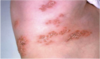
A

shingles

deratomes

97
Q

what is this an example of?

A

Toxic Epidermal Necrolysis (TEN)

98
Q

what is this an example of?

A

vitiligo

99
Q

what is this an example of?

A

wheal

100
Q

What is this and what was most likely the cause?

A

ulcer

Stasis ulcer of venous insufficiency, syphilitic chancre

101
Q

extra credit: is it possible for a patient to appear cyanotic with a low hemoglobin? why/why not?

A

no, in order for the blue pigmentation to appear, the patient must have a high enough hemoglobin. If there aren’t enough cells, then the pigmentation won’t be visible