Bones Flashcards

1
Q

Bones

A

Bone is specialised a type of connective tissue. It has a unique histological appearance, which enables it to carry out its numerous functions:

Haematopoeisis – the formation of blood cells from haematopoietic stem cells found in the bone marrow.

Lipid and mineral storage – bone is a reservoir holding adipose tissue within the bone marrow and calcium within the hydroxyapatite crystals.

Support – bones form the framework and shape of the body.

Protection – especially the axial skeleton which surrounds the major organs of the body.

2
Q

Types of Bone Cells

A

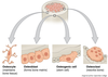
Osteoblasts – Synthesises uncalcified/unmineralised ECM called osteoid. This will later become calcified/mineralised to become bone.

Osteocytes – As the osteoid mineralises, the osteoblasts become entombed between lamellae in lacunae where they mature into osteocytes. They then monitor the minerals and proteins to regulate bone mass.

Osteoclasts – Derived from monocytes and resorb bone by releasing H+ ions and lysosomal enzymes. They are large and multinucleated cells.

3
Q

Structure of Bone

A

Under the microscope, bone can be divided into two types:

Woven bone (primary bone) – Appears in embryonic development and fracture repair, as it can be laid down rapidly. It consists of osteoid (unmineralised ECM), with the collagen fibres arranged randomly. It is a temporary structure, soon replaced by mature lamellar bone.

Lamellar bone (secondary bone) – The bone of the adult skeleton. It consist of highly organised sheets of mineralised osteoid. This organised structure makes it much stronger than woven bone. Lamella bone itself can be divided into two types – compact and spongy.

In both types of bone, the external surface is covered by a layer of connective tissue, known as the periosteum. A similar layer, the endosteum lines the cavities within bone (such as the medullary canal, Volkmann’s canal and spongy bone spaces).

Lamellar bone can be divided into two types. The outer is known as compact bone – this is dense and rigid. The inner layers of bone are marked by many interconnecting cavities, and is called spongy bone.

4
Q

Compact Bone

A

Compact bone forms the outer ‘shell’ of bone. In this type of bone, the lamellae are organised into concentric circles, which surround a vertical Haversian canal (which transmits small neurovascular and lymphatic vessels). This entire structure is called an osteon, and is the functional unit of bone.

The Haversian canals are connected by horizontal Volkmann’s canals – these contain small vessels that anastamose (join together) the arteries of the Haverisan canals.

Osteocytes are located between the lamellae, within small cavities (known as lacunae). The lacunae are interconnected by a series of interconnecting tunnels, called canaliculi.

5
Q

Spongy Bone

A

Spongy bone makes up the interior of most bones, and is located deep to the compact bone. It contains many large spaces – this gives it a honeycombed appearance.

The bony matrix consists of a 3D network of fine columns, which crosslink to form irregulartrabeculae. This produces a light, porous bone, that is strong against multidirectional lines of force. The lightness afforded to spongy bone is crucial in allowing the body to move. If the only type of bone was compact, they would be too heavy to mobilise.

The spaces between trabeculae are often filled with bone marrow. Yellow bone marrow refers to adipocytes and red bone marrow consists of haematopoietic stem cells.

This type of bone does not contain any Volkmann’s or Haversian canals.

6
Q

Embryonic Development of Bones

A

Ossification is the term for the formation of bone. There are two ways that bone can ossify during embryonic development. Each way has its own name: intramembranous ossification and endochondral ossification. Because of these two ways that bone can develop, two types of bone can be described to exist based on the way they developed embryonically: intramembranous bones and endochondral bones. The intramembranous bones are always flat bones, but the endochondral bones include the long, short, and irregular bones.

7
Q

Growth of Bones

A

Long bones lengthen at the epiphyseal plate with the addition of bone tissue and increase in width by a process called appositional growth.

The epiphyseal plate, the area of growth composed of four zones, is where cartilage is formed on the epiphyseal side while cartilage is ossified on the diaphyseal side, thereby lengthening the bone.

Each of the four zones has a role in the proliferation, maturation, and calcification of bone cells that are added to the diaphysis.

The longitudinal growth of long bones continues until early adulthood at which time the chondrocytes in the epiphyseal plate stop proliferating and the epiphyseal plate transforms into the epiphyseal line as bone replaces the cartilage.

Bones can increase in diameter even after longitudinal growth has stopped.

Appositional growth is the process by which old bone that lines the medullary cavity is reabsorbed and new bone tissue is grown beneath the periosteum, increasing bone diameter.

8
Q

Vertebral Column

A

The most important functions of the vertebral column are as follows:

Protection: it encloses the spinal cord, shielding it from damage.

Support: it carries the weight of the body above the pelvis (below the pelvis, the lower limbs take over).

Axis: the vertebral column forms the central axis of the body.

Movement: it has roles in both posture and movement.

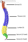
The vertebral column can be separated into five different regions. Each region is characterised by a different vertebral structure. Before looking, at individual structures, we first need to look at the general form of a vertebra.

9
Q

Vertebral Structure

A

Although vertebrae do have significant differences in size and shape between groups, they have the same basic structure. Each vertebrae consists of avertebral body, situated anteriorly, and a posterior vertebral arch.

Pedicles: There are two of these, one left and one right. They point posteriorly, meeting the flatter laminae.
Lamina: The bone between the transverse and spinal processes.
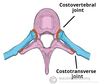
Transverse processes: These extend laterally and posteriorly away from the pedicles. In the thoracic vertebrae, the transverse processes articulate with the ribs.
Articular processes: At the junction of the lamina and the pedicles, superior and inferior processes arise. These articulate with the articular processes of the vertebrae above and below.
Spinous processes: Posterior and inferior projection of bone, a site of attachment for muscles and ligaments.

10
Q

Cervical Vertebrae

A

There are seven cervical vertebrae in the human body. They have three main distinguishing features:

The spinous process bifurcates into two parts, and so is known as a bifid spinous process.
There are two transverse foramina, one in each transverse process. These conduct the vertebral arteries.
The vertebral foramen is triangular in shape

There are some cervical vertebrae that are unique. C1 and C2 (called the atlas and axis respectively), are specialised to allow for the movement of the head.

The C7 vertebrae has a much longer spinous process, which does not bifurcate.

11
Q

Thoracic Vertebrae

A

The twelve thoracic vertebrae are medium sized, and increase in size as they move down the back. Their main function is to articulate with ribs, producing the bony thorax.

Each thoracic vertebrae has two ‘demi facets‘ on each side of its vertebral body. These articulate with the head of the respective rib, and the rib inferior to it. On the transverse processes of the thoracic vertebrae there is a costal facet for articulation with its respective rib.

The spinous processes are slanted inferiorly and anteriorly. This offers increased protection to the spinal cord, preventing an object like a knife entering the spinal canal through the intervetebral discs.

In contrast to the cervical vertebrae, the vertebral foramen is circular.

12
Q

Lumbar Vertebrae

A

These are the largest of the vertebrae, of which there are five. They act to support the weight of the upper body, and have various specialisations to enable them do this.

Lumbar vertebrae have very large vertebral bodies, which are kidney shaped. They lack the characteristic features of other vertebrae, with no foramen transversarium, costal facets, or bifid spinous processes.

However, like the cervical vertebral, they have a triangular shaped vertebral foramen.
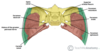
13
Q

Sacrum and Coccyx

A

The sacrum is a collection of five fusedvertebrae. It is described as a upside downtriangle, with the apex pointing inferiorly. On the lateral walls of the sacrum are facets, for articulation with the pelvis at the sacro-iliac joints.

The coccyx is a small bone, which articulates with the apex of the sacrum. It is recognised by its lack of vertebral arches. Due to the lack of vertebral arches, there is no vertebral canal, and so the coccyx does not transmit the spinal cord.
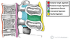
14
Q

Vertebral Joints

A

For every vertebrae, there are five articulations. The vertebral bodies indirectly articulate with each other, and the articular processes also form joints.

The vertebral body joints are cartilaginous joints, designed for weight bearing. The articular surfaces are covered by hyaline cartilage, and are connected by a fibrocartilage intervertebral disk. There are two ligaments that strengthen these joints; the anterior and posteriorlongitudinal ligaments. The anterior longitudinal ligament is thick and prevents hyperextension of the vertebral column. The posterior longitudinal ligament is weaker and prevents hyperflexion.

The joints between the articular facets are called facet joints. These allow for some gliding motions between the vertebrae. They are strengthened by various ligaments:

Ligamentum Flavum: extends from lamina to lamina.
Interspinous and Supraspinous ligaments: These join the spinous processes together. The interspinous ligaments attach between processes, and the supraspinous ligaments attach to the tips.
Intertransverse ligaments: extends between transverse processes.
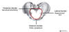
15
Q

Hip Bone

A

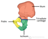
The hip bones have three articulations:

Sacroiliac joint – articulation with sacrum.
Pubic symphysis – articulation with the corresponding hip bone.
Hip joint – articulation with the head of femur.

The hip bone is made up of the three parts – the ilium, pubis and ischium. Prior to puberty, the triradiate cartilage separates these constituents. At the age of 15-17, the three parts begin to fuse.

Their fusion forms a cup-shaped socket known as the acetabulum, which becomes complete at 20-25 years of age. The head of the femurarticulates with the acetabulum to form the hip joint.

16
Q

Ilium

A

The superior part of the hip bone is formed by theilium, the widest and largest of the three parts. The body of the ilium forms the superior part of theacetabulum. Immediately above the acetabulum, the ilium expands to form the wing (or ala).

The wing of the ilium has two surfaces. The inner surface is concave, and known as the iliac fossa, providing origin to the iliacus muscle. The external surface is convex, and provides attachments to the gluteal muscles. Hence it is known as the gluteal surface.

The superior margin of the wing is thickened, forming the iliac crest. It extends from the anterior superior iliac spine to the posterior superior iliac spine.

Muscles attaching to the Ilium: –

Gluteal muscles attach to the external surface of the Ilium at the anterior, posterior and inferior gluteal lines.
The iliacus muscle attaches medially at the iliac fossa.

17
Q

Pubis

A

The pubis is the most anterior portion of the hip bone. It consists of a body and superior and inferior rami (branches).

The body is located medially, articulating with its opposite pubic body, at the pubic symphysis.

The superior rami extends laterally from the body, forming part of the acetabulum. The inferior rami projects towards, and joins the ischium. Together, the two rami enclose part of the obturator foramen, through which the obturator nerve, artery and vein pass through to reach the lower limb.

18
Q

Ischium

A

The posterioinferior part of the hip bone is formed by the ischium. Much like the pubis, it is composed of a body, an inferior and a superior ramus.

The inferior ischial ramus combines with the inferior pubic ramus forming the ischiopubic ramus which encloses part of the obturator foramen. The posterorinferior aspect of the ischium forms the ischial tuberosities and when sitting, it is these tuberosities on which our body weight falls.

On the posterior aspect of the ischium there is an indentation known as the greater sciatic notch, with the ischial spine at its most inferior edge.

The important ligaments attach to the ischium:

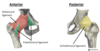
The sacrospinous ligament runs from the the ischial spine to the sacrum, thus creating the greater sciatic foramen through which lower limb neurovasculature (including the sciatic nerve) and the piriformis muscle transends.

The sacrospinous ligament and thesacrotuberous ligament run from the sacrum to the ischial tuberosity, forming the lesser sciatic foramen.

19
Q

Joints

A

A joint is defined as the point at which two or more bones make contact (or articulate). In this article we shall look at the structure and classification of different types of joints.

Joints can be classified by the type of tissue present. Using this method, we can split the joints of the body into fibrous, cartilaginous and synovial joints.

20
Q

Fibrous Joints

A

A fibrous joint is where the bones concerned are bound by fibrous tissue. Fibrous joints can further subclassified into sutures, gomphmoses and syndesmoses.

Sutures

Immovable joint (called a synarthrosis)
Only found between the bones of the skull.
There is limited movement until about 20 years of age, after which they become fixed.

Gomphmoses

Immovable joint (called a synarthrosis)
Where the teeth articulate with their sockets in the maxillae (upper teeth) or the mandible(lower teeth)
The fibrous connection that binds the tooth and socket is the periodontal ligament

Syndesmoses

Slightly movable joins (called an amphiarthrosis)
Usual structure is bones held together by together by an interosseous membrane
Middle radio-ulnar and middle tibiofibular joint are key examples

21
Q

Cartilaginous Joints

A

In cartilaginous joints, the bones are attached by fibrocartilage or hyaline cartilage. They be split into primary and secondary cartilgainous joints.

Synchondroses

Also known as primary cartilaginous joints, they only involve hyaline cartilage.
The joints can be immovable (synarthroses) or slightly movable (amphiarthroses)
The joint between the diaphysis and epiphysis of a growing long bone is asynchondrosis. This is interesting in that it is a temporary joint with no movement.

Symphyses

Also known as a secondary cartilaginous joint, it can involve fibrocartilage or hyaline cartilage
The joints are slightly movable (amphiarthroses)
An example of a symphysis is the pubic symphysis.

22
Q

Synovial Joints

A

A synovial joint is a joint filled with synovial fluid. These joints tend to be fully moveable (known as diarthroses), and are the main type of joint found around the body. They are defined by the arrangement of their articular surfaces, and types of movement they allow.

Hinge

Permits flexion and extension
Elbow joint is a notable example

Saddle

Concave and convex joint surfaces
E.g. Metatarsophalangeal joint
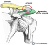
Plane

Permit gliding or sliding movements
E.g. Acromioclavicular joint

Pivot

Allows rotation; a round bony process fits into a bony ligamentous socket
E.g. Atlantoaxial joint & proximal radio-ulnar joint

Condyloid

Permits flexion & extension, adduction, abduction & circumduction
E.g. Metacarpophalangeal joint

Ball & Socket

Permits movement in several axis; a rounded head fits into a concavity
E.g. Glenohumeral Joint.
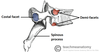
23
Q

Ribs

A

The typical rib consists of a head, neck and body:

The head is wedge shaped, and has two articular facets separated by a wedge of bone. One facet articulates with the numerically corresponding vertebrae, and the other articulates with the vertebrae above.

The neck contains no bony prominences, but simply connects the head with the body. Where the neck meets the body there is a roughed tubercle, with a facet for articulation with the transverse process of the corresponding vertebrae.

The body, or shaft of the rib is flat and curved. The internal surface of the shaft has a groove for the neurovascular supply of the thorax, protecting the vessels and nerves from damage.

24
Q

Atypical Ribs

A

Ribs 1, 2, 11 and 12 can be describes as ‘atypical’ – they have features that are not common to all the ribs.

Rib 1 is shorter and wider than the other ribs. It only has one facet on its head for articulation with its corresponding vertebrae (there isn’t a thoracic vertebrae above it). The superior surface is marked by two grooves, which make way for the subclavian vessels.

Rib 2 is thinner and longer than rib 1, and has two articular facets on the head as normal. It has a roughened area on its upper surface, where the serratus anterior muscle attaches.

Ribs 11 and 12 have no neck, and only contain one facet, which is for articulation with the corresponding vertebrae.

25
Q

Rib Articulations

A

Posterior

All of the twelve ribs articulate posteriorly with thevertebrae of the spine. Each rib forms two joints:

Costotransverse joint – Between the tubercle of the rib, and the transverse costal facet of the corresponding vertebrae.
Costovertebral joint – Between the head of the rib, superior costal facet of the corresponding vertebrae, and the inferior costal facet of the vertebrae above.

Anterior

The anterior attachment of the ribs vary:

Ribs 1-7 attach independently to the sternum.
Ribs 8 – 10 attach the costal cartilages superior to them.
Ribs 11 and 12 do not attach anywhere, and end in the abdominal muscles. Because of this, they are sometimes called ‘floating ribs’.

26
Q

Calvarium and Cranium

A

The cranium (also known as the neurocranium), is formed by the superior aspect of the skull. It encloses and protects the brain, meninges and cerebral vasculature.

Anatomically, the cranium can be subdivided into a roof (known as the calvarium), and a base:

Calvarium: Comprised of the frontal, occipital and two parietal bones.

Cranial base: Comprised of six bones – the frontal, sphenoid, ethmoid, occipital, parietal and temporal bones. These bones are important as they provide an articulation point for the 1st cervical vertebra (atlas), as well as the facial bones and the mandible (jaw bone).

27
Q

Bones of the Facial Skeleton

A

The facial skeleton (also known as the viscerocranium) supports the soft tissues of the face. In essence, they determine our facial appearance.

It consists of 14 individual bones, which fuse to house the orbits of the eyes, nasal and oral cavities, as well as the sinuses. The frontal bone, typically a bone of the calvaria, is sometimes included as part of the facial skeleton.

Facial Bones:

Zygomatic (2) – Forms the cheek bones of the face, and articulates with the frontal, sphenoid, temporal and maxilla bones.

Lacrimal (2) – The smallest bones of the face. They form part of the medial wall of the orbit.

Nasal (2) – Two slender bones, located at the bridge of the nose.

Inferior nasal conchae (2) – Located within the nasal cavity, these bones increase the surface area of the nasal cavity, thus increasing the amount of inspired air that can come into contact with the cavity walls.

Palatine (2) – Situated at the rear of oral cavity, and forms part of the hard palate.

Maxilla (2) – Comprises part of the upper jaw and hard palate.
Vomer – Forms the posterior aspect of the nasal septum.

Mandible (jaw bone) – Articulates with the base of the cranium at the temporomandibular joint (TMJ).

28
Q

Sutures of the Skull

A

Sutures are a type of fibrous joint that are unique to the skull. They are immovable, and fuse completely around the age of 20.

The main sutures in adulthood are:

Coronal suture which fuses the frontal bone with the two parietal bones.

Sagittal suture which fuses both parietal bones to each other.

Lambdoid suture which fuses the occipital bone to the two parietal bones.

In neonates, the incompletely fused suture joints give rise to membranous gaps between the bones, known as fontanelles. The two major fontanelles are the frontal fontanelle (located at the junction of the coronal and sagittal sutures) and theoccipital fontanelle (located at the junction of the sagittal and lambdoid sutures).

29
Q

Lateral Surface of the Skull

A

The temporal bone contributes to the lower lateral walls of the skull. It contains the middle and inner portions of the ear, and is crossed by the majority of the cranial nerves. The lower portion of the bone articulates with themandible, forming the temporomandibular joint of the jaw.

The temporal bone itself is comprised of five constituent parts. The squamous, tympanic and petromastoid parts make up the majority of the bone, with the zygomatic and styloid processes projecting outwards.

30
Q

Squamous Part of Temporal Bone

A

Also known as the squama temporalis, this is the largest part of the temporal bone. It is flatand plate-like, located superiorly. The outer facing surface of the squamous bone is convex in shape, forming part of the temporal fossa.

The lower part of the sqaumous bone is the site of origin of the temporalis muscle

The bone articulates with the sphenoid bone anteriorly, and parietal bone laterally.

31
Q

Zygomatic Process of Temporal Bone

A

The zygomatic process arises from the lower part of the squama temporalis. It projects anteriorly, articulating with the temporal process of the zygomatic bone. These two structures form the zygomatic arch (palpable as ‘cheek bones’).

One of the zygomatic processes’ attachments to the temporal bone forms the articular tubercle – the anterior boundary of the mandibular fossa, part of the temporomanidbular joint

The masseter muscles attaches some fibres to the lateral surface of the zygomatic process.

32
Q

Tympanic Part of Temporal Bone

A

This portion of the temporal bone is located posteriorly. It can be split into a mastoid and petrous parts. On a lateral view of the temporal bone, such as figure 1.1 above, only the mastoid part is visible.

There are two items of note on the mastoid. The first is the mastoid process, a inferior projection of bone, palpable just behind the ear. It is a site of attachment for many muscles, such as thesternocleidomastoid.

Also of clinical importance are the mastoid air cells. These are hollowed out areas within the temporal bone. They act as an reservoir of air, equalising the pressure within the middle ear in the case of auditory tube dysfunction. The mastoid air cells can also become infected, known asmastoiditis.

The petrous part is pyramidal shaped, and lies at the base of temporal bone. It contains the inner ear.

33
Q

Styloid Process of Temporal Bone

A

The styloid process located immediately underneath the opening to the auditory meatus. It acts as an attachment point for muscles and ligaments, such as the stylomandibular ligament of the TMJ.

34
Q

Cranial Base: Anterior

A

The anterior cranial fossa consists of three bones: the frontal bone, ethmoidbone and sphenoid bone.

It is bounded as follows:

Anteriorly and laterally it is bounded by the inner surface of the frontal bone.

Posteriorly and medially it is bounded by the limbus of the sphenoid bone. The limbus is a bony ridge that forms the anterior border of the prechiasmatic sulcus (a groove running between the right and left optic canals).

Posteriorly and laterally it is bounded by the lesser wings of the sphenoid bone (these are two triangular projections of bone that arise from the central sphenoid body).

The floor consists of the frontal bone, ethmoid bone and the anterior aspects of the body and lesser wings of the sphenoid bone

The frontal bone is marked in the midline by a body ridge, known as the frontal crest. It projects upwards, and acts as a site of attachment for the falx cerebri (a sheet of dura mater that divides the two cerebral hemispheres).

In the midline of the ethmoid bone, the crista galli (latin for cock’s comb) is situated. This is an upwards projection of bone, which acts as another point of attachment for the falx cerebri.

On either side of the crista galli is thecribriform plate. It is a sheet of bone which contains numerous small foramina – these transmit olfactory nerve fibres (CN I) into the nasal cavity. It also contains two larger foramen:

Anterior ethmoidal foramentransmits the anterior ethmoidal artery, nerve and vein.
Posterior ethmoidal foramentransmits the posterior ethmoidal artery, nerve and vein.

The anterior aspect of the sphenoid bone lies within the anterior cranial fossa. From the central body, the lesser wings arise. The rounded ends of the lesser wings are known as the anterior clinoid processes. They serve as a place of attachment for the tentorium cerebelli (a sheet of dura mater that divides the cerebrum from the cerebellum).

35
Q

Cranial Base: Middle

A

The middle cranial fossa is located, as its name suggests, centrally in the cranial floor. It is said to be “butterfly shaped”, with a middle part accommodating the pituitary gland and two lateral parts accommodating the temporal lobes of the brain.

The middle cranial fossa consists of three bones – the sphenoid bone and the two temporal bones.

Its boundaries are as follows:

Anteriorly and laterally it is bounded by the lesser wings of the sphenoid bone. These are two triangular projections of bone that arise from the central sphenoid body.

Anteriorly and medially it is bounded by the limbus of the sphenoid bone. The limbus is a bony ridge that forms the anterior border of the chiasmatic sulcus (a groove running between the right and left optic canals).

Posteriorly and laterally it is bounded by the superior border of the petrous part of the temporal bone.

Posteriorly and medially it is bounded by the dorsum sellae of the sphenoid bone. This is a large superior projection of bone that arises from the sphenoidal body.

The floor is formed by the body and greater wing of the sphenoid, and the squamous and petrous parts of the temporal bone.

The central part of the middle cranial fossa is formed by the body of the sphenoid bone. It contains the sella turcica (latin for Turkish saddle), which is a saddle-shaped bony prominence. It acts to hold and support the pituitary gland, and consists of three parts:

The tuberculum sellae (horn of the saddle) is a vertical elevation of bone. It forms the anterior wall of the sella turcica, and the posterior aspect of the chiasmatic sulcus. (a groove running between the right and left optic canals).

The hypophysial fossa or pituitary fossa (seat of the saddle) sits in the middle of the sella trucica. It is a depression in the body of the sphenoid, which holds the pituitary gland.

The dorsum sellae (back of the saddle) forms the posterior wall of the sella turcica. It is a large square of bone, pointing upwards and forwards. It separates the middle cranial fossa from the posterior cranial fossa.

The sella turcica is surrounded by the anterior and posterior clinoid processes. The anterior clinoid processes arise from the sphenoidal lesser wings, while the posterior clinoid processes are the superolateral projections of the dorsum sellae. They serve as attachment points for thetentorium cerebelli, a membranous sheet that divides the brain.

The optic canals are situated anteriorly in the middle cranial fossa. They transmit the optic nerves (CN II) and ophthalmic arteries into the orbital cavities. The optic canals are connected by the chiasmatic sulcus, a depressed groove running transversely between the two.

Immediately lateral to the central part of the middle cranial fossa are four foramina:

The superior orbital fissure opens anteriorly into the orbit. It transmits the oculomotor nerve (CN III), trochlear nerve (CN IV), opthalmic branch of the trigeminal nerve (CN V1), abducens nerve (CN VI), opthalmic veins and sympathetic fibres.

The foramen rotundum opens into the pterygopalatine fossa and transmits the maxillary branch of the trigeminal nerve (CN V2).

The foramen ovale opens into the infratemporal fossa, transmitting the mandibular branch of the trigeminal nerve (CN V3) and accessory meningeal artery.

The foramen spinosum also opens into the infratemporal fossa. It transmits the middle meningeal artery, middle meningeal vein and a meningeal branch of CN V3.

The temporal bone is marked by three major foramina:

Hiatus of the greater petrosal nerve – transmits the greater petrosal nerve (a branch of the facial nerve), and the petrosal branch of the middle meningeal artery.

Hiatus of the lesser petrosal nerve – transmits the lesser petrosal nerve (a branch of the glossopharyngeal nerve).

Carotid canal – located posteriorly and medially to the foramen ovale. This is traversed by the internal carotid artery, which ascends into the cranium to supply the brain with blood. The deep petrosal nerve also passes through this canal.

At the junction of the sphenoid, temporal and occipital bones is the foramen lacerum. In life, this foramen is filled with cartilage, which is pierced only by small blood vessels.

36
Q

Cranial Base: Posterior

A

The posterior cranial fossa is comprised of three bones: the occipital bone and two temporal bones.

It is bounded as follows:

Anteriorly and medially it is bounded by the dorsum sellae of the sphenoid bone. This is a large superior projection of bone that arises from the body of the sphenoid.

Anteriorly and laterally it is bounded by the superior border of the petrous part of the temporal bone.

Posteriorly it is bounded by the internal surface of the squamous part of the occipital bone.

The floor consists of the mastoid part of the temporal bone and the squamous, condylar and basilar parts of the occipital bone.

The internal acoustic meatus is an oval opening in the posterior aspect of the petrous part of the temporal bone. It transmits the facial nerve (CN VII), vestibulocochlear nerve (CN VIII) and labrynthine artery.

A large opening, the foramen magnum, lies centrally in the floor of the posterior cranial fossa. It is the largest foramen in the skull. It transmits the medulla of the brain, meninges, vertebral arteries, spinal accessory nerve (ascending), dural veins and anterior and posterior spinal arteries. Anteriorly an incline, known as the clivus, connects the foramen magnum with the dorsum sellae.

The jugular foramina are situated either side of the foramen magnum. Each transmits the glossopharyngeal nerve, vagus nerve, spinal accessory nerve (descending), internal jugular vein, inferior petrosal sinus, sigmoid sinus and meningeal branches of the ascending pharyngeal and occipital arteries.

Immediately superior to the anterolateral margin of the foramen magnum is thehypoglossal canal. It transmits the hypoglossal nerve through the occipital bone.

Posterolaterally to the foramen magnum lies the cerebellar fossae. These are bilateral depressions that house the cerebellum. They are divided medially by a ridge of bone, theinternal occipital crest.

37
Q

External Cranial Base

A
38
Q

Bony Skeleton of Nasal Cavity

A

The external skeleton extends the nasal cavities onto the front of the face. It is partly formed by the nasal and maxillary bones, which are situated superiorly. The inferior portion of the nose is made up of cartilages; lateral, major alar, minor alar, and the cartilaginous septum. The lateral and major alar cartilages are the largest, and contribute the most to the shape of the nose here. The minor alar cartilages vary in number, there are usually 3 or 4 on each side.

The internal nasal septum separates the nasal cavity into two nostrils. The bones that contribute to the nasal septum can be divided into:

Paired bones: Nasal, maxillary and palatine bones
Unpaired bones: Ethmoid and vomer bones.

In addition to the bones of the nose, the septal and greater alar cartilages also contitute part of the nasal septum.

The ethmoid contributes to the central portion of nasal septum. It is one of the most complex bones in the human body, and its structure is beyond the scope of this article. The anterior and posterior parts are formed by the septal cartilage and vomer bone respectively.

The floor of the nasal cavity is formed by the hard palate, separating it from the oral cavity. The hard palate consists of the palatine bone posteriorly, and the palatine process of themaxilla anteriorly. The cribiform plate of the ethmoid bone forms the posterior roof.

39
Q

Orbit

A

The borders and anatomical relations of the bony orbit are as follows:

Roof (superior wall) – Formed by the frontal bone and the lesser wing of the sphenoid. The frontal bone separates the orbit from the anterior cranial fossa.

Floor (inferior wall) – Formed by the maxilla, palatine and zygomatic bones. The maxilla separates the orbit from the underlying maxillary sinus.

Medial wall – Formed by the ethmoid, maxilla, lacrimal and sphenoid bones. The ethmoid bone separates the orbit from the ethmoid sinus.

Lateral wall – Formed by the zygomatic bone and greater wing of the sphenoid.

Apex – Located at the opening to the optic canal, the optic foramen.

Base – Opens out into the face, and is bounded by the eyelids. It is also known as the orbital rim.

The bony orbit contains the eyeballs and their associated structures:

Extra-ocular muscles – These muscles are separate from the eye. They are responsible for the movement of the eyeball and superior eyelid.

Eyelids – These cover the orbits anteriorly.

Nerves: Several cranial nerves supply the eye and its structures; optic, oculomotor, trochlear, trigeminal and abducens nerves.

Blood vessels: The eye receives blood primarily from the ophthalmic artery. Venous drainage is via the superior and inferior ophthalmic veins.

Any space within the orbit that is not occupied is filled with orbit fat. This tissue cushions the eye, and stabilises the extraocular muscles.

The optic canal transmits the optic nerve and ophthalmic artery.

The superior orbital fissure transmits the lacrimal, frontal, trochlear (CN IV), oculomotor (CN III), nasociliary and abducens (CN VI) nerves. It also carries the superior ophthalmic vein.

The inferior orbital fissure transmits the maxillary nerve (a branch of CN V), the inferior ophthalmic vein, and sympathetic nerves.

There are other minor openings into the orbital cavity. The nasolacrimal canal, which drains tears from the eye to the nasal cavity, is located on the medial wall of the orbit. Other small openings include thesupraorbital foramen and infraorbital canal – they carry small neurovascular structures.

40
Q

Atlanto-Occipital Joint

Atlanto-Axial Joint

A

Atlas

The atlas (C1) differs from the other cervical vertebrae in that it has no vertebral body and nospinous process. It also has an articular facet anteriorly, which articulates with the dens of the axis.

The atlas also has lateral masses on either side of the vertebral arch, which provide an attachment for the transverse ligament of the atlas.

The posterior arch has a groove for the vertebral artery and C1 spinal nerve.

Axis

The axis (C2) is easily identifiable due to its dens (odontoid process) which extends superiorly from the anterior portion of the vertebra. The dens articulates with the articular facet of the atlas, in doing so creating the medial atlanto-axial joint. This allows for rotation of the head independently of the torso.

Joints

The joints of the cervical spine can be divided into two groups – those that are present throughout the vertebral column, and those unique to the cervical spine.

Present throughout Vertebral Column

There are two different joints present throughout the vertebral column:

Between vertebral bodies – adjacent vertebral bodies are joined by intervertebral discs, made of fibrocartilage. This is a type of cartilaginous joint, known as a symphysis.

Between vertebral arches – formed by the articulation of superior and inferior articular processes from adjacent vertebrae. It is a synovial type joint.

Unique to Cervical Spine

The atlanto-axial and atlanto-occipital joints are unique to the cervical spine. The atlanto-axial joints are formed by the articulation between the atlas and the axis:

There are two lateral atlanto-axial joints which are formed by the articulation between the inferior facets of the lateral masses of C1 and the superior facets of C2. These are plane type synovial joints.

The medial atlanto-axial joint is formed by the articulation of the dens of C2 with the articular facet of C1. This is a pivot type synovial joint.

The atlanto-occipital joints consist of an articulation between the spine and the cranium. They occur between then superior facets of the lateral masses of the atlas and the occupital condyles at the base of the cranium. These are condyloid type synovial joints, and permit flexion at the head i.e. nodding.

Ligaments

There are six major ligaments to consider in the cervical spine. The majority of these ligaments are present throughout the entire vertebral column.

Present throughout Vertebral Column

Anterior and posterior longitudinal ligaments: Long ligaments that run the length of the vertebral column, covering the vertebral bodies and intervertebral discs.

Ligamentum flavum: Connects the laminae of adjacent vertebrae.

Interspinous ligament: Connects the spinous processes of adjacent vertebrae.

Unique to Cervical Spine

Nuchal ligament: A continuation of the supraspinous ligament. It attaches to the tips of the spinous processes from C1-C7, and also provides the proximal attachment for the rhomboids and trapezius.

Transverse ligament of the atlas: Connects the lateral masses of the atlas, and in doing so anchors the dens in place.

(Note: Some texts consider the interspinous ligament to be part of the nuchal ligament).

41
Q

TemporoMandibular Joint

A

The temporomandibular joint consists of articulations between three surfaces; the mandibular fossa and articular tubercle (from the squamous part of the temporal bone), and the head ofmandible.

This joint has a unique mechanism; the articular surfaces of the bones never come into contact with each other – they are separated by an articular disk. The presence of such a disk splits the joint into two synovial joint cavities, each lined by a synovial membrane. The articular surface of the bones are covered by fibrocartilage, not hyaline cartilage.

There are three extracapsular ligaments. They, like all ligaments, act to stablise the TMJ, preventing joint injury

Lateral ligament – It runs from the beginning of the articular tubule to the mandibular neck. It is a thickening of the joint capsule, and acts to prevent posterior dislocation of the joint.
Sphenomandibular ligament – Originates from the sphenoid spine, and attaches to the mandible
Stylomandibular ligament – A thickening of the fascia of the parotid gland. Along with the facial muscles, it supports the weight of the jaw.

Movements at this joint are produced by the muscles of mastication, and the hyoid muscles. The two divisions of the temporomandibular joint have different functions.

Protrusion and Retraction The upper part of the joint allows protrusion and retraction of the mandible – the anterior and posterior movements of the jaw. The lateral pterygoid muscle is responsible for protrusion, and the geniohyoid and digastric muscles perform retraction.

Elevation and Depression. The lower part of the joint permits elevation and depression of the mandible; opening and closing the mouth. Depression is mostly caused by gravity.However, if there is resistance, the digastric, geniohyoid, and mylohyoid muscles assist. Elevation is very strong movement, caused by the contraction of the temporalis, masseter, and medial pterygoid muscles.

42
Q

Shoulder Joint

A

The shoulder joint is formed by the articulation of the head of the humerus with the glenoid cavity (or fossa) of the scapula. This gives rise to the alternate name for the shoulder joint – the glenohumeral joint.

Both the articulating surfaces are covered with hyaline cartilage – which is typical for a synovial type joint.

The head of the humerus is much larger than the glenoid fossa, giving the joint inherent instability. To reduce the disproportion in surfaces, the glenoid fossa is deepened by a fibrocartilage rim, called the glenoid labrum.

Glenohumeral ligaments (superior, middle and inferior) – Consists of three bands, which runs with the joint capsule from the glenoid fossa to the anatomical neck of the humerus. They act to stablise the anterior aspect of the joint.

Coroacohumeral ligament – Attaches the base of the coracoid process to the greater tubercle of the humerus. It supports the superior part of the joint capsule.

Transverse humeral ligament – Spans the distance between the two tubercles of the humerus. It holds the tendon of the long head of the biceps in the intertubecular groove.

The other major ligament is thecoracoacromial ligament. Unlike the others, it is not a thickening of the joint capsule. It runs between the acromion and coracoid process of the scapula, forming the coraco-acromial arch. This structure overlies the shoulder joint, preventing superior displacement of the humeral head.

Neurovascular Supply

Arterial supply to the glenohumeral joint is via the anterior and posterior circumflex humeralarteries, and the suprascapular artery. Branches from these arteries form an anastamotic network around the joint.

The joint is supplied by the axillary, suprascapular and lateral pectoral nerves. These nerves are derived from roots C5 and C6 of the brachial plexus. Thus, an upper brachial plexus injury (Erb’s palsy) will affect shoulder joint function.

Movements

As a ball and socket synovial joint, there is a wide range of movement permitted:

Extension (upper limb backwards in sagittal plane)
Produced by the posterior deltoid, latissimus dorsi and teres major.

Flexion (upper limb forwards in sagittal plane)
Produced by the biceps brachii (both heads), pectoralis major, anterior deltoid and corocobrachialis.

Abduction (upper limb away from midline in coronal plane)
The first 0-15 degrees of abduction is produced by the supraspinatus. The middle fibres of the deltoid are responsible for the next 15-90 degrees. Past 90 degrees, the scapula needs to be rotated to achieve abduction – that is carried out by the trapezius and serratus anterior.

Adduction (upper limb towards midline in coronal plane)
Produced by contraction of pectoralis major, latissimus dorsi and teres major.

Medial Rotation (rotation towards the midline, so that the thumb is pointing medially)
Produced by contraction of subscapularis, pectoralis major, latissimus dorsi, teres major and anterior deltoid.
Lateral Rotation (rotation away from the midline, so that the thumb is pointing laterally)
Produced by contraction of the infraspinatus and teres minor.
43
Q

AcromioClavicular Joint

A

The acromioclavicular joint is a plane type synovial joint. It is located where the lateral end of the clavicle articulates with the acromion of the scapula. The joint can be palpated during a shoulder examination; 2-3cm medially from the ‘tip’ of the shoulder (formed by the end of the acromion).

The acromioclavicular joint consists of an articulation between the lateral end of the clavicle and theacromion of the scapula. It has two atypical features:

The articular surfaces of the joint are lined withfibrocartilage (as opposed to hyaline cartilage).

The joint cavity is partially divided by an articular disc – a wedge of fibrocartilage suspended from the upper part of the capsule.

There are three major ligaments present in the acromioclavicular joint:

Acromioclavicular – runs horizontally from the acromion to the lateral clavicle. It covers the joint capsule, reinforcing its superior aspect.

Conoid – runs vertically from the coracoid process of the scapula to the conoid tubercle of the clavicle.

Trapezoid – runs from the coracoid process of the scapula to the trapezoid line of the clavicle.

Collectively, the conoid and trapeziod ligaments are known as the coracoclavicular ligament. It is a very strong structure, effectively suspending the weight of the upper limb from the clavicle.

44
Q

SternoClavicular Joint

A

The sternoclavicular joint is a saddle type synovial joint (sometimes called a double-plane joint) between the clavicle and the manubrium of the sternum. It is the only attachment of the upper limb to the axial skeleton.
The sternoclavicular joint consists of the sternal end of the clavicle, the manubrium of the sternum, and part of the 1st costal cartilage. The articular surfaces are covered withfibrocartilage (as opposed to hyaline cartilage, present in the majority of synovial joints). The joint is separated into two compartments by a fibrocartillaginous articular disc.

The ligaments of the sternoclavicular joint provide much of its stability. There are four major ligaments:

Sternoclavicular ligaments (anterior and posterior) – these strengthen the joint capsuleanteriorly and posteriorly.

Interclavicular ligament – this spans the gap between the sternal ends of each clavicle and reinforces the joint capsule superiorly.

Costoclavicular ligament – the two parts of this ligament (often separated by a bursa) bind at the 1st rib and cartilage inferiorly and to the anterior and posterior borders of theclavicle superiorly. It is a very strong ligament and is the main stabilising force for the joint, resisting elevation of the pectoral girdle.

The sternoclavicular and interclavicular ligaments can be considered to be thickenings of the joint capsule.

Arterial supply to the sternoclavicular joint is from the internal thoracic artery and thesuprascapular artery.

The joint is supplied by the medial supraclavicular nerve (C3 and C4) and the nerve to subclavius (C5 and C6).

The sternoclavicular joint has a large degree of mobility. There are several movements that require joint involvement:

Elevation of the shoulders – shrugging the shoulders or abducting the arm over 90º
Depression of the shoulders – drooping shoulders or extending the arm at the shoulder behind the body
Protraction of the shoulders – moving the shoulder girdle anteriorly
Retraction of the shoulders – moving the shoulder girdle posteriorly
Rotation – when the arm is raised over the head by flexion the clavicle rotates passively as the scapula rotates. This is transmitted to the clavicle by the coracoclavicular ligaments

45
Q

Elbow Joint

A

The elbow is the joint connecting the proper arm to the forearm. It is marked on the upper limb by the medial and lateral epicondyles, and the olecranon process. Structually, the joint is classed as a synovial joint, and functionally as a hinge joint.

It consists of two separate articulations:

Trochlear notch of the ulna and the trochlea of the humerus
Head of the radius and the capitulum of the humerus

(nb: The proximal radioulnar joint is found within same joint capsule of the elbow, but most literature considers it as a separate articulation)

The orientation of the bones forming the elbow joint produces a hinge type synovial joint, which allows for extension and flexion of the forearm:

Extension: Triceps brachii and anconeus

Flexion: Brachialis, biceps brachii, brachioradialis

Like all synovial joints, the elbow joint has a capsule enclosing the joint. This in itself is strong and fibrous, strengthening the joint. The joint capsule is thickened medially and laterally to form collateral ligaments, which stablise the flexing and extending motion of the arm.

The radial collateral ligament is found on the lateral side of the joint, extending from the lateral epicondyle, and blending with the anular ligament of the radius (a ligament from the proximal radioulnar joint).

The ulnar collateral ligament originates from the medial epicondyle, and attaches to thecoronoid process and olecrannon of the ulna.

46
Q

RadioUlnar Joints

A

The radioulnar joints are two locations in which the radius and ulna articulate in the forearm:

Proximal radioulnar joint: This is located near the elbow, and is an articulation between the head of the radius, and the radial notch of the ulna.

Distal radioulnar joint: This is located near the wrist, and is an articulation between the ulnar notch of the radius, and the ulnar head.

Both of these joints are classified as pivot joints, responsible for pronation and supination of the forearm.

The proximal radioulnar joint is located immediately distal to theelbow joint, and is enclosed with in the same articular capsule. It is formed by an articulation between the head of the radius and the radial notch of the ulna.

The radial head is held in place by the anular radial ligament, which forms a ‘collar’ around the joint. The anular radial ligament is lined with a synovial membrane, reducing friction during movement.

Movement is produced by the head of the radius rotating within the anular ligament. There are two movements possible at this joint; pronation and supination.

Pronation: Produced by the pronator quadratus and pronator teres.
Supination: Produced by the supinator and biceps brachii.

This distal radioulnar joint is located just proximally to the wrist joint. It is an articulation between the ulnar notch of the radius, and the ulnar head.

In addition to anterior and posterior ligaments strengthening the joint, there is also a fibrocartilaginous ligament present, called the articular disk. It serves two functions:

Binds the radius and ulna together, and holds them together during movement at the joint.
Separates the distal radioulnar joint from the wrist joint.

Like the proximal radioulnar joint, this is a pivot joint, allowing for pronation and supination. The ulnar notch of the radius slides anteriorly over the head of the ulnar during such movements.

Pronation: Produced by the pronator quadratus and pronator teres

Supination: Produced by the supinator and biceps brachii

47
Q

Wrist Joints

A

The wrist joint is formed by:

Distally – The proximal row of the carpal bones (except the pisiform).

Proximally – The distal end of the radius, and the articular disk (see below).

The ulna is not part of the wrist joint – it articulates with the radius, just proximal to the wrist joint, at the distal radioulnar joint. It is prevented from articulating with the carpal bones by a fibrocartilginous ligament, called the articular disk, which lies over the superior surface of the ulna.

Ligaments: There are four ligaments of note in the wrist joint, one for each side of the joint

Palmar radiocarpal – It is found on the palmar (anterior) side of the hand. It passes from the radius to both rows of carpal bones. Its function, apart from increasing stability, is to ensure that the hand follows the forearm during supination.

Dorsal radiocarpal – It is found on the dorsum (posterior) side of the hand. It passes from the radius to both rows of carpal bones. It contributes to the stability of the wrist, but also ensures that the hand follows the forearm during pronation.

Ulnar collateral – Runs from the ulnar styloid process to the triquetrum and pisiform. Works in union with the other collateral ligament to prevent excessive lateral joint displacement.

Radial collateral – Runs from the radial styloid process to the scaphoid and trapezium. Works in union with the other collateral ligament to prevent excessive lateral joint displacement.

All the movements of the wrist are performed by the muscles of the forearm.

Flexion – Produced mainly by the flexor carpi ulnaris, flexor carpi radialis, with assistance from the flexor digitorum superficialis.

Extension – Produced mainly by the extensor carpi radialis longus and brevis, and extensor carpi ulnaris, with assistance from the extensor digitorum.

Adduction – Produced by the extensor carpi ulnaris and flexor carpi ulnaris

Abduction – Produced by the abductor pollicis longus, flexor carpi radialis, extensor carpi radialis longus and brevis.

48
Q

Hip Joint

A

The hip joint consists of an articulation between thehead of femur and acetabulum of the pelvis.

The acetabulum is a cup-like depression in the lateral side of the pelvis (much like the glenoid fossa of the scapula). The head of femur is hemispherical, and fits completely into the concavity of the acetabulum.

Both the acetabulum and head of femur are covered in articular cartilage, which is thicker at the places of weight bearing.

LIGAMENTS:

Intracapsular

The only intracapsular ligament is the ligament of head of femur. It is a relatively small ligament that runs from the acetabular fossa to the fovea of the femur. It encloses a branch of the oburator artery, which comprises a small proportion of the hip joint blood.

Extracapsular

There are three extracapsular ligaments. They are continuous with the outer surface of the hip joint capsule.

Iliofemoral: Located anteriorly. It originates from the ilium, immediately inferior to the anterior inferior iliac spine.The ligament attaches to the intertrochanteric line in two places, giving the ligament a Y shaped appearance. It prevents hyperextension of the hip joint.

Pubofemoral: Located anteriorly and inferiorly. It attaches at the pelvis to the iliopubic eminance and obturator membrane, and then blends with the articular capsule. It prevents excessive abduction and extension.

Ischiofemoral: Located posteriorly. It originates from the ischium of the pelvis and attaches to the greater trochanter of the femur. It prevents excessive extension of the femur at the hip joint.

Listed below are the movements of the hip joint, and the principle muscles responsible for those movements:

Flexion: Iliosoas, rectus femoris, sartorius
Extension: Gluteus maximus, semimembranous,semitendinosus and biceps femoris
Abduction: Gluteus medius, gluteus minimus and the deep gluteals (piriformis, gemelli etc)
Adduction: Adductors longus, brevis and magnus,pectineus and gracillis
Lateral rotation: Biceps femoris, gluteus maximus, and the deep gluteals (piriformis, gemelli etc)
Medial rotation: Gluteus medius and minimus, semitendinosus and semimembranosus

49
Q

Knee Joint

A

The knee joint consists of two articulations:

Tibiofemoral – The medial and lateral condyles of the femur articulating with the tibia.
Patellofemoral – The anterior and distal part of the femur articulating with the patella.

The tibiofemoral joint is the weightbearing joint of the knee.

The patellofemoral joint allows the tendon of thequadriceps femoris (the main extensor of the knee) to be inserted directly over the knee, increasing the efficiency of the muscle. Both joint surfaces are lined with hyaline cartilage, and enclosed within a single joint cavity.

The patella is formed inside the tendon of the quadriceps femoris; its presence minimises wear and tear on the tendon.

The medial and lateral menisci are fibrocartilage structures in the knee that serve two functions:

To deepen the articular surface of the tibia, thus increasing stability of the joint.
To act as shock absorbers.

They are C shaped, and attached at both ends to the intercondylar area of the the tibia.

In addition to the intercondylar attachment, the medial meniscus is fixed to the tibial collateral ligament and the joint capsule. Any damage to the tibial collateral ligament results in tearing of the medial meniscus.

The lateral meniscus is smaller and does not have any extra attachments, rendering it fairly mobile.

  1. Patellar ligament – A continuation of the quadriceps femoris tendon distal to the patella. It attaches to the tibial tuberosity.
  2. Collateral ligaments – These are two strap-like ligaments. They act to stablise the hinge motion of the knee, preventing any medial or lateral movement

Tibial (medial) collateral ligament – A wide and flat ligament, found on the medial side of the joint. Proximally, it attaches to the medial epicondyle of the femur, distally it attaches to the medial surface of the tibia.

Fibular (lateral) collateral ligament – Thinner and rounder than the tibial collateral, this attaches proximally to the lateral epicondyle of the femur, distally it attaches to a depression on the lateral surface of the fibular head.

  1. Cruciate Ligaments – These two ligament connect the femur and the tibia. In doing so, they cross each other, hence the term ‘cruciate’ (latin for like a cross)

Anterior cruciate ligament – attaches at the anterior intercondylar region of the tibia and ascends posteriorly to attach to the femur, in the intercondylar fossa. It prevents anterior dislocation of the tibia onto the femur.

Posterior cruciate ligament – attaches at the posterior intercondylar region of the tibia, and ascends anteriorly to attach to the femur in the intercondylar fossa. It prevents posterior dislocation of the tibia onto the femur.

There are four main movements that the knee joint permits:

Extension: Produced by the quadriceps femoris, which inserts into the tibial tuberosity.

Flexion: Produced by the hamstrings, gracilis, sartorius and popliteus.

Lateral rotation: Produced by the biceps femoris.

Medial rotation: Produced by five muscles; semimembranosus, semitendinosus, gracilis, sartorius and popliteus.

50
Q

Ankle Joint

A

The ankle joint is formed by three bones; the tibia and fibula of the leg, and the talus of the foot:

The tibia and fibula are bound together by strong tibiofibular ligaments, producing a bracket shaped socket, which is covered in hyaline cartilage. This socket is known as a mortise.

The body of the talus fits snugly into the mortise formed by the bones of the leg. The articulating part of the talus is wedge shaped. It is wider anteriorly, and thinner posteriorly. During dorsiflexion, the anterior part of the bone is held in the mortise, and the joint is more stable (vice versa for plantarflexion).

There are two sets of ligaments, which originate from each malleolus. The medial ligament (or deltoid ligament) is attached to the medial malleolus. It consists of four separate ligaments, which fan out from the malleolus, attaching to the talus, calcaneus and navicular bones. The primary action of the medial ligament is to resist over-eversion of the foot.

The lateral ligament originates from the lateral malleolus. It resists over-inversion of the foot. It is comprised of three distinct and separate ligaments:

Anterior talofibular: Spans between the lateral malleolus and lateral aspect of the talus.
Posterior talofibular: Spans between the lateral malleolus and the posterior aspect of the talus.
Calcaneofibular: Spans between the lateral malleolus and the calcaneus.

The ankle joint is a hinge type joint, with movement only possible in one plane. Thus,plantarflexion and dorsiflexion are the only movements that occur at the ankle joint. Eversion and inversion are produced at the other joints of the foot, such as the subtalar joint.

Plantarflexion – Produced by the muscles in the posterior compartment of the leg; gastrocnemius, soleus, plantaris and posterior tibialis.

Dorsiflexion – Produced by the muscles in the anterior compartment of the leg; tibialis anterior, extensor hallucis longus and extensor digitorum longus.

51
Q

Pelvic Girdle

A

There are four articulations within the pelvis:

Sacroiliac Joints (x2) – Between the ilium of the hip bones, and the sacrum
Sacrococcygeal symphysis – Between the sacrum and the coccyx.
Pubic symphysis – Between the pubis bodies of the two hip bones.

The superior portion of the pelvis is known as the greater pelvis (or false pelvis). It provides support for the lower abdominal viscera (ileum and sigmoid colon), and has no obstetric relevance.

The inferior portion of the pelvis is known as the lesser pelvis (or “true” pelvis). Within which resides the pelvic cavity and pelvic viscera.

The junction between the greater and lesser pelvis is known as the pelvic inlet. The outer bony edges of the pelvic inlet are called the pelvic brim.

The pelvic inlet marks the boundary between the greater pelvis and lesser pelvis. Its size is defined by its edge, the pelvic brim.

The borders of the pelvic inlet:

Posterior: The sacral promontory (the superior portion of the sacrum).
Lateral: The arcuate line on the inner surface of the ilium, and the pectineal line on the superior ramus.
Anterior: The pubic symphysis.

The pelvic inlet determines the size and shape of the birth canal, with the prominent ridges key areas of muscle and ligament attachment.

Some alternative descriptive terminology can be used in describing the pelvic inlet:

Linea Terminalis – Refers to the combined pectineal line, arcuate line and sacral promontory.
Iliopectineal line – Refers to the combined arcuate and pectineal lines.

The pelvic outlet is located at the end of the lesser pelvis, and the beginning of the pelvic wall.

Its borders are:

Posterior: The tip of the coccyx
Lateral: The ischial tuberosities and the inferior margin of the sacrotuberous ligament
Anterior: The pubic arch (the inferior border of the ischiopubic rami).

The angle beneath the pubic arch is known as the sub-pubic angle and is of a greater size in women.

The sacroiliac joint is a composite joint that has both a syndesmotic junction and a synovial capsule. The syndesmosis occurs where strong anterior and posterior sacroiliac ligaments bind the os coxa to the sacrum. In addition to these sacroiliac ligaments, iliolumbar, sacrospinous, and sacrotuberous ligaments also stabilize the os coxae on the sacrum.