Module Four Flashcards

Thickened epithelium of primary epithelial band

Thickened epithelium of primary epithelial brand

Schematic representation of the change in plane of cleavage during formation of the band and subsequently of the dental lamina

Coronal section through the anterior portion of the developing head. The dental and vestibular laminae at higher magnification.

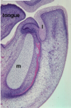
Bud stage. Human, frontal section (10th week). Several areas of tooth development. There has been a downgrowth of the oral epithelium into the underlying mesenchyme (arrows). The two mandibular tooth buds are probably early bud stage (Frame #4), but the maxillary tooth bud may already be at early cap stage (Frame #5). Mandibular bone (pink) can be seen lateral to Meckel’s cartilages (m), and the palatal shelves are fusing with the nasal septum (ns).

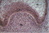
Bud stage. This light micrograph shows the bud stage of tooth development. Note that there has been a down growth of the oral epithelium (e) into the underlying mesenchyme (m), and the epithelium is separated from the connective tissue by a distinct basement membrane. Inferior to the epithelial bud there appears to be the begining “condensation” of mesenchymal cells (arrow), which will eventual become the dental papilla and the dental follicle. None of the distinctive characteristics of the dental organ have yet appeared, i.e. there has been no histodifferentiation.
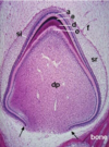
Cap stage. Mandible, sagittal section. Tooth bud at cap stage. Note Meckel’s cartilage (m) and mandibular bone (pink) formation. The tooth bud appears to be connected to the labial lamina (l), but that is only an artifact due to the plane of sectioning. Cells in the center of the labial lamina will die and thus free the lip from the alveolar ridge.

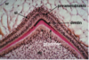
Cap stage. This is a light micrograph of another cap stage tooth bud. The dental follicle is not very distinct, but the dental papilla (dp) is a dense collection of mesenchymal cells underlying the inner enamel epithelium (i). The other portions of the dental organ include the outer enamel epithelium (o) and the stellate reticulum (sr), which is just begining to develop. The junction between the inner and outer enamel epithelia is called the cervical loop (arrows).
Cap stage. This is higher power micrograph of a cap stage tooth bud showing the dental papilla (dp), the inner (i) and outer (o) enamel epithelia and the stellate reticulum (sr). The inner enamel epithelial cells will eventually differentiate into ameloblasts and stratum intermedium.

Advanced cap stage tooth germ showing the position of the enamel knot.

Photomicrograph of the early bell stage of tooth development. Further epithelial proliferation from the dental lamina at its deepest extremity forms the tooth bud of the successional tooth germ. This situation occurs only in relation to primary or deciduous tooth germs.

Bell stage. One of the characteristics of the bell stage is the differentiation of the inner enamel epithelium (iee) into two different cell types. The columnar cells next to the basement membrane (pink line) will become ameloblasts. Note the two mitotic figures (arrows) that demonstrate one of the other features of this stage in tooth development. During bell stage the shape of the tooth is being determined by the differential proliferation of inner enamel epithelial cells. The flattened layer of cells between the inner enamel epithelium and the stellate reticulum (sr) is called the stratum intermedium (si). The function of the cells of the stratum intermedium is not precisely known, but they may play a role in Ca+2 and PO4-3 transport and concentration during the maturation phase of amelogenesis. The dental papilla is separated from the inner enamel epithelium by an acellular zone (az) and a basement membrane (bm).

Bell stage. Human fetus, sagittal section (16 weeks). Two incisor tooth buds (arrows) are shown in this section. Each shows the characteristic features of bell stage. Morphodifferentiation has occured, due to differential proliferation of the inner enamel epithelium, and this has resulted in the formation of the shape of the tooth. The outline formed by the inner enamel epithelium represents the future dentinooenamel junction and not the surface topography of the fully formed crown. Other landmarks include the hard palate (H), the frontal (F) and vomer (V) bones, the nasal capsule (NC), the vertical plate of the ethmoid (NS) and the mandible (M).

Toothbud, late bell stage at the beginning of the appositional phase, showing deposition of predentin (PD) and enamel matrix (EM) at the cusp tip. At this stage the toothbud is no longer connected to the oral epithelium (OE). The dental follicle (DF) surrounds the enamel organ, and the dental papilla (P) is completely enclosed within the “bell” of the enamel organ. Blood vessels have penetrated the dental papilla, and are visible in the follicular connective tissue. The stellate reticulum (SR) has become reduced, bring the blood vessels of the dental follicle in close proximity to the ameloblasts. The cervical loop (c) defines the position of the anatomical cervix of the tooth.
Toothbud, appositional phase. At this stage cytodifferentiation has occured at the cusp tip, with the development of odontoblasts (o) and ameloblasts (a). The formation of dentin (d, dark pink) initiates the terminal differentiation of the ameloblasts and thereby enamel matrix (e, dark purple) formation. The lighter pink material is unmineralized predentin. In this specimen it is possible to see all of the phases of cytodifferentiation by following changes in the inner enamel epithelium and dental papilla/dental pulp (dp) from the cervical loop at the tips of the epithelial diaphragm (arrows) toward the cusp tip. Also note the stellate reticulum (sr) has collapsed near the cusp tip bringing the dental follicle cells into close proximity with the ameloblasts and the stratum intermedium (si).
Toothbud, appositional phase. This is a higher magification view of the beginning of apposition. The stellate reticulum (sr), stratum intermedium (si), preameloblasts of the enamel organ, and the odontoblasts (o) in the dental papilla (dp) are clearly differentiated. At this stage the odontoblasts have begun to form unmineralized dentin matrix (red) which is just beginning to mineralize (black) at the cusp tip. Very quickly the preameloblasts will undergo their final differentiation to become secretory ameloblasts.

Toothbud, appositional phase. At this point the ameloblasts have completely differentiated at the cusp tip. The stellate reticulum (sr), stratum intermedium (si), preameloblasts and odontoblasts (o) are also clearly visible. In this micrograph the unmineralized predentin matrix (p) stains light pink and dentin (d)dark pink. Enamel matrix is the thin line of dark purple material at the cusp tip. If you look carefully on the left slope of the cusp tip, you can see the indentations of Tomes’ processes in the newly formed enamel matrix.