Module Five Flashcards
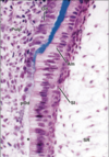
Changes in the dental papilla associated with initiation of dentin formation. An acellular zone (*) separates the undifferentiated cells of the dental papilla (preodontoblasts, pOd) from the differentiating inner enamel epithelium (ameloblasts, Am).

Preodontoblasts develop into tall and polarized odontoblasts (Od) with the nucleus away from the matrix they deposit at the interface with ameloblasts. The matrix first accumulates as an unmineralized layer, predentin (PD), which gradually mineralizes to form mantle dentin (D). Odp, odontoblast process; SI, stratum intermedium; SR, stellate reticulum.

Characteristic deposition of first collagen fibers to form coronal mantle predentin. Large-diameter collagen fibers (Collagen) intermingle with aperiodic fibrils (arrows) associated with the basal lamina supporting the EE. mv, matrix vesicle.

Predentin stains distinctively from dentin

Dentin formation. Odontoblasts (o) are seen next to the layer of predentin (p) in which globular profiles of mineralizing dentin are seen. These calcifying foci coalesce, but the globular pattern is discernible as the more densely staining areas within the zone of mineralized dentin. w=subodontoblastic cell-free zone of Weil. Quiescent period.

Primary dentin. The pattern of dentinal tubules and areas of globular dentin (g) are visible here. Globular dentin refers to the lumpy appearance in such an area of dentin. md=zone of mantle dentin that appears uniform in appearance.

Higher magnification of primary dentin showing globular dentin. Areas of normally mineralized dentin are interspersed among areas of hypomineralized dentin. Globular dentin is most commonly seen just below the mantle dentin. Dentinal tubules (arrow) run through the normal and hypomineralized (asterisk) areas.

Interglobular dentin. A=ground section B and C are demineralized sections. Spherical borders in interglobular areas indicate failure of calcospherite fusion. Dentinal tubules pass through interglobular dentin, but no peritubular dentin is present in these areas.
Odontoblast processes (Odp) run in canaliculi called dentinal tubules (arrowheads)

Dentinal tubules. A and B=predentin and C and D=dentin. Although no dentinal tubules (dt) occur in predentin, each Odp is surrounded by intertwined collagen fibrils (Coll) that outline the future dentinal tubule. The fibrils run circumferentially and perpendicular to this process. In healthy dentin, each tubule is occupied by a process or its ramifications. The dentinal tubule is delimited by a layer of peritubular dentin (arrowheads) that is poor in collagen and more mineralized than the rest of the dentin. The dentin between tubules is referred to as intertubular dentin (iD).

Lines of Von Ebner. Fine incremental deposition von ebner lines in dentin.
Primary Dentin. The diagonal light lines are dentinal tubules which are separated from one another by the blue-staining dentin matrix. The dark blue striations oriented perpendicular to the dentinal tubules are the lines of von ebner. These lines are thought to represent changes in the orientation of collagen fibers that occur within the dentin matrix every four to five days. These changes in direction may contribute to elasticity and strength of dentin.

Primary S-curves. Represent the organization of the dentinal tubules in coronal dentin. The odontoblasts are forced to take an apically-curved path as they lay down dentin due to the constriction of pulpal space as dentin matrix is added to the pulpal wall.

Secondary Dentin. At the end of crown formation, the rate of dentinogenesis slows but the odontoblasts continue to make dentin. This secondary dentin resembles primary dentin in structure and biochemistry. Sometimes it is possible to distinguish a line that demarcates between primary and secondary dentin (arrows). Over time the production of secondary dentin will reduce the size of the pulp chamber and the pulp horns.

Tertiary dentin. When the dentinal tubules are opened through carious activity, fracture or restorative preparation, the odontoblasts again start to rapidly produce dentin matrix. The dentin formed is called reactive dentin. If the insult is too great, the odontoblasts will die and be replaced by newly differentiated pulpal cells that will take their place. The tertiary dentin is this case is called repairative dentin. Pulp is attempting to seal open tubules. Tertiary dentin is very irregular and may or may not have a tubular structure. Arrow shows boundary between secondary and tertiary.

Odontoblastic processes branch near the DEJ

Odontoblastic processes. Odontoblasts have been gently pulled away from predentin. Mesh-like arrangement of collagen fibers in the predentin

Tomes Granular layer at the CEJ. The lamellar appearance of the acellular cementum (c) is apparent. Note that the cervical margin of cementum overlaps the cervical enamel (e). The tubular structure of dentin (d) is visible and the granular layer of tomes (g) can be seen subjacent to the cementum.

Dentinal Tubules (devoid of odontoblastic processes). Small arrow=an opening of a secondary tubule that can be seen in the longitudinal tubule.

Cross-section of dentin demonstrating the presence of dentinal tubules. Red dots=odontoblastic processes.

Pulpal wall of dentin. Stubs of odontoblast processes=arrow protruding from tubules. Uneven contours of pulpal wall due to dentin formation.
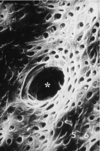
Pulpal wall of dentin. Odontoblasts and processes removed to reveal predentin. Asterisk=opening of dentinal tubule

Dentoenamel Junction. Dentin with its tubules (arrow) surface has pointed projections (triangle) which fit into the corresponding depressions in the enamel.