(5) hypo & hyperventilation notes Flashcards
Effect of changes in VA on alveolar and arterial Po2 and Pco2.
The normal situation, VA = 5 L/min (Fig. 1)
Hypoventilation (Figs. 1 & 2)-
Hypoventilation (Figs. 1 & 2)
Causes a decrease and increase, respectively, in alveolar Po2 and Pco2
Causes a decrease in arteriolar Po2 (hypoxemia) and increase in arteriolar Pco2 (hypercapnia)

Hypoventilation causes
Depression of respiratory center by drugs, trauma.
Weakness of respiratory muscles by injury, paralysis
High work of breathing obesity decreased compliance, increased airway resistance.

Hypoventilation. Signs
Hypercapnia – only caused by hypoventilation
Hypoxemia
CNS depression
Hyperventilation causes and signs
Causes an increase and decrease, respectively, in alveolar Po2 and Pco2
Causes an increase in arteriolar Po2 (hyperoxemia) and decrease in arteriolar Pco2 (hypocapnia).
Causes (Table 2).
Signs (Table 2).
Hypocapnia
Hyperoxemia
increased CNS excitability.
The adequacy of alveolar ventilation is measured in terms of arterial Pco2. Normal alveolar ventilation means that Paco2 equals 40mmHg. Hyperventilation (overventilation) means that Paco2 is less than 40mmHg.
Hypoventilation (underventilation), which is the more common condition encountered in patients with lung diseases, means that Paco2 is more than 40mmHg.
Diffusion blocks

Shunt-anatomical(Fig.3). Normally effect is minor and caused by
1) Small amount of venous blood enters arterial system without passing ventilated alveoli.
2) Bronchial artery enters vein; thebesian veins carrying venous coronary blood directly to left ventricle.
Disease - significant shunts as with septal defects (right-to-left shunts, see West p. 68).

Calculating shunt flow

Shunt-anatomical-Manifestations
- In normal conditions effect is negligible (A-a difference of 1-2mmHg decline on Po2)
- In more sever conditions: Hypoxemia
- Little or no effect on Pco2; usually in some cases hypoxemia may stimulate VA and person may be mildly hypocapneic.
- Giving 100% O2 will not correct hypoxemia caused by shunting.
Ventilation - Perfusion inequalitie
Most common cause of hypoxemia is
is mismatching of ventilation and perfusion.
In patients with cardiopulmonary disease. the most common cause of systemic arterial hypoxemia is
uneven matching of alveolar ventilation to alveolar blood flow.
The following extreme example illustrates the importance of ventilation and perfusion matching. A 2-year oId child is brought to the hospital emergency room because he inhaled a peanut into the bronchus of the left lung, and ventilation of that lung was blocked. Unfortunately, the child was born with a very narrow right pulmonary artery and,
therefore, nearly alI pulmonary blood flow goes to the left lung. This congenital defect did not seriously bother the child before he aspirated the peanut, but now it threatens his life. Ail of the fresh air goes to the right lung, whereas nearly aII of the lung’s blood flow goes to the left lung. Little or no useful gas exchange occurs
C. VA/Q < 1 (Fig. 5) - Net consequences
Hypoxemia – due to shunt
Reflex hypoxic vasoconstriction results.
Create physiological shunt (wasted circulation).
Tendency to increase Paco2
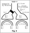
In pulmonary diseases venous admixture occurs commonly because ventilation is low relative to perfusion in many lung units. Low ventilation/perfusion units effectively shunt some of the blood around the alveolar gas (wasted blood flow).
On the other hand, high ventilation/perfusion does not cause venous admixture, only wasted ventilation.
VA/Q > 1 (Fig. 6) - Net consequences
- Hypoxemia
2. Physiological dead space increase (wasted ventilation)
a. Creates hypoventilation situation and leads to reduced PaO2.
3. Tendency to increase Paco2

The intensive-care physician often uses a positive-pressure machine to ventilate the patient with diseased lungs. If the pressure that inflates the lungs is too high, some of the lung may be put into Zone 1, where there is no blood flow.
Indeed, the healthiest part of the lung may be the most affected; thus, blood flow may be shifted to more diseased parts of the lung. Hence, the ventilator may make ventilation/ perfusion mismatching worse, instead of better.
VA/Q - further considerations
- In normal lung (Fig. 7)
- Effect on gas exchange (Fig. 8)

The anesthetist is sometimes faced with a dilemma. In order to ventilate the lung, it may be necessary to use a machine that inflates the lungs be delivering positive pressure at the nose and mouth. That raises alveolar pressure, which compresses the capillaries.
Pulmonary vascular resistance is increased, but if there is lung disease, the pressure effect may not be uniformly distributed. This non-uniformity may markedly affect the matching of perfusion to ventilation and disturb not only the work of the right ventricle but also systemic arterial oxygen concentration.
Net consequences of mismatched VA/Q
- Hypoxemia
- No effect on arterial Pco2 since VA/Q inequalities do affect CO2 exchange but effects are minimal and any increase in Pco2 will stimulate respiratory center to increase VT and F. Thus,
compensation for any increase in Pco2 due to VA/Q inequality.
Alveolar Gas equation
For predicting alveolar Po2 under conditions where no CO2 is breathed. Normally,

Summing up
Causes of hypoxemia
Causes of hyperoxemia -
Cause of hypercapnia
Cause of hypocapnia
A. Causes of hypoxemia
- Hypoventilation
- Diffusion blocks
- Anatomical shunts
- VA/Q inequalities
Causes of hyperoxemia - hyperventilation
Cause of hypercapnia - hypoventilation
Cause of hypocapnia - hyperventilation.
