6. Nanotheranostics Flashcards
(30 cards)
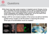
What is nanotheranostics?
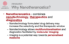
Nanotheranostics - combines nanotechnology, therapeutics and diagnostics.

What are the (potential) advantages of nanotheranostics?
– Nanotechnology formulated drug delivery may increase the selectivity and the therapeutic window
– Nanotechnology allows multifunctionalisation and diagnostics facilitated by molecular imaging
– Imaging is a potential way towards personalised medicine
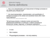
What is meant by “personalised medicine”?
Using a clinical biomaker to indentify patients who most benefit from treatment ie, identification of responder-patients, non-responder patients, and patients who experience limiting toxicity.

Mention 5 techniques used for molecular imaging.
- Fluorescence
- Magnetic resonance imaging (MRI)
- X-ray Computed tomography (CT)
- Nuclear imaging (PET/SPECT)
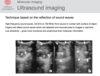
- Ultrasonic imaging
What are the key aspects in definitions of molecular imaging?
- in vivo
- non-invasive
- biological processes (not only anatomical)
What is PET?
positron emission tomography, an imaging technique using radionuclide imaging
11C, 18F, 68Ga, 64Cu
Describe the technique of PET.
Information from Wikipedia:
As the radioisotope undergoes positron emission decay (also known as positive beta decay), it emits a positron, an antiparticle of the electron with opposite charge. The emitted positron travels in tissue for a short distance (typically less than 1 mm, but dependent on the isotope[11]), during which time it loses kinetic energy, until it decelerates to a point where it can interact with an electron.[12] The encounter annihilates both electron and positron, producing a pair of annihilation (gamma) photons moving in approximately opposite directions. These are detected when they reach a scintillator in the scanning device, creating a burst of light which is detected by photomultiplier tubes or silicon avalanche photodiodes (Si APD). The technique depends on simultaneous or coincident detection of the pair of photons moving in approximately opposite directions

Describe the principles of the SPECT imaging technique.

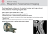
Describe the technique of MRI imaging.
Describe the technique of X-ray CT.

Describe the technique of optical imaging. Give two examples.
Describe how ultrasound imaging works.
Describe 5 imaging methods, the type of radiation from which an image is generated, nad the advantages and disadvantages of the technique.

Mention aspects to address when designing a nanotheranostics in oncology.

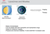
Liposomes and micelles can be loaded and functionalised.
1) In general terms, with what can it be loaded?
2) What do you need to consider, when developing liposomes and micelles for imaging?
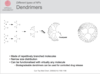
How can you achieve controlled release of a drug using a dendrimer?
Use a biodegradable dendrimer.
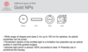
Describe 4 different shapes which exist for gold nanoparticles.
sphere, rod, shell, cage.

- How can you tune the optical properties of Au NPs?
- How can the optical properties be used in medicine?
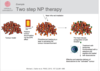
Give an example of a 2 step therapy using NPs for the treatment of tumors.
Which properties of iron nanoparticles make them useful in nanotheranostics?

Mention properties of fullerenes and nanotubes which are useful in nanotheranostics.

Describe limitations of current cancer therapy and how nanothechnology can provide potential solutions to these.
- barriers
- non-specific delivery
- multi-drug resistance
- monitoring of effect

Mention biological barriers for NPs and how you can design a NP to circumvent the barrier.

How do NPs cross cellular membranes?