6.1 Special Senses (Pt 1 The Eye) Flashcards
(34 cards)
Name 4 common causes of blindness
1) ARMD (age related macular degeneration)
2) glaucoma
3) cataracts
4) diabetic retinopathy
What are the 2 segments of the eye and what ‘fluid’ is found in each?
1) posterior segment: behind the lens- filled with vitrious humour
2) anterior segment: in-front of the lens- filled with aqueous humour
What are the 3 layers of the eye?
1) fibrous (sclera and cornea)
2) vascular/ uvea (choroid, cillary body, iris and pupil containing BVs, pigmented layers)
3) neural (retina containing nerve fibres for optic nerve)
Where is the ‘blind spot’?
A small area lateral to the centre of the visual field where there is no visual perception
Corresponds to the optic disc (where the optic nerve passes through the surface of the retina). Here there are no photoreceptors
What is phototransduction?
The process in which light energy is translated into electrical energy
What is the role of photoreceptors?
List the two types and describe each
They code the image formed on the retina into APs
Rods:
- Sensitive to low level light – night vision
- All areas of retina except fovea
- black and white
Cones:
- Highest density at fovea
- 3 different photopigments- red, green and blue
- Daytime vision
How an image is focussed onto retina is known as what?
Refraction
What are the clinical terms for near and long-sightedness?
Near: Myopia (can see nearby objects clearly but not in distance)
Long: Hypermetropia (able to see well at distance but not nearby)
What 2 things cause Hypermetropia and where is the image formed?
How do glasses fix this?
If eyeball is too short or the lens is to flat then the focuss will be focussed behind the retina
Correct by putting in a convex lens which will allow light rays to converge and form on the retina instead of behind it
What 2 things cause Myopia and where is the image formed?
How do glasses fix this?
If the eyeball is too long or the lens too curved the Image is formed in front of the retina
Correct by putting in a concave lens which will allow light rays to diverge (converge later) and form on the retina instead of behind it
Name five components of a vision assessment and the tools that might be used to test them + two more things that can be tested for bonus!
1) Visual Acuity: snellen chart
2) Colour vision: Ishihara test
3) Pupillary reflexes
4) Blind spot
5) Ophthalmoscopy: visualise retina and optic nerve
6) Other: test visual fields and eye movements, ocular alignment
What is meant by visual acuity?
Describe the snellen chart and what value is normal vs abnormal
How well we can resolve fine detail
Snellen chart compares how well we see compared to the average person. Normal vision is 6:6 (stand 6m away from image, and still be able to see image, compared to the average person who can see at 6m)
6:9- 6:12 is short sighted
What does ophthalmoscopy allow for the visualisation of?
What must we do to patient to get a ‘good look’?
Visualisation of the vitreous and retina: to get a good look we MUST dilate the pupil
If the vitreous appears as ‘black blobs’ on an ophthalmoscopy, what does this indicate?
Vitreous hemorrhage
What is swelling of the optic nerve on an ophthalmoscopy indicative of?
Papilloedema: sign of raised ICP
Give 3 abnormalities of the retina that may be seen on an ophthalmoscopy
Detachment, vessels, haemorrhages
What is the Fovea?
Region of highest density of photoreceptors in retina
What is ‘accommodation’ and what are the 3 ‘processes’ involved
Accommodation is when we change from looking far away to looking close up. It involves
1) constriction of the pupil as the eye fixes on near objects
2) thickening of lens due to constriction of ciliary muscles. This changes the optic power to maintain a clear image on the retina
3) convergence of both eyeballs to focus an image as its distance varies
What nerve controls accommodation?
The PNS efferent branch of the oculomotor nerve (CN III)
What is Presbyopia and why does it occur?
How can this be corrected?
Age failure of accommodation with age (starts around 45yrs and complete by 60yrs)
The lens becomes stiffer with age resulting in a decrease in accommodation/focusing. This means close objects are no longer focused onto the retina
This is corrected with convex lens’ of increasing strength
How is presbyopia corrected for?
Convex lens’ of increasing strength
Describe the optic pathway
1) starts at the eye: optic nerve travels from eye to optic chaism where the inner fibres cross over
2) fibres then travel in optic tract to the lateral geniculate nucleus where they synapse
3) from here we get optic radiation which goes to the primary visual cortex in the occipital nerve

Where does the temporal vs nasal visual field come from on the eye?
Which fibres cross over?
The temporal visual field comes from the nasal side of the retina and the Nasal visual field comes from the temporal side of the retina
Fibres from nasal area/side cross over BUT fibres from temporal side of the retina stay on the same side
Explain what type of visual field defect would be seen if there was a lesion at 2, 3 or 5
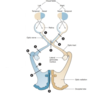
2) lesion affecting optic nerve (R). We will lose our nasal retina (temporal visual field) and the temporal retina (nasal visual field). Complete visual field loss on the right side
3) lesion affecting the optic chiasm. The nasal retina on both sides crosses over at the optic chiasm, so we will lose our nasal retina (temporal visual field). This is a loss of the temporal visual field on both sides (bitemporal heminopia)
5) lesion affecting the optic tract (L). We will lose the temporal retina (nasal visual field) on the left and nasal retina (temporal visual field) on the right (homonimis heminopia)

