S4) The Leg Flashcards
What is the tibia?
The tibia is the main bone of the leg, expanding at the proximal and distal ends and articulating with the knee and ankle joints respectively

Describe the features of the proximal end of the tiba
- Medial and lateral condyles (form the tibial plateau)
- Intercondylar eminence (two tubercles & a roughened area)
- Tibial tuberosity (anterior surface)

Describe the proximal articulations of the tibia with the femur
- Medial and lateral condyles articulate with condyles of femur
- Tibial intercondylar tubercles fit into the intercondylar fossa of the femur

Describe the posterior surface of the shaft of the tibia.
- Marked by a ridge of bone called the soleal line which runs inferomedially, blending with the medial border of the tibia
- Marked by a groove where the tibialis posterior muscle attaches

Describe the features of the anterior and lateral borders of the shaft of the tibia
- Anterior border – marked by tibial tuberosity
- Lateral border (aka interosseous border) – attachment site for interosseous membrane which binds the tibia and fibula

Describe the features of the distal region of the tibia
- Medial malleolus – bony projection continuing inferiorly & articulates with the tarsal bones to form part of the ankle joint
- Fibular notch – found laterally, where the fibula is bound to the tibia

What is the fibula?
The fibula is a thin bone of the leg found laterally to the tibia and does not articulate with the femur at the knee joint but rather acts as an attachment for muscles

Describe the features of the distal and proximal ends of the fibula
- Proximal: enlarged head with a facet for articulation with the lateral condyle of the tibia
- Distal: lateral surface continues inferiorly producing a bony projection (lateral malleolus - more prominent)

Identify the four muscles in the anterior compartment of the leg
- Tibialis anterior
- Extensor digitorum longus
- Extensor hallucis longus
- Fibularis tertius
Describe the neurovascular supply of the anterior compartment of the leg
- Arterial supply via the anterior tibial artery
- Innervation via deep fibular nerve

Describe the anatomical location and function of the tibialis anterior muscle

- Location: located on lateral surface of tibia
- Function: dorsiflexion, inversion

State the origin and attachment of the tibialis anterior muscle

- Origin: lateral surface of the tibia
- Attachment: medial cuneiform and base of metatarsal I

Describe the anatomical location and function of the extensor digitorum longus muscle

- Location: lies lateral and deep to the tibialis anterior
- Function: dorsiflexion, extension of the lateral four toes

State the origin and attachment of the extensor digitorum longus muscle

- Origin: lateral condyle of tibia and medial surface of fibula
- Attachment: tendon splits into four, each inserting onto a toe

Describe the anatomical location and function of the extensor hallucis longus

- Location: deep to the extensor digitorum longus and tibialis anterior
- Function: great toe extension, dorsiflexion

State the origin and attachment of extensor hallucis longus

- Origin: medial surface of the fibular shaft
- Attachment: base of the distal phalanx of the great toe

Describe the anatomical location and function of the fibularis tertius muscle

- Location: arises from the most inferior part of the extensor digitorum longus (not present in all individuals)
- Function: eversion, dorsiflexion

State the origin and attachment of the fibularis tertius muscle

- Origin: medial surface of the fibula
- Attachment: metatarsal V (diverges from EDL at dorsum of foot)

Identify the two muscles in the lateral compartment of the leg
- Fibularis longus
- Fibularis brevis

Describe the structure, function and innervation of the fibularis longus muscle
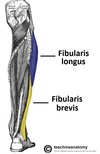
- Structure: larger and more superficial muscle within the compartment
- Function: plantarflexion, eversion, supports the lateral and transverse foot arches
- Innervation: Superficial fibular nerve

State the origin and attachment of the fibularis longus muscle

- Origin: superior and lateral surface of fibula and lateral tibial condyle (moves posterior to the lateral malleolus)
- Attachment: medial cuneiform and base of metatarsal I (crosses under foot)

Describe the structure, function and innervation of the fibularis brevis muscle

- Structure: deeper and shorter than fibularis longus
- Function: eversion
- Innervation: superficial fibular nerve

State the origin and attachment of the fibularis brevis muscle

- Origin: inferolateral surface of the fibular shaft (travels posteriorly to the lateral malleolus)
- Attachment: tubercle on metatarsal V (past over calcaneus and cuboid)

Identify the two types of muscles in the posterior compartment of the leg
- Superficial muscles
- Deep muscles
The two layers are separated by a band of fascia
The superficial muscles of the posterior leg all insert into the calcaneus of the foot via the calcaneal tendon.
Identify and describe the two bursae associated with the calcaneal tendon
- Subcutaneous calcaneal bursa – lies between the skin and the tendon
- Retrocalcaneal bursa – lies between the tendon and the calcaneus

Identify the superficial muscles of the posterior leg
- Soleus
- Gastrocnemius
- Plantaris
Describe the structure, function and innervation of the gastrocnemius muscle

- Structure: most superficial muscle in posterior leg, two heads – medial and lateral
- Function: knee flexion, plantarflexion
- Innervation: tibial nerve

State the origin and attachment of the gastrocnemius muscle

- Origin: lateral femoral condyle and medial femoral condyle
- Attachment: calcaneus

Describe the structure, function and innervation of the plantaris muscle

- Structure: small muscle with a long tendon (absent in 10% of people)
- Function: plantarflexion, knee flexion
- Innervation: tibial nerve

State the origin and attachment of the plantaris muscle

- Origin: lateral supracondylar line of the femur
- Attachment: blends with calcaneal tendon

Describe the structure, function and innervation of the soleus muscle

- Structure: large, flat, deep to the gastrocnemius
- Function: plantarflexion
- Innervation: tibial nerve

State the origin and attachment of the soleus muscle

- Origin: soleal line of the tibia and proximal fibular area
- Attachment: calcaneus

Describe the structure, function and innervation of the popliteus muscle

- Structure: lies superiorly, behind the knee joint, forming the base of the popliteal fossa
- Function: lateral rotation of the femur on the tibia (‘unlocking’ the knee)
- Innervation: tibial nerve

State the origin and attachment of the popliteus muscle

- Origin: posterior surface of the proximal tibia
- Attachment: lateral condyle of femur, lateral meniscus of the knee joint

Describe the structure, function and innervation of tibialis posterior muscle

- Structure: deepest of the four muscles, lies between flexor digitorum longus and flexor hallucis longus
- Function: inversion, planterflexion, maintains medial foot arch
- Innervation: tibial nerve

State the origin and attachment of the tibialis posterior muscle

- Origin: interosseous membrane between the tibia and fibula, and posterior surfaces of the two bones
- Attachment: plantar surfaces of the medial tarsal bones

Describe the structure, function and innervation of flexor digitorum longus

- Structure: smaller muscle than flexor hallucis longus, located medially
- Function: flexes the lateral four toes
- Innervation: tibial nerve

State the origin and attachment of the flexor digitorum longus muscle

- Origin: medial surface of the tibia
- Attachment: plantar surfaces of the lateral four digits

Describe the structure, function and innervation of the flexor hallucis longus muscle

- Structure: lies on lateral side of leg (counter-intuitive as it acts on great toe)
- Function: great toe flexion
- Innervation: tibial nerve

State the origin and attachment of the flexor hallucis longus muscle

- Origin: posterior surface of the fibula
- Attachment: plantar surface of the phalanx of the great toe

What is the popliteal fossa?
- The popliteal fossa is a diamond-shaped area found on the posterior side of the knee
- It is the main path in which structures move from the thigh to the leg

Identify the four borders of the popliteal fossa as well as the muscles which form them
- Superomedial border: semimembranosus
- Superolateral border: biceps femoris
- Inferomedial border: medial head of the gastrocnemius
- Inferolateral border: lateral head of the gastrocnemius and plantaris

Identify the neurovascular structures found in the popliteal fossa

Describe the structure/relative location of the neurovascular structures in the popliteal fossa

- Most superficial — tibial and common fibular nerves
- Deepest — popliteal artery
- Small saphenous vein pierces the popliteal fascia to empty into the popliteal vein

Describe the anatomical course of the obturator artery
⇒ Arises from internal iliac artery in the pelvic region
⇒ Descends via the obturator canal to enter the medial thigh
⇒ Bifurcates into two branches: anterior & posterior branches

Describe the anatomical course of the popliteal artery
⇒ Descends down the posterior thigh & gives off genicular branches
⇒ Moves through the popliteal fossa
⇒ Exits between the gastrocnemius and popliteus
⇒ Divides into anterior and posterior tibial arteries (lower border or popliteus)

Describe the anatomical course of the posterior tibial artery
⇒ Continues inferiorly along the surface of the deep muscles
⇒ During descent, fibular artery rises & moves laterally
⇒ Main artery enters sole of the foot via the tarsal tunnel (with tibial nerve)

Describe the anatomical course of the anterior tibial artery
⇒ Passes anteriorly between the tibia and fibula, through a gap in the interosseous membrane
⇒ Moves inferiorly down the leg
⇒ Runs into the foot and becomes the dorsalis pedis artery

The dorsalis pedis artery mainly supplies the tarsal bones and the dorsal aspect of the metatarsals as well as the toes (deep plantar arch).
Describe its anatomical course through the foot
⇒ Passes over the dorsal aspect of the tarsal bones
⇒ Moves inferiorly, towards the sole of the foot
⇒ Anastamoses with the lateral plantar artery to form the deep plantar arch

The posterior tibial supplies the plantar side of the foot as well as the toes (deep plantar arch).
Describe its anatomical course through the foot
⇒ Enters the sole of the foot through the tarsal tunnel
⇒ Divides into lateral and medial plantar arteries

Describe the venous drainage of the foot
- The dorsal venous arch drains into the superficial veins & some penetrate deep into the leg, forming the anterior tibial vein
- The medial and lateral plantar veins combine to form the posterior tibial and fibular veins

Describe the venous drainage of the leg
- Posterior tibial vein enters the leg posteriorly to the medial malleolus
- Anterior tibial, posterior tibial and fibular veins unite on the posterior surface of the knee to form the popliteal vein
- Popliteal vein enters the thigh via the adductor canal

Describe the venous drainage of the thigh
- Popliteal vein enters the thigh and becomes the femoral vein
- The profunda femoris vein also drains blood from thigh muscles & empties into the distal section of the femoral vein
- The femoral vein leaves the thigh running underneath the inguinal ligament and becomes the external iliac vein

The superficial veins of the lower limb run in the subcutaneous tissue.
Describe the anatomical course of the great saphenous vein
⇒ Formed by the dorsal venous arch of the foot & dorsal vein of the great toe
⇒ Ascends up the medial side of the leg
⇒ Passing anteriorly to medial malleolus & posteriorly to the medial condyle
⇒ Terminates by draining into the femoral vein (inferior to inguinal ligament)

The superficial veins of the lower limb run in the subcutaneous tissue.
Describe the anatomical course of the small saphenous vein.
⇒ Formed by the dorsal venous arch of the foot & dorsal vein of the little toe
⇒ Moves up the posterior side of the leg
⇒ Passes posteriorly to lateral malleolus & laterally to the calcaneal tendon
⇒ Moves between two heads of the gastrocnemius muscle
⇒ Empties into the popliteal vein (in popliteal fossa)

The lymphatic vessels of the lower limb can be divided into two major groups; superficial vessels and deep vessels.
Describe the distribution of the medial group of superficial lymphatic vessels
- Originate on the dorsal surface of foot
- Travel up the anterior and posterior aspects of the medial lower leg, with the great saphenous vein
- Ends in the groin by draining into the sub-inguinal group of the inguinal lymph nodes

The lymphatic vessels of the lower limb can be divided into two major groups; superficial vessels and deep vessels.
Describe the distribution of the lateral group of superficial lymphatic vessels
- Arise from the lateral surface of foot
- Either accompany small saphenous vein to enter popliteal nodes, or ascends in front of the leg and cross join the medial group

The lymphatic vessels of the lower limb can be divided into two major groups; superficial vessels and deep vessels.
Describe the distribution of the deep lymphatic vessels
- Found in 3 main groups: anterior tibial, posterior tibial and peroneal
- Following the corresponding artery respectively, and entering the popliteal lymph nodes

Where are the inguinal nodes found?
The inguinal nodes are found in the upper aspect of the femoral triangle and are 1—20 in number

Describe the anatomical location and drainage of the superficial inguinal lymph nodes of the lower limb
- Location: form a line directly below the inguinal ligament
- Drainage: receive lymph from the penis, scrotum, perineum, buttock and abdominal wall

Describe the anatomical location and drainage of the superficial sub-inguinal lymph nodes of the lower limb
- Location: found on each side of the proximal section of the great saphenous vein
- Drainage: receive lymph from the superficial lymphatic vessels of the lower leg

Describe the structure, anatomical location and drainage of the popliteal lymph nodes
- Structure: small in size, usually between five and seven in number
- Location: found imbedded in fat reserves in the popliteal fossa
- Drainage: receive lymph from the lateral superficial vessels

Identify the nerve roots of the common peroneal nerve

L4 – S3
Describe the anatomical course of the common peroneal nerve

⇒ Begins at apex of the popliteal fossa (sciatic nerve bifurcation)
⇒ Follows the medial border of the biceps femoris
⇒ Gives rise to two cutaneous branches over lateral head of gastrocnemius
⇒ Wraps around the neck of the fibula
⇒ Passes between the attachments of fibularis longus muscle
⇒ Divides into superficial fibular and deep fibular nerves

Describe the motor functions of the common peroneal nerve

- Innervates the short head of the biceps femoris muscle
- Innervates muscles of the lateral compartment of the leg (superficial fibular nerve)
- Innervates muscles of the anterior compartment of the leg (deep fibular nerve)
Describe the sensory functions of the common peroneal nerve

- Sural communicating nerve innervates the skin over the lower posterolateral leg
- Lateral sural cutaneous nerve Innervates the skin over the upper lateral leg
- Superficial fibular nerve innervates the skin of the anterolateral leg and dorsum of the foot
- Deep fibular nerve innervates the skin between first and second toes

Identify the nerve roots of the superficial fibular nerve
L4-S1

Identify the nerve roots of the deep fibular nerve
L4 and L5

Identify the nerve roots of the tibial nerve

L4-S3
Describe the anatomical course of the tibial nerve

⇒ Arises at the apex of the popliteal fossa (sciatic nerve bifurcation)
⇒ Travels through popliteal fossa, then posterior to tibia
⇒ Giving off branches to posterior leg muscles
⇒ Gives rise to branches which contribute to the sural nerve
⇒ Passes posterioinferiorly to medial malleolus, through tarsal tunnel
⇒ Tibial nerve terminates by dividing into sensory branches

Describe the motor functions of the tibial nerve

- Innervates the deep muscles in the superficial compartment of the leg
- Innervates the deep muscles in the posterior compartment of the leg
Describe the sensory functions of the tibial nerve

- Sural nerve innervates the skin of the posterolateral leg and lateral foot
- Medial calcaneal branches innervate the skin over the heel
- Medial plantar nerve innervates the plantar surface of the medial 3½ digits and the associated sole area
- Lateral plantar nerve innervates the plantar surface of the lateral 1½ digits and the associated sole area

What is foot drop?
- Foot drop is a the manifestation of a condition caused by damage to the common peroneal nerve (/deep fibular) often due to fractures of the fibula
- The patient can’t dorsiflex the foot and hence the foot appears permanently plantarflexed (footdrop) producing a characteristic high steppage gait
