S9) The Hand Flashcards
Identify and describe the bones of the wrist and hand
- Carpal bones – set of eight irregularly shaped bones located in the wrist area
- Metacarpals – five bones, each related to a digit
- Phalanges – each finger has three phalanges (thumb has two)

Identify the carpal bones according to their organisation proximally and distally
- Proximal row: scaphoid, lunate, triquetrum, pisiform
- Distal row: trapezium, trapezoid, capitate, hamate

Describe the proximal and distal articulations of the carpal bones
- Proximal: scaphoid and lunate articulate with the radius
- Distal: carpal bones articulate with the metacarpals

Describe the arrangement of the metacarpal bones
- Metacarpal I – thumb
- Metacarpal II – index finger
- Metacarpal III – middle finger
- Metacarpal IV – ring finger
- Metacarpal V – little finger

Describe the proximal and distal articulations of the metacarpal bones
- Proximal: metacarpals articulate with the carpal bones
- Distal: metacarpals articulate with the proximal phalanges

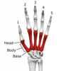
Describe the structure of the metacarpal bones in the hand
- Each metacarpal consists of a base, shaft and a head
- The medial and lateral surfaces of the metacarpals are concave, allowing attachment of the interossei muscles
The phalanges are the bones of the fingers.
Describe their structure
- Four fingers have proximal, middle and distal phalanges
- Thumb has proximal and distal phalanges

Describe the distribution of the radial and ulnar arteries in the hand
- Radial artery – contributes mainly to supply of the thumb and the lateral side of the index finger
- Ulnar artery – contributes mainly to the supply of the rest of the digits, and the medial side of the index finger
Describe the anatomical course of the ulnar artery in the hand
- The ulnar artery moves into the hand anteriorly to the flexor retinaculum, and laterally to the ulnar nerve
- It forms the superficial palmar arch and the deep palmar branch
- The superficial palmar arch gives rise to palmar digital arteries and then anastamoses with a branch of the radial artery

Where is the superficial palmar arch found?
The superficial palmar arch is found superficial to the flexor tendons in the hand and deep to the palmar aponeurosis

Describe the anatomical course of the radial artery in the hand
- The radial artery enters the hand dorsally through the floor of the anatomical snuffbox and turns medially to move between the heads of the adductor pollicis
- The radial artery then anastamoses with the deep palmar branch of the ulnar artery, forming the deep palmar arch, which gives rise to five common digital arteries

What is the Allen’s test?
- The Allen test is a worldwide test used to determine whether the patency of the radial or ulnar artery is normal
- It is performed prior to radial cannulation or catheterisation, because placement of such a catheter often results in thrombosis
- Thus, the test is used to reduce the risk of ischaemia to the hand

How does one interpret the result of the Allen’s test?

A positive Allen’s test means that the patient does not have dual blood supply to the hand, which is a negative indication for catheterisation or removal of the radial arteries
What are the two types of muscles of the hand?
- The extrinsic muscles
- The intrinsic muscles
Where are the two diffferent types of muscles of the hand located?
- The extrinsic muscles are located in the anterior and posterior compartments of the forearm
- The intrinsic muscles of the hand are located within the hand itself
What do the two diffferent types of muscles of the hand do?
- Extrinsic muscles – they control crude movements and produce a forceful grip
- Intrinsic muscles – they are responsible for the fine motor functions of the hand
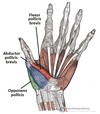
What are the thenar muscles?
The thenar muscles are three short muscles located at the base of the thumb

Identify the 3 thenar muscles

State the structure, function and innervation of the thenar muscles
- Structure: muscle bellies produce a bulge (thenar eminence)
- Function: fine movements of the thumb
- Innervation: median nerve

Describe the structure and function of the opponens pollicis muscle

- Structure: largest and deepest of the thenar muscles
- Function: opposes the thumb

State the origin and attachment of opponens pollicis

- Origin: tubercle of the trapezium and associated flexor retinaculum
- Attachment: lateral margin of the metacarpal I

Describe the structure and function of the abductor pollicis brevis muscle

- Structure: anterior to the opponens pollicis and proximal to the flexor pollicis brevis
- Function: thumb abduction

State the origin and attachment of the abductor pollicus brevis

- Origin: tubercles of the scaphoid and trapezium and associated flexor retinaculum
- Attachment: lateral side of proximal phalanx of the thumb

Describe the structure and function of the flexor pollicis brevis muscle

- Structure: most distal of the thenar muscles
- Function: flexes the MCPJ of thumb

State the origin and attachment of the flexor pollicis brevis
- Origin: tubercle of the trapezium and associated flexor retinaculum
- Attachment: base of the proximal phalanx of the thumb

What are the hypothenar muscles?
The hypothenar muscles are three short muscles located at the base of the little finger

Identify the 3 hypothenar muscles


Describe the structure, function and innervation of the opponens digiti minimi muscle

- Structure: lies deep to the other hypothenar muscles
- Function: opposes little finger
- Innervation: ulnar nerve

State the origin and attachment of the opponens digiti minimi muscle

- Origin: hook of hamate and associated flexor retinaculum
- Attachment: medial margin of metacarpal V

Describe the structure, function and innervation of the abductor digiti minimi muscle

- Structure: most superficial of all hypothenar muscles
- Function: abducts the little finger
- Innervation: ulnar nerve

State the origin and attachment of the abductor digiti minimi muscle

- Origin: pisiform and the tendon of the flexor carpi ulnaris
- Attachment: base of the proximal phalanx of the little finger

Describe the structure, function and innervation of the flexor digiti minimi brevis muscle

- Structure: lies laterally to the abductor digiti minimi
- Function: flexes the MCPJ of little finger
- Innervation: ulnar nerve

State the origin and attachment of the flexor digiti minimi muscle

- Origin: hook of hamate and adjacent flexor retinaculum
- Attachment: base of the proximal phalanx of the little finger

What are the lumbricals?
The lumbricals are 4 intrinsic muscles of the hand that flex the MCPJs and extend the IPJs

Describe the function and innervation of the lumbricals

- Function: MCPJ flexion, IPJ extension at each finger
- Innervation:
I. Medial two lumbricals – ulnar nerve
II. Lateral two lumbricals – median nerve

State the origin and attachment of the lumbricals

- Origin: tendon of the flexor digitorum profundus
- Attachment: pass dorsally and laterally to inserts into the extensor hood

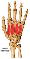
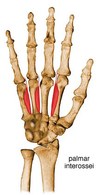
What are the interossei muscles?
- The interossei muscles are 7 intrinsic muscles of the hand located between the metacarpals
- They can be divided into two groups: the dorsal and palmar interossei

Describe the structure, function and innervation of the dorsal interossei muscles

- Structure: most superficial of all dorsal muscles (4 muscles)
- Function: finger abduction at the MCPJ
- Innervation: ulnar nerve

State the origin and attachment of the dorsal interossei muscles

- Origin: lateral and medial surfaces of the metacarpals
- Attachment: extensor hood and proximal phalanx of each finger
Describe the structure, function and innervation of the palmar interossei muscles

- Structure: located anteriorly on the hand (3 muscles)
- Function: finger adduction MCPJ
- Innervation: ulnar nerve

State the origin and attachment of the palmar interossei muscles

- Origin: medial or lateral surface of a metacarpal
- Attachment: extensor hood and proximal phalanx of same finger
Describe the structure, function and innervation of the palmaris brevis muscle

- Structure: small, thin muscle, found superficially in the subcutaneous tissue of the hypothenar eminence
- Function: wrinkles the skin of the hypothenar eminence and deepens the curvature of the hand (improving grip)
- Innervation: ulnar nerve

State the origin and attachment of the palmaris brevis muscle

- Origin: palmar aponeurosis and flexor retinaculum
- Attachment: dermis of the skin on the medial margin of the hand

Describe the structure, function and innervation of the adductor pollicis muscle

- Structure: large triangular muscle with two heads (radial artery passes between to form deep palmar arch)
- Function: thumb adduction
- Innervation: ulnar nerve

State the origin and attachment of the adductor pollicis muscle

- Origin:
I. One head – metacarpal III
II. Other head – capitate and adjacent areas of metacarpals II and III
- Attachment: base of the proximal phalanx of the thumb

Describe the cutaneous distribution of the ulnar nerve in the dorsum and palm of the hand
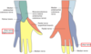
Describe the cutaneous distribution of the median nerve in the palm and dorsum of the hand

Describe the cutaneous distribution of the radial nerve in the palm and dorsum of the hand

What is a boxer’s fracture?
- A boxer’s fracture is a fracture of one of the metacarpal bones of the hand
- The fracture occurs transversely across the neck of the bone, after the patient strikes an object with a closed fist

What is Dupuytrens contracture?
- Dupuytren’s contracture is a clinical condition where 1/more fingers become permanently bent in a flexed position due to the thickening of the connective tissue in the palm
- It begins as small hard nodules under the skin of the palm then worsens over time until the fingers can no longer be straightened

What is reflex sympathetic dystrophy?
- Reflex sympathetic dystrophy (CRPS 1) is a clinical syndrome of the SNS with unknown cause characterised by pain, swelling, and vasomotor dysfunction of an extremity
- It is associated with injury to the nerves, trauma, surgery, CVD and infection

What are the 4 X-ray features of osteoarthritis?
- Joint space narrowing
- Subchondral sclerosis (thin layer of increased bone density)
- Osteophytes (bony spurs)
- Subchondral cysts (fluid filled sacs)

What is a scaphoid fracture?
- A scaphoid fracture is a fracture of one of the carpal bones, presenting as tenderness over the anatomical snuffbox and often has delayed presentation in X-rays (notable swelling)
- It needs to be reduced quickly and untreated, can lead to avascular necrosis

What is ulnar claw?
- Ulnar claw is a clinical condition resulting from the long term damage of the ulnar nerve which presents as hyperextension of MCPJs (little and ring fingers) and flexion of the IPJs
- The interossei muscles and the medial lumbricals are paralysed but the two muscles in the forearm are unaffected (laceration occurs at the wrist)

What is the hand of benediction?
- The hand of benediction is a clinical condition which occurs as a result of prolonged compression or injury of the median nerve at the forearm or elbow
- The thenar eminence is wasted, due to atrophy of the thenar muscles and if the patient tries to make a fist, only the little and ring fingers can flex completely
