Abdomen - Hernia Flashcards

1
Q

In a patient with an abdominal bulge, be able to determine whether a hernia is present and the type of hernia. In addition, be able to distinguish between abdominal wall hernias and diastasis recti.

A
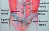
  • A ventral hernia is a defect in the abdominal wall fascia with protrusion of abdominal or preperitoneal contents.
  • Spontaneously occurring midline hernias: epigastric hernias (located between the xyphoid and umbilicus), umbilical hernias (located at the umbilicus), and hypogastric hernias (located below the umbilicus). Spigelian hernias occur off of midline and lateral to the rectus in the lower abdomen (between the iliac crest and 6 cm cephalad to the iliac crest).
  • Incisional hernias occur at the location of prior surgical incisions.
  • Diastasis recti is not a true hernia but rather stretching of the linea alba with bulging at the medial edge of the rectus muscle.
2
Q

Be able to distinguish between indications for emergent and elective operative intervention for ventral hernias.

A
  • Emergent:
    • Acute incarceration w/ inability to reduce hernia
    • Hernia content strangulation
    • Bowel obstruction with signs of bowel ischemia
    • Leakage of ascites through skin in pt w/ cirrhosis
  • Elective:
    • Pain or discomfort
    • Hi risk for obsx: chronic incarcx, intermittent incarcx
    • Interference with daily activities or quality of life
3
Q

Identify contraindications to operative ventral hernia repair

A
  • Relative contraindications:
    • Obesity
    • Active tobacco use
    • Pregnancy
    • Poor surgical candidate with multiple comorbidities
  • Absolute contraindications
    • Inability to withstand general anesthesia if required
4
Q

In regards to hernias, be able to determine clinical factors that would favor laparoscopic versus open repair

A

Factors that favor laparoscopic repair are smaller defects, older age, and lack of need to reapproximate the linea alba.

Factors that favor open repair are larger defects, multiple prior operations with concern for dense adhesions not amenable to laparoscopic lysis of adhesions, and inability to tolerate pneumoperitoneum.

5
Q

Name the layers of the abdominal wall

A

Encountered laterally from anterior to posterior:

  1. Skin
  2. Subcutaneous tissue
  3. Camper and Scarpa fasciae
  4. External oblique muscle
  5. Internal oblique muscle
  6. Transversus abdominis muscle and aponeurosis
  7. Transversalis fascia
  8. Preperitoneal fat
  9. Peritoneum
6
Q

Describe the appropriate perioperative patient positioning, and prophylaxis in a patient undergoing ventral hernia repair (open or laparoscopic).

A
  • Open procedure: supine with arms out.
  • Laparoscopic procedure: supine with arms tucked.
  • SCD stockings for VTE ppx w/ chemoproppx as indicated by patient/surgical risk factors.
  • Appropriate abx within 1 hour of incision.
    • Gram + coverage is sufficient for a clean case (ancef).
  • If a longer duration case is anticipated or the operative field includes the space of Retzius (retropubic space), a Foley catheter should be placed.
7
Q

To obtain informed consent, describe the relevant risks, benefits, and alternatives to operative intervention for ventral hernia repair.

A
  • Risks: bleeding, wound infection, mesh infection if mesh is used, injury to surrounding structures including bowel, port site hernia if repaired laparoscopically, hernia recurrence, seroma, chronic or persistent pain, skin necrosis, and abdominal compartment syndrome.
  • Benefits: fixing the hernia defect, decreasing risk of future incarceration or strangulation, and potential for decreased pain.
  • Alternatives: abdominal binder or abdominal support.
8
Q

Be able to discuss the recurrence rate of primary repair versus mesh repair for ventral hernias.

A
  • Small primary (≤ 2-3 cm) w/ suture repair: 5% to 7%.
  • Larger primary (> 2-3 cm) w/ suture repair: 10% and 50%.
  • Incisional hernias w/ suture repairs: 10% to 50%
    • Recurrence decreases by > 50% w/ mesh.
  • Risk factors for recurrence: smoking, obesity, DM, immunosuppression, malnutrition, corticosteroids, chemotherapy/radiation, and CTDs.
9
Q

In a patient with an infected prosthetic mesh, be able to describe the criteria for operative intervention.

A
  • Nonoperative management: antibiotics and local wound care.
  • Mesh infections involving macroporous mesh made from polypropylene or polyester may be treated with local measures, mesh removal may be required.
  • Nonoperative strategies unlikely to be successful in treating microporous or polytetrafluoroethylene (PTFE) prosthetic infections.
  • Failure of nonoperative management is demonstrated by ongoing signs and symptoms of mesh infection.
10
Q

In an open ventral hernia repair, decide on the material and location for mesh placement.

A
  • Options: underlay (retrorectus, pre vs intraperitoneal), sublay, inlay (mesh placed as a bridge between the fascial edges), or onlay (mesh placed above the fascia).
  • ideal: preperitoneal, sublay, fascia closed
  • Intra-peritoneal mesh needs non-adhesive barrier
  • Options: polyester, polypropylene, and PTFE vs biologic
  • Prosthetics: macro- or micro-porous (macro resists infection)
  • Biologic meshes can be cross-linked or non–cross-linked.
  • Several synthetic absorbable meshes are now available
  • Biologic and synthetic absorbable meshes: contaminated fields to decrease the risk of long-term mesh infections.
11
Q

Describe the approaches to component separation to gain additional mobilization of the abdominal wall.

A
  • Approaches to component separation: anterior release, posterior release, transversus abdominis release (TAR), separation of internal obl and transversus.
  • Anterior release: creation of subcutaneous flaps, division of the external oblique aponeurosis lateral to the semilunar line, and development of the plane between the external oblique aponeurosis and the internal oblique muscle laterally. Mesh reinforcement can be placed in an underlay or overlay position.
  • Posterior release: opening the retrorectus space medially and creating the posterior plane between the rectus muscle and the posterior rectus sheath. The posterior rectus sheath is incised again medial to the laterally perforating neurovascular bundles. The underlying transversus abdominis muscle can be divided (TAR), and dissection is carried laterally to mobilize the transversalis fascia and peritoneum off of the overlying muscle.
12
Q

In a laparoscopic ventral hernia repair, describe the principles of port size and location of placement.

A
  • Ports should be placed well laterally to the location of the hernia defect to allow appropriate working space and placement of mesh.
  • Ports (5 mm) can be used for laparoscopic instruments and the tacking device. A 10- to 12-mm port is often needed for mesh insertion.
13
Q

In a laparoscopic ventral hernia repair, decide on the appropriate mesh size and describe the appropriate sequence in mesh fixation to the anterior abdominal wall.

A
  • Defect is measured intraperitoneally for most accuracy. Use spinal needles through the abd wall along defect edge w/ a ruler intra-abd.
  • Mesh size: allow for a 3- to 5-cm overlap on all sides.
  • Mesh orientation/fixation: transfascial sutures placed in the four quadrants of the mesh prior to insertion, or use a mesh deployment/positioning device.
  • Mesh tacking: transfascial sutures brought through the abd wall, mesh tacked in at 1-cm intervals along the edge, then a second inner row of tacks is placed.
14
Q

In a patient with an infected prosthetic mesh, be able to describe the key steps of the operative procedure.

A
  • Incision and gaining access to the peritoneal cavity
  • LoA: free intra-abdominal contents from mesh, gain working room laterally
  • Removal of infected mesh and non-absorbable sutures
  • Assessment of ability to close fascia or need for component release
  • Closure of the abdominal wall with or without the aid of a biologic mesh
15
Q

During an open ventral hernia repair, assess the tension on the abdominal wall

A

The ability to close the fascia can be assessed by grasping the fascial edges on each side with Kocher forceps and pulling the fascial edges to the midline.

Abdominal compartment syndrome is indicated by end-organ dysfunction, including impaired ventilation, cardiac output, and renal function. Intraoperatively, the risk of abdominal compartment syndrome may be indicated by increased peak ventilator pressures when closing the abdominal wall.

16
Q

During ventral hernia repair in a contaminated field, describe the strategies of repair and expected outcomes.

A
  • Repair of enterotomy includes primary repair for a smaller or single enterotomy or bowel resection with primary anastomosis for larger or multiple enterotomies.
  • Strategies for abdominal wall closure include primary closure, placement of absorbable mesh, or placement of biologic mesh (NOT synthetic non-absorbable)
17
Q

Describe potential complications after ventral hernia repair

A
  • Complications include ileus, postoperative pain, sepsis, intestinal obstruction or fistualization, necrotizing soft tissue infection, seroma formation, hernia recurrence, and mesh infection.
  • Recurrence rates range from 0% to 30% for prosthetic repairs.
  • Factors that increase the risk of prosthetic mesh infections include presence of infection prior to surgery, skin ulceration over hernia, enterotomy during hernia repair, obesity, and incarcerated or obstructed bowel within the hernia.
18
Q

Describe the spectrum of clinical manifestations of an infected prosthesis.

A
  • Systemic: fevers, chills, leukocytosis, tachycardia, and hypotension.
  • Sugrical site infection: erythema, purulent drainage, and chronic sinus tracts.
  • Imaging and diagnostic findings: contrasted CT scan demonstrating fluid collection, with signs of infection including rim enhancement and gas, as well as positive culture from surgical site fluid collection in continuity with mesh.
  • It is important to note that patients can develop postoperative seromas or fluid collections that are not infected, and both clinical examination findings and diagnostic maneuvers are required to differentiate between infected and noninfected fluid collections.
19
Q

A 58-year-old chronic alcoholic has an umbilical hernia and ascites of recent onset. He has never been treated with diuretics or salt restriction. On examination, he has massive ascites with a large umbilical hernia, with thin skin at the apex. There is a slow ooze of clear, odorless fluid from the hernia. Therapy now should be:

A

Bedrest, IV abx, aggressive diuresis, hernia repair during this admission

  • Leaking abdominal ascites is urgent and requires aggressive management.
  • Admit pt: risk of bacterial peritonitis and/or hernia rupture.
  • Repair in cirrhotics w/ uncontrolled ascites: high M/M - 8.3%, 16.6%
  • Control the ascites prior to repair: diuresis, sodium, fluid restriction
  • Bedrest: remove undue strain on the weak and leaking site
  • Intravenous antibiotics: prevent bacterial peritonitis
  • If operation must be undertaken emergently (true rupture), or diuretic therapy fails to control the ascites, combined umbilical herniorrhapy with a peritoneal-venous shunt can achieve a stable repair with relatively low morbidity.
20
Q

Epidemiologic information about umbilical hernias: men vs women, incarceration, content, spontaneous closure, association w/ ascites

A
  • Umbilical hernias are common - just as common if not more than incisional
  • More common in women than men (3:1)
  • Present more commonly with incarceration in men
  • Content is usually pre-peritoneal fat or omentum.
  • Spontaneous closure in children under 5 and pregnant patients.
    • Persistent hernia beyond 5, sx hernias s/p delivery - repair
  • Associated w/ ascites - repair for rupture, redness/ulceration of skin, incarceration.
  • If a patient has ascites and a non-incarcerated hernia - address the ascites.
21
Q

Describe rectus sheath anatomy in reference to the arcuate line

A

Above the arcuate line, the posterior rectus sheath is composed of the posterior lamella of the internal oblique aponeurosis with the transversus abdominis muscle and aponeurosis.

Below the arcuate line, there is no posterior sheath.

22
Q

Describe the composition of the linea semilunaris and linea alba

A

The aponeurosis of the external oblique, internal oblique, and transversus abdominis fuse lateral to the rectus muscle to form the linea semilunaris and medial to the rectus muscle to form the linea alba.

23
Q

Describe the relationship between the major muscles with their blood and nerve supplies.

A
  • The nerves to the lateral muscles and rectus abdominis arise from the seventh through twelfth intercostal nerves and first and second lumbar nerves.
  • Run in a plane between the internal oblique muscle and the transversus abdominis and pierce through the lateral rectus sheath.
  • Blood supply to rectus muscles: superior epigastric artery, internal thoracic artery, inferior epigastric artery, lower intercostal arteries.
  • Blood supply to lateral abdominal wall muscles: lower three to four intercostal arteries, the deep circumflex iliac artery, and the lumbar arteries.
24
Q

Indications for surgery in a spigelian hernia

A

lower abdominal wall swelling, pain lateral to the rectus abdominis muscle, and small bowel obstruction

25
Q

What complication/morbidity is spigelian hernia often associated with?

A

bowel incarceration, should be repaired on diagnosis

26
Q

Describe a spigelian hernia? What makes a physical exam difficult in some of these hernias?

A

Defects occur through the Spigelian aponeurosis, composed of the transverse abdominal muscle, which lies between the semilunar line and the lateral edge of the rectus muscle. Most are in the Spigelian belt, the region between the umbilicus and the anterior superior iliac spines.

Spigelian hernias may not penetrate all layers of the abdominal wall. Consequently, they can be difficult to detect on examination; a clear fascial defect may not be palpable.

27
Q

How does an obturator hernia present?

A

Obturator hernias are rare (< 0.1% of all hernias) but occur more commonly in women, particularly the elderly.

Patients may present with pain, numbness, or discoloration of the medial thigh. On examination, 25% to 50% of patients have the Howship-Romberg sign (eg, obturator distribution pain with extension, adduction, and medial rotation of the thigh).

28
Q

What are the lumbar triangles that a lumbar hernia can present through?

A

95% of lumbar hernias occur in the superior or inferior lumbar triangle.

The superior lumbar triangle is the space bounded superiorly by the twelfth rib, medially by the quadratus lumborum, and laterally by the internal oblique.

The inferior lumbar triangle is defined medially by the latissimus dorsi, laterally by the external oblique, and inferiorly by the iliac crest.

29
Q

How do you manage a parastomal hernia? Describe the operation?

A
  • Consider weight loss and smoking cessation prior to surgery to optimize postoperative outcomes.
  • Determine whether the ostomy should be re-sited or reversed - convert the parastomal to an incisional hernia.
  • Both open and laparoscopic parastomal hernia repair adhere to the general principles of hernia repair: reduction of the hernia contents, excision of the hernia sac, tension-free closure of the fascial defect (if possible), possible mesh
  • Mesh placement, in a retrorectus or intraperitoneal underlay position, reduces recurrence.
  • In emergent or contaminated cases, suture repair alone +/1 absorbable/biologic mesh placement may be preferred.
  • A “keyhole” mesh can be fashioned, allowing passage of the stoma conduit through the mesh.
  • A modified Sugarbaker-type mesh repair can be used, where the ostomy is lateralized using a noncircumferentially secured underlay mesh.
30
Q

The inguinal ligament spans from what to what?

A

ASIS to the pubic tubercle

31
Q

What is the inguinal ligament made from?

A

External oblique aponeurosis

32
Q

Describe the femoral canal boundaries

A

Femoral vein lateral, iliopubic tract superior, Cooper’s ligament inferior, lacunar ligament lateral

33
Q

How do you differentiate between a femoral and an inguinal hernia on exam?

A

femoral hernia is inferior to inguinal ligament

34
Q

One month after an open repair of an inguinal hernia, a patient presents to clinic with intense pain under the incision, radiating to the scrotum. It is made worse with movement and coughing. There is no obvious mass, and you note no signs of infection, but even light palpation of the wound causes the patient significant pain. What are the possible diagnoses? How will you proceed?

A
  • This is either a cut or entrapped nerve.
  • If absorbable sutures, possible resolution when dissolves.
  • Local anesthetic to block inguinal nerve can dx and tx.
  • Can have IR do neurolysis.
  • Can operate under local to find the entrapped nerve.
  • Can remove mesh at next op if still no resolution.
  • The best way to treat this complication is to prevent it.
  • During the dissection, keep the ilioinguinal nerve out of the field. Also, take care when closing the external oblique muscle not to catch the nerve in a suture.
35
Q

If you are seeing a young athletic male in your clinic, how would your approach to a symptomatic inguinal hernia be different than for an 80-year-old male?

A

With a young athletic male, a laparoscopic approach may provide a faster return to work or sports. It also permits checking of both sides, and there may be a faster recovery (the resident should be able to discuss the literature).

In the older patient, you can perform a hernia repair as an open operation and with local anesthesia, avoiding the risks of general anesthesia.

36
Q

A 60-year-old man in otherwise good health presents with a long-standing, massive inguinal hernia extending into the scrotum. It cannot be fully reduced. What are the potential operative challenges? What types of anesthetic are suitable?

A
  • This is likely to be a sliding hernia.
  • Not ideal for local anesthesia d/t traction on the peritoneum.
  • Can divide the sac, leave distal end open and within the scrotum.
  • In this type of hernia, the cord structures are often splayed out over the hernia sac, making the vasculature to the testis prone to injury. If there is any question of blood supply to the testis after repair, an ultrasound of the testicle should be obtained to look for adequate blood flow.
37
Q

Six years after Shouldice repair of a right groin hernia, a patient reports a bulge in the same inguinal region, which you confirm on physical examination. Where do hernias typically recur? What would you recommend?

A

Most hernias recur medial to the spermatic cord.

Perform a Lichtenstein repair (repair with mesh) or a laparoscopic repair with mesh.

38
Q

Describe a sliding inguinal hernia.

A
  • A retroperitoneal organ slides through the internal ring, dragging a sac of peritoneum with it.
  • On opening the sac and looking into the peritoneal cavity, the retroperitoneal organ forms part of the circumference of the “sac” and cannot be reduced independently of the sac.
  • This is most easily repaired by en-masse reduction of the sac and organ, followed by floor repair.
39
Q

You are seeing a patient with an incarcerated femoral hernia. The skin over the hernia mass is warm and erythematous. The patient has a fever and an elevated white blood cell count. What is your plan for treatment?

A
  • Sepsis gets fluid resuscitation, cultures, abx, and source control.
  • Approach through inguinal incision, open inguinal canal floor (transversalis fascia) and peritoneum, assess the small bowel.
  • Release the lacunar ligament if small bowel will not reduce.
  • McVay repair to cover the defect if a bowel resection is needed.
  • Mesh should not be used in the face of infection.
40
Q

What is the differential diagnosis for an infra-inguinal groin mass? What methods can you use to determine if a mass in the inguinal region is a femoral hernia?

A
  • Use a careful history and physical.
  • US to make sure no vascular problem and to look for signs of incarcerated bowel, such as air in the mass.
  • Although not usually necessary, CT may be helpful but is the most expensive modality.
41
Q

A 50-year-old obese woman presents with a firm right groin mass and bilious vomiting. Her plain abdominal films show a small bowel obstruction. What will your approach be?

A
  • If no surgery hx, likely SBO d/t hernia.
  • Approach through an inguinal incision.
  • Open hernia sac, evaluate bowel viability.
  • If the bowel is questionable or nonviable, it may occasionally be possible to do bowel resection through a groin incision, but a small midline incision is often required.
42
Q

A 78-year-old man with an incarcerated right inguinal hernia refuses to have general anesthesia. What are your options?

A

Discuss open surgery that can be done under local anesthesia.

Consider epidural/spinal anesthesia.

43
Q

A 58-year-old man has a long-standing large inguinal hernia extending into the scrotum. At surgery, a significant length of his left colon can be seen through the wall of the hernia sac. How will you manage the repair?

A
  • Beware of pulling on the bowel and tearing the colon mesentery. If you tear the colon or make part of it ischemic (requiring a resection), you cannot place a permanent mesh.
  • ID the vas deferens and vascular supply of the testicle.
  • If you cannot fully reduce the sac, divide it, leave the distal end open, and remove the proximal portion.
  • It will probably be a sliding hernia, so resect the sac back to a convenient point, close the proximal sac, and reduce en masse.
  • Enlarge the opening in the abdominal wall if needed to reduce.
44
Q

While performing an open repair of an inguinal hernia, you encounter brisk bleeding after placing a suture through the shelving edge of the inguinal ligament. When you pull the stitch tight, the bleeding stops. What will you do?

A

Do not leave the stitch in. You should remove it, as a pseudoaneurysm could develop if this is arterial.

Apply pressure to the area.

Rarely, it may be necessary to explore the vessel and repair with fine Prolene suture.

45
Q

During an inguinal hernia repair on a middle-aged man, you identify the vas, the testicular artery and vein. You complete your dissection, place your mesh and close as usual. The patient wakes up with excruciating testicular pain on the ipsilateral side. What will you do?

A
  • This is either nerve entrapment or an ischemic testicle.
  • Obtain immediate ultrasound to rule out ischemia. If it is ischemia, re-operate immediately and check for the site of occlusion/thrombosis. Obtain consent for an orchiectomy.
  • This could be thrombosis from retraction, pressure, or the mesh.
  • Attempt to characterize the pain. If it appears to be related to nerve entrapment, re-operate, remove your sutures, and identify the nerve. Pay special attention to the external oblique closure.
46
Q

Describe the location of the ilioinguinal and genitofemoral nerves as seen during an open hernia repair.

A

The ilioinguinal nerve runs between internal oblique and transverses abdominis and follows the path of the cord.

The genital branch of the genitofemoral nerve is found at the lower edge of the cremasteric muscle sheath, running parallel to the inguinal ligament.

47
Q

During a totally extraperitoneal repair, you are struggling to complete the operation secondary to scarring. You are not sure you have completely reduced the hernia sac. What are your options?

A

The best option is to convert to a standard “open” hernia repair done through an inguinal incision.

If you are familiar with the technique, you can convert to a transabdominal approach, although this will probably lead to the same difficulties.

48
Q

Describe the difference between the “triangle of doom” and the “triangle of pain.” What can you do to avoid these two areas? What landmarks will you use?

A

The “triangle of doom” is bounded laterally by the gonadal vessels and medially by the vas deferens, with the apex at the internal ring and the base formed by the peritoneum. The triangle contains the external iliac vessels and the genital and femoral branches of the genitofemoral nerve. No dissection should be performed there.

The “triangle of pain” is bounded by the spermatic vessels medially and the iliopubic tract laterally, and is lateral to the triangle of doom. This triangle contains the lateral femoral cutaneous nerve and the femoral branch of the genitofemoral nerve. Staples should not be used in this area because nerve entrapment can cause neuralgia, nor should electrocautery be used in this area.

49
Q

During a laparoscopic femoral hernia repair, you injure the femoral vein, and it starts to bleed profusely. What will you do?

A
  • 4x4s into the area to tamponade the bleeding.
  • Alert Anesthesia.
  • Perform a lower quadrant transverse “kidney transplant” incision on the same side as the injury to gain retroperitoneal exposure and access to this area.