CKD Flashcards

1
Q

define CKD

A

Impaired renal function for >3 months based on abnormal structure/function, or GFR <60ml/min for >3 months with/out evidence of kidney damage.

2
Q

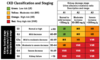
outline stages of CKD

A
  • note that for stages 1 and 2 evidence of kidney damage is needed! E.g. proteinuria, or abnormalities on scanning
  • stages 3-5 are defined by GFR alone
  • stages 1-3 are common, whereas 4-5 are not
3
Q

staging : G and A

A
4
Q

starting dialysis

A
  • there is an argument to leave it for as long as possible
  • most patient need ≤8.8 ml/min
5
Q

management of CKD in GP

A
  • commonly co exists with eg heart failure and diabetes - screen high risk groups
  • urinalysis
    • proteinuria must be quantified - spot urine sample
    • increases the risk of progression to CKD
  • referal
  • identify and treat CV risk factors
6
Q

which GN typically progresses to CKD

A

IgA nephropathy - 25% in 10-30 years

7
Q

causes

A
  • Diabetes: type 2>1
  • Glomerulonephritis: commonly IgA nephropathy
  • Unknown
  • Hypertension or renal vascular disease
  • Artery walls are thickened and narrowed, meaning less blood and oxygen gets to the kidneys. Ischaemic injury and glomerulosclerosis – diminished ability to filter blood.*
  • Falling GFR activates RAAS, causing further hypertension.*
  • Vicious cycle.*
  • Renal artery stenosis
  • Ischaemia/hypertensive nephrosclerosis
  • Microangiopathic e.g. HUS/TTP
  • Polycystic kidneys
  • Pyelonephritis and reflux nephropathy
  • Small vessel vasculitis
  • Tubulointerstitial:
    • Nephritis – uveitis
    • ADPKD
    • Reflux nephropathy
  • Post-renal (obstructive)
8
Q

risk of CKD after AKI

A
  • After AKI some tubules are lost, meaning that the other nephrons hyperfilter in order to take up the efforts of the lost nephrons. This compensation means the creatinine returns to normal but as the remaining nephrons are working harder they burn out quicker
  • Therefore, there is a risk of CKD developing or progressing, and patients must be informed of this after AKI
  • Patients must be monitored for the development or progression of CKD after AKI episode for 2-3 years, even if the serum creatinine has returned to baseline
9
Q

first line management of a person with kidney damage presenting with loin to groin pain

A

insert a catheter - may relieve obstruction if it is below bladder level

10
Q

screening

A

important, all high risk patients are screened

11
Q

test criteria

A

minimum of 2 samples at lest 90 days apart

12
Q

eGFR

A
  • estimates GFR
  • MDRD4 equation: takes into account serum creatinine, sex, age and ethnicity
  • correction factor for women and black race
13
Q

muscle masse effects on creatinine measuremente of GFR

A
  • Over-estimates GFR is muscle mass is low
  • Under-estimates if muscle mass high
  • Only valid if serum creatinine is stable
14
Q

how does pregnancy effect GFR

A

it tends to increase during pregnancy

15
Q

cystatin C

A
  • protein that can be used to estimate GFR
  • useful when a previous kidney function test was inconclusive or needs confirmed, or in overweight, elderly of muscly patients
16
Q

eGFR 3a, eGFR cystatin C >60ml/min and no other evidence of kidney disease - CKD?

A

no

17
Q

define accelerated progression of CKD

A
  • Sustained decrease in GFR ≥25% and a change in GFR category within 12 months OR
  • Sustained decrease in GFR of 15ml/min/1.73m2 per year
18
Q

risk factors for CKD progression

A
  • CVD
  • PROTEINURIA – more likely to progress
  • YOUNGER – longer to progress
  • AKI
  • Hypertension
  • Diabetes
  • Smoking
  • African, Afro-Caribbean or Asian family origin
  • Chronic use of NSAIDs
  • Untreated urinary outflow tact obstruction
19
Q

what are the 4 principles of management

A
  1. Slow progression
  2. Reduce CV risk
  3. Identify and treat complications of CKD
  4. Prepare for RRT
20
Q
  1. slow progession
A

control BP and reduce proteinuria

  • BP: aim for <140/90 mmHg, or <130/80mmHg in those who also have DM/ACR>70
  • ACEi
    • reduce proteinuria and BP
  • good glycaemic control in DM
  • stop smoking
21
Q

what is expected when initially commencing an ACEi

A
  • reduce filtration pressure - small fall in GFR and rise in creatinine
  • fine if GFR decrease <25% and serum creatinine increase <25%
  • watch out for hyperkalaemia
22
Q
  1. reduce CV risk
A

BP, proteinuria, smoking, statins

  • offer atorvastatin 20mg
23
Q

symptoms of advanced uraemia

A
  • Yellow tinge
  • Involuntary muscle twitching
  • Encephalopathic: flapping tremor and confusion
  • Pericardial rub or haemorrhagic pericardial effusion (pericarditis)
  • Kussmaul breathing due to metabolic acidosis
    • The kidney normally excretes H+ ions
    • increased anion gap
  • bleeding
  • uremic frost
24
Q

why does uraemia cause bleeding

A
  • Uremia acts as an antiplatelet, less clot formation, meaning patients bleed and bruise more easily
25
Q

uremic frost

A
  • Established renal failure for years, trying to excrete/secrete toxins through skin pores – urea concentration increases in sweat. Evaporation of sweat with high urea concentration causes urea to crystallize and deposit on the skin.
  • Stale urine smell to skin
  • Extreme end
  • Have to progress very slowly to get to this point e.g. congenital
26
Q

at what GFR level do specific symptoms tend to occur

A

<20 ml/min - v late

27
Q

general symptoms of CKD

A
  • non-specific - tiredness, poor appetite, sleep disturbance, itch
  • Impaired urinary concentrating ability – symptoms occur earlier e.g. nocturia
28
Q

symptoms of anaemia

A
  • fatigue
  • muscle weakness
  • tachycardia
  • aortic flow murmur
29
Q

consequences of CKD

A
  • Local e.g. pain, haemorrhage, infection
  • Urinary e.g. haematuria, proteinuria, nocturia, oliguria
  • Impaired salt and water handling e.g. fluid overload and hypertension
  • Electrolyte abnormalities e.g. hyperkalaemia
    • Normally excrete potassium, can cause cardiac arrhythmias
  • Acid-base disturbance
  • ESRD
30
Q

CKD and CVD

A
  • share some common risk factors
  • CKD makes one susceptible to atherosclerosis and CV calcification from changes that normally wouldn’t.
  • CVD is 10-20 times higher in those with ESRD
31
Q

vitamin D and calcium in CKD

A
  • kidneys produce 1-25 OH vitamin D and are involved in the absorption of calcium
  • hypocalcemia leads to 2y hyperparathyroidism
  • Hyperparathyroidism maintains normal serum calcium at the expense of the bones: bone resorption: pain, fractures etc.
  • can progress to 3y hyperparathyroidism if one PT gland becomes autonomous
32
Q

phosphate levels in CKD

A
  • are high - not excreted
  • this also increases PTH secretion and has bone effects
  • phosphate is not removed efficiently by dialysis
33
Q

what gene is implicated in CKD and is involved in vitamin D and Calcium metabolism

A

FGF23

34
Q

what effect does high calcium and phosphate have on vessels and valves

A
  • calcify them
  • associated increased BP and CV risk
35
Q

clinical manifestations of bone abnormalities

A
  • Osteitis fibrosa cystica - high PTH
  • Adynamic: reduction in cellular activity (OC and OB) in bone, may be due to over treatment with vitamin D
  • Osteomalacia due to low vitamin D
  • Osteoporosis
  • Osteosclerosis
36
Q

dietary restrictions

A
  • restrict phosphate - eg dairy , chocolate
  • salt reduction, potassium restriction if elevated, fluid restriction
37
Q

active vitamin D medication

A

alfacacidol - active vitamin D that doesnt need metabolised by the kidneys

38
Q

phosphate binders

A
  • reduce gut absorption of phosphate
  • taken with food
  • calcium based eg Adcal
  • aluminium eg Alucaps - need aluminium monitoring
  • non-calcium based eg Sevelamer
39
Q

EPO and CKD

A
  • EPO is released in the kidney in response to hypoxia (decreased renal blood flow), it stimulates RBC production in bone marrow.
  • Levels fall in CKD, resulting in anaemia.
40
Q

who should be screened for anaemia

A

use Hb levels

  • At least annually with CKD G3
  • At least twice a year with CKD G4-5 not on dialysis
41
Q

at what GFR level is CKD likely to be the cause of anaemia

A
  • Becomes apparent at GFR <35 ml/min – CKD is likely to be the cause at this level, if no other cause, e.g. blood loss, folic acid/vitamin B12 deficiency, identified
  • diabetics are more at risk of CKD causing anaemia at a higher GFR
42
Q

target Hb

A
  • 100-120g/L
  • Lower levels of Hb are acceptable if the Hb fails to rise despite adequate iron replacement and erythropoietin therapy
43
Q

investigation of anaemia

A
  • renal function and baseline investigations
    • Hb
    • exclude other causes
    • determine iron status: check ferritin and iron stores (ferritin >100 (in absence of acute inflammatory response) and TSats >20%)
    • CRP
44
Q

what should not be used for anaemia investigation

A

EPO levels

45
Q

what can ferritin levels tell us

A
  • Low ferritin can be used to diagnose absolute iron deficiency, but cannot exclude iron deficiency if normal/high
46
Q

management of anaemia

A
  • Iron replete – can give iron supplements (usually IV iron)
  • ESA should not be initiated in the presence of absolute iron deficiency
  • If still anaemic:
    • ESA: (erythropoiesis stimulating agent): ARANESP, EPREX. Use if Hb <100-110g/dl despite no iron/haematinic deficiencies and in those who are likely to benefit
47
Q

how often is ESA given

A

every week/fortnight

48
Q

what happens to iron stores as EPO works

A

they are depleted - need regular top ups