Lecture 12 and (13?) : Oral Cavity and Submandibular gland Flashcards
If someone has an ulcer in the mouth for more than a week, what must you do?
Do a thorough examination of the oral cavity (90% are cancers)
Note: every single part of the oral cavity will be in the exam
Note: every single part of the oral cavity will be in the exam
Describe the surface anatomy of the oral cavity (FLOOR) ** EXAM
- Sublingual fold overlying sublingual gland
- Sublingual caruncles
- Opening ducts from sublingual gland
- Submandibular gland
- 1 opening at the bottom/base of the frenulum (of tongue)
- Frenulum of tongue
- Lingual vein (don’t need to know different lingual veins)
- Lingual artery
- Lingual Nerve

____ is a small fold of mucous membrane extending from the floor of the mouth to the midline of the underside of the tongue.
frenulum
Submandibular gland opening is at the base of the frenulum of the tongue.

What surface anatomy can you see underneath the tongue?
- Frenulum of tongue
- Lingual vein (don’t need to know different lingual veins)
- Lingual artery
- Lingual Nerve

Describe the surface anatomy of the back of the oral cavity
- Palatoglossal arch
- Palatophrayngeal arch
- Paltine tonsil
- May not be easily identifiable
- Can be identified by looking at the other 2 arches.
- Soft palate
- Uvula

What are the 2 structures you can identify the palatine tonsils by?
1) Palatoglossal arch
2) Palatopharyngeal arch

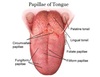
Describe the anatomy of the Tongue
- Different papillion on the tongue
- Vallate papillae (v shaped)
- Pharyngeal part of tongue (at the back)
- Foramen cecum (where the pharyngeal part of the tongue meet)
- Where the thyroid develops
- Filiform papillae
- Fungiform papillae
What makes up the walls of the oral cavity?
The oral cavity consists of a roof (the hard and soft palate),
floor (the geniohyoid, myohyoid and tongue)
lateral walls (fascia and a buccinator).
It opens anteriorly to the oral fissure and
posteriorly to the oropharyngeal isthmus.
Describe the Skeletal framework of the oral cavity
Roof
- Palatine process of the maxilla
- Horizontal plate of the palatine bone
- Sphenoid bone that contributes to the roof of the oral cavity
Floor
- Either side by the mandible ramus
- Lower part by the body of the mandible

What makes up the lateral walls of the oral cavity?
The lateral walls of the oral cavity are formed by the cheeks.
Each one consists of fascia and a t_hin layer of skeletal muscle (buccinator)_ sandwiched between skin and oral mucosa.
The buccinator muscle is one of the muscles of the facial expression and it is in the same plane as the superior constrictor muscle of the pharynx.
These two muscles provide continuity between walls of the oral and the pharyngeal cavities.
The muscle holds the cheeks against the alveolar arches and keeps food between the teeth when chewing. As with other muscles of facial expression it is innervated by CN VII.

Describe the floor of the oral cavity
Floor of the oral cavity is formed by:
- Paired mylohyoid muscle (1) connected in midline by a raphe. They attach to the hyoid bone, posterior part of the mandible. The most superficial part of the oral cavity (to get into oral cavity, have to cut this muscle).
- Nerve Innervation: V3
- Paired geniohyoid muscles (2) (deep to mylohyoid) originates from hyoid muscle and goes to inferior mental spines.
- Nerve Innervation: C1 (C1 also innervates thyrohyoid muscles, embryologically one muscle)
- Tongue (composed of extrinsic and intrinsic muscles) anteriorly attaches to superior mental spinal process.
-
Extrinsic muscles: muscles which originate outside the tongue, e.g. palatine, hyoid (glossus = tongue)
- Palatoglossus (3), styloglossus (4), hyoglossus (5) and genioglossus (6)
-
Intrinsic muscle: within tongue
- Superior longitudinal, vertical, transverse and inferior longitudinal
- (Believed to be) Mylohyoid muscle (but is now thought to be wrong)
- Posterior and lateral part of the mylohyoid muscle has no attachment
-
Raphe
- Mylohyoid meet at the raphe
-
Geniohyoid
- Deep to the mylohyoid
*
- Deep to the mylohyoid

Describe the “gateway” in the oral cavity
RIght at the back of the mylohyoid, there is a gateway filled with facia. This is called the Triangular Aperture.
Formed by:
1) Mylohyoid muscle
2) Superior constrictor
3) Middle constrictor
Some important structures go through the oral cavity through here.

Describe the floor of the oral cavity
1- the paired mylohyoid muscle (1)
connected in midline by a raphe.
2- the paired geniohyoid muscles (2).
3- the tongue [composed of extrinsic and
intrinsic muscles].
-
Extrinsic muscles (E): palatoglossus
(3) , styloglossus (4), hyoglossus (5) and - *genioglossus** (6).
- Intrinsic muscle: superior longitudinal,
vertical, transverse and inferior
longitudinal.
*Floor Of Oral Cavity
Floor of the oral cavity is formed by:
- Paired mylohyoid muscle (1) connected in midline by a raphe. They attach to the hyoid bone, posterior part of the mandible. The most superficial part of the oral cavity (to get into oral cavity, have to cut this muscle).
* Nerve Innervation: V3 - Paired geniohyoid muscles (2) (deep to mylohyoid) originates from hyoid muscle and goes to inferior mental spines.
* Nerve Innervation: C1 (C1 also innervates thyrohyoid muscles, embryologically one muscle) - Tongue (composed of extrinsic and intrinsic muscles) anteriorly attaches to superior mental spinal process.
-
Extrinsic muscles: muscles which originate outside the tongue, e.g. palatine, hyoid (glossus = tongue)
- Palatoglossus (3), styloglossus (4), hyoglossus (5) and genioglossus (6)
-
Intrinsic muscle: within tongue
- Superior longitudinal, vertical, transverse and inferior longitudinal

Describe the Muscles of the Tongue
2 groups
Intrinsic muscles (Origin and insertion lies within the tongue)
- Vertical
- Transverse
- Superior longitudinal
- Inferior longitudinal
Extrinsic muscles (Origin and insertion is outside of the tongue)
- Palatoglossus
- Styloglossus
- Hyoglossus
- Genioglossus

Name the extrinsic muscles
Palatoglossus (from the palate)
Styloglossus
Hyoglossus (from hyoid bone)
Genioglossus

Describe the Internal muscles of the tongue
- Vertical
- Transverse
- Superior longitudinal
- Inferior longitudinal

What muscles of the tongue are not innervated by the hypoglossal nerve?
Extrinsic: Palatoglossus muscle
(from the palate so is innervated by CNX)

Describe the Roof of the oral cavity
The roof of the oral cavity or palate
consisted of an anterior hard palate
(1) and posterior soft palate (2).
The hard palate is covered by
mucosa and is formed by the
palatine process of the maxillae
(anterior 3/4) and the horizontal
plates of the palatine bones
(posterior 1/4).

The soft palate hangs off the posterior edge of the
hard palate. Much of its bulk is made of _____ and
________ within the mucous membrane of its oral
surface. It consists of ______________
The soft palate hangs off the posterior edge of the
hard palate. Much of its bulk is made of mucous and
serous glands within the mucous membrane of its oral
surface.
It consists of an aponeurosis and 5 pairs of
muscles and acts as a valve during the co-ordinated
process of swallowing to prevent reflux of material
into the nasopharynx.
Muscles of the soft palate
What is the difference between geniohyoid and genioglossus
Geniohyoid goes down from the mental spine of mandible to the hyoid bone
Genioglossus goes from the mental spine up to the body of the tongue.

What is the hyoglossus muscle?
- The structure indicated is the hyoglossus muscle of the tongue.
- The hyoglossus muscle is one of the extrinsic muscles of the tongue.
- The muscles involved with the tongue consist of intrinsic muscles which lie within the tongue itself, and the extrinsic muscles which attach to the tongue and are responsible for depression, elevation, protraction and retraction of the tongue.
- There are four sets of extrinsic tongue muscles:
- Genioglossus
- Hyoglossus
- Palatoglossus
- Styloglossus
- Origin: Greater horn and body of hyoid bone
- Insertion: Lateral aspect of tongue
- Action: Depression and retraction of tongue
- Innervation: Hypoglossal nerve (cranial nerve XII)

Label


Where are the 2 ‘potential spaces’
What runs through these spaces?
1) Deep to the mylohyoid and lateral/superfiicla to hyoglossus
-a) Hypoglossal nerve (goes through via the ‘gateway’)
-b) Lingual nerve
-c) Deep lingual vein
(hypoglossal and lingual nerve are in the same plane from lateral-plane. But lingual is higher than the hypoglossal nerve)
2) Deep to hyoglossus and lateral to genioglossus
-a) Lingual artery
-b) Dorsal lingual vein
(goes deep to the hypoglossus)

_______ vein is the vein you are looking at at the base of the togue
Deep Lingual Vein

Deep Lingual vein runs in the _____ potential space, running with the _______
Dorsal lingual vein runs with the ______and runs _________________
Deep Lingual vein runs in the 1st potential space, running with the hypoglossal nerve
Dorsal lingual vein runs with the artery and runs deep to the hyoglossus

Where does the glossopharngeal nerve run?
Between Internal and External carotid
Goes through the aperature
Sits deep to the hyoglossus muscle.

What nerves run in the potential spaces on either side of the hyoglossus?
1) Lingual nerve (1st)
2) Hypoglossal (1st potential space)
3) Glossophargneal nerve (2nd potential space) –> wrong?

If you are preforming surgery (e.g. removing submandubular gland) and you have reached the hyoglossus, it means that you have damaged the…..
Important landmark
damaged: CN12 (hypoglossal nerve)
and Lingual nerve

What is the submandibular gland
The submandibular glands are bilateral salivary glands located in the face.
It has a superficial and deep part.
Wraps around the free edge of the mylohyoid.
Therefore if you are surgically trying to get to the submandibular gland, you must think about the 1st and 2nd potential space and the risk to the hypoglossal and lingual nerves
Their mixed serous and mucous secretions are important for the lubrication of food during mastication to enable effective s_wallowing_ and aid digestion.

What structures run across the submandibular gland?
There are a number of structures running across the gland:
1) facial vein,
2) facial artery
3) lymph nodes.

The taste of the posterior 1/3 of the tongue comes from ________
Glossophargneal nerve

Describe the nerve (sensory and motor) supply of the tongue
Sensory
- Anterior 2/3
- General sensation: lingual nerve
- Special sensation: chroda tympani
- Posterior 1/3
- General sensation: glossopharyngeal
- Special sensation: glossophargneal
Motor
- Hypoglossal nerve
- Intrinsic muscles
- Genioglossus
- Hyoglossus
- Styloglossus
- Vagus nerve
- Palatoglossus

The roof of the oral cavity or palate consisted of________ and ______
The roof of the oral cavity or palate consisted of an anterior hard palate (1) and posterior soft palate (2).

All muscles that form the oft palate are innervated by ___________________
All muscles of the soft palate are innervated by the pharyngeal plexus (CN X) except for the tensor veli palatine (CN V3).

Name the muscles of the soft palate
Tensor veli palatini (1) has two components (connected by a narrow tendon at pterygoid hamulus):
- A vertically oriented part originating from the base of the skull and the cartilage of the pharyngotympanic tube;
- A horizontal fibrous palatine aponeurosis which fans out to meet its opposite number.
Its function is to tense the soft palate and open the pharyngotympanic tube.
Levator veli palatini (2) arises from the apex of the petrous temporal bone and from the pharyngotympanic tube. It passes down to insert into the palatine aponeurosis.
It pulls the soft palate up and back to shut off the nasopharynx.
Palatopharyngeus (3) takes origin from the superior surface of the palatine aponeurosis and adjacent hard palate, and arches down posteriorly on the inner aspect of the pharynx to the thyroid cartilage. The palatine tonsil sits just anterior it.
- The part of the muscle fixed to the hard palate elevates the larynx and pharynx during swallowing.
- The part attached to the palatine aponeurosis helps to narrow the oropharyngeal isthmus.
Palatoglossus (4) arises from the inferior surface of the palatine aponeurosis and passes down in front of the palatine tonsil to insert into the side of the tongue.
It helps narrow the oropharyngeal isthmus and elevate the tongue.
Musculus uvulae (5) passes back from the hard palate and fuses with its counterpart in the uvula. They elevate and retract the uvula helping levator veli palatine close the pharyngeal isthmus.

The arch closer to the back of the oral cavity and closer to the pharynx is the
Paltopharyngeal arch

Describe the Innervation of the Soft Palate (NOT muscle)
- Innervation (examinable!): Lesser and greater palatine n. through lesser and greater palatine foramen (V3)

Where are the greater and lesser Palatine foramen
Innervation and Arterial Supply of Soft Palate
Innervation (examinable!): Lesser and greater palatine n. through lesser and greater palatine foramen (V3)

Describe the Lmyphatic Drainage of the Oral cavity
Oral cavity mainly drains into zone 1, posterior part drains into zone 2
