7 - The Motor System Flashcards
(40 cards)
How many neurones is the motor system made up of and where do these neurones originate from?
2 unlike 3 in the sensory
- Upper motor neurone from primary motor cortex in CNS
- Lower motor neurone from ventral horn of spinal cord or nuclei in brainstem, both in CNS
- LMN projects onto skeletal muscle
Where are upper motor neurones NOT found?
- Cerebellum
- Basal ganglia
Where in the spinal cord do motor neurones run?
- Corticospinal tract
(85% lateral to distal mucles for fine motor control, 15% anterior to proximal postural)

Where are lower motor neurones found and what is their function?
- Ventral horn of spinal cord or motor-nuclei in brain stem
- Cause muscle contraction
- Involved in spinal reflexes, sensory neurones from muscle spindles synapse on LMN in ventral horn of spinal cord

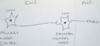
How does the patella reflex work?
- Spindle muscle fibres detect stretch so sensory neurone activated and synapses on LMN in L3
- Sensory neurone activates L3 LMN so contraction of quadriceps
- Sensory neurone also branches off and synapses with inhibitory interneurone at L5 to cause hamstring relaxation

How can lower motor neurones be damaged?
CNS and PNS lesions as body in CNS but axons in PNS!!!
What are signs of a lower motor neurone lesion?
- Hypotonia: due to loss of muscle activation
- Areflexia: no LMN to complete reflex arc
- Dennervation Muscle atropy: LMN supplies trophic factors to muscle so lost
- Fasiculations:due to upregulation of muscle nAChRs to try to compensate for denervation
- Weakness/Flaccid paralysis
- Fibrillations
- Weakness: dennervation

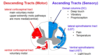
What is the connection like between most upper and lower motor neurones?
Via inhibitory interneurones mostly net inhibition until spindles send excitatory sensory neurones to overcome the inhibitory, e.g wanting to stand up
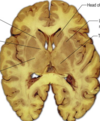
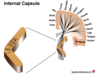
What is the internal capsule and what are the different parts of it?
- Genu: UMNs that supply face
- Anterior Limb
- Posterior limb
White matter structure situated in the inferomedial part of each cerebral hemisphere of the brain carrying motor and sensory information. Encapsulates the thalamus
Carpal tunnel of hand

What is a peduncle?
White matter connecting a hemisphere
What is the pathway of upper motor neurones from the motor cortex to the lower motor neurones for spinal nerves?
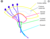
- Corona radiata
- Internal capsule
- Cerebral peduncle in the midbrain
- Pons
- Medullary pyramids
- Decussation of the pyramids (in the caudal medulla)
- Lateral corticospinal tract (in the lateral funiculus of the cord)
- Ventral horn
- Synapse (directly but usual indirectly via inhibitory interneurones) on LMNs
Where are upper motor neurones found and what is their function?
- Cell body and axon in primary motor cortex (pre-central gyrus) and synapse onto LMN at nuclei or ventral horn
- Excite LMNs by direct synapses or inhibit LMNs by projecting onto inhibitory interneurones
- Net effect is inhibitory so lesion of UMN leads to hyperactivity of muscles

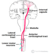
How do upper motor neurones travel when they are travelling to spinal nerves as their target?
- Cell bodies lateral/medial in motor cortex depending on which spinal nerve/homunculus
- Axons descend through corona radiata then through the internal capsule (space between lentiform nucleus and thalamus)
- Radiations that create the corona radiata come very close and descend through the cerebral peduncle in the brainstem, the pons and medullary pyramids
- At the pyramids there is decussation
- Axons descend in lateral corticospinal tract to ventral horn of required spinal level where they will synapse with LMN

How do upper motor neurones travel when they are travelling to cranial nerves (using the facial nerve as an example) as their target?
- UMN cell body is lateral in motor cortex as going to facial nerve nucleus where they will synapse with LMN
- UMNs supplying the upper face (top eyelid above) prject contralterally and ipsilaterally
- UMNs supplying the lower face project contralaterally only

If there was a stroke affecting the left upper motor neurone in the facial area of the homunculus, what would be the motor loss to the muscles of facial expression and why?
- Loss of muscles on lower half of right face due to decussation and the upper right face being supplied by right UMN too
- FOREHEAD SPARING of occipitofrontalis
- Whereas if you had Bell’s palsy this is a lesion to the facial nerve so complete paralysis

How could you tell if ipsilateral facial muscle weakness was due to an MCA stroke or Bell’s palsy?
- Bell’s palsy is lesion of facial nerve so all muscles affected, e.g cannot raise eyebrow. LMN lesion
- Stroke may be forehead sparing so can still raise eyebrow. UMN lesion
What is the corticobulbar/corticonuclear tract?
Two-neuron white matter motor pathway connecting motor cortex to medullary pyramids, and are primarily involved in carrying the motor function of the non-oculomotor cranial nerves
What is the ventral corticospinal tract?
- 15% of UMN do not decussate in the inferior medullary pyramids, they continue ipsilaterally until the level of the LMN and then they decussate
- For proximal postural muscles
- Other 85% of UMNs decussate at pyramids and travel in lateral corticospinal tract for distal musculature
What is the golgi tendon reflex?
- Proprioceptive sensoryreceptor organ that senses changes in muscle tension and activated in high tension to protect the bone and muscles
- Inhibits the lower motor neuron in a reflex to protect bone and muscle (biceps curl example) and excites the antagonising muscle

What are signs of an upper motor neurone lesion?
- Hypertonia: loss of descending inhibition by inhibitory interneurones
- Spasticity: LMN still in tact but no inhibition
- Hyperreflexia: still LMN
- Clasp Knife rigidity: golgi tendon reflex
- Weakness
- Disue atropy not due to loss of trophic factors
- Babinski’s reflex (Extensor plantar response)
- Acute flaccid paralysis: spinal shock
What would the upper and lower limbs look like in an upper motor neurone lesion?
Upper: spastic flexed
Lower: spastic extended
STILL HAVE REFLEXES
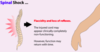
What is spinal shock?
- In the acute phase (days) of UMN lesion
- Flaccid paralysis with areflexia (like in LMN lesions) but then after a few weeks tone increases (becoming hypertonia) and reflexes become exaggerated (hyperreflexia)
- Related to neuroplasticity in the spinal cord and LMN shutting down following UMN lesion
What is Babinski’s sign?
Present in babies and UMN lesions

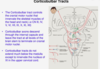
In the lateral corticospinal tract what is the topography of the trunk, arm and legs?
Arm most medially then trunk then leg laterally