Introduction Flashcards
CSF fluid is produced by what and in what specific locations throughout the ventricular system?
CSF is produced by the choroid plexus, which is located:
- Body and temporal horn of each lateral ventricle.
- Roof of third ventricle.
- Roof of fourth ventricle.
There is NO choroid plexus in the cerebral aqueduct or occipital or frontal horns of the lateral ventricles.
What is cytotoxic edema?
- Cytotoxic edema is cell swelling caused by damaged molecular sodium-potassium ATPase ion pumps. It can affect both gray and white matter.
- Cytotoxic edema is caused by cell death, most commonly due to infarct. Water ions trapped inside swollen cells feature reduced diffusivity.
What is vasogenic edema?
- Vasogenic edema is interstitial edema caused by increased capillary permeability.
- It is seen primarily in the white matter, as there is more interstitial space. Vasogenic edema is caused most commonly by neoplasm, infection, or infarct.
What is interstitial edema?
- Interstitial edema is caused by imbalances in CSF flow, most commonly due to obstructive hydrocephalus.
- Presents on imaging as periventricular fluid, often called “transependymal flow of CSF”, even though it is unlikely that CSF actually flows across the ependymal cells.
Herniation Patterns
Subfalcine Herniation
- Subfalcine herniation is seen when the cingulate gyrus slides underneath the falx.
- May rarely cause compression of the anterior cerebral artery ACA against the falx, resulting in infarction.
- Contralateral hydrocephalus may result from foramen of Monro obstruction, resulting in ventricular entrapment.
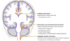
Ventricular Anatomy and CSF Flow
- The ventricular system consists of two lateral ventricles and midline third and fourth ventricles.
- The foramen of Monro connects the lateral ventricles with the third ventricle.
- The cerebral aqueduct of Sylvius connects the third ventricle with the fourth ventricle.
- The fourth ventricle continues inferiorly as the central canal of the spinal cord. The fourth ventricle also drains into the subarachnoid space and basal cisterns via three foramina:
- Paired foramina of Luschka (Luschka is lateral). Single foramen of Magendie (Magendie is medial).

What are the four recesses of the third ventricle?
- Chiasmatic (supraoptic recess)
- Infundibular recess
- Suprapineal recess
- Pineal recess

What are some causes of communicating hydrocephalus?
- Subarachnoid hemorrhage
- Meningitis (ie TB and Cocci)
- Tumor infiltration of leptomeninges (ie leptomeningeal carcinomatosis, disseminated oligodendroglial-like leptomeningeal tumor of childhood)
- Post-op complication
- Normal Pressure Hydrocephalus
What can cause obstructive hydrocephalus?
- Congenital aqueduct stenosis
- Obstructive tumor or mass (ie tectal plate glioma or colloid cyst
Downward (Uncal) Transtentorial Herniation
Downward transtentorial hernia on results in inferomedial displacement of the medial temporal lobe uncus through the tentorial notch, causing compression on the brainstem and adjacent structures.
- The ipsilateral cranial nerve III oculomotor nerve may be compressed, leading to pupillary dilation and CN III palsy (eye is “down and out”).
- Compression of the ipsilateral posterior cerebral artery PCA may cause medial temporal/ occipital infarct.
- Upper brainstem Duret hemorrhages are caused by shearing of perforating vessels due to downward force on the brainstem.
- Compression of the contralateral cerebral peduncle against Kernohan’s notch causes a hemiparesis ipsilateral to the herniated side.

Upward Transtentorial Herniation
Upward transtentorial herniation is superior transtentorial hernia on of the cerebellar vermis due to posterior fossa mass effect. The main complication of upward transtentorial herniation is obstructive hydrocephalus from aqueductal compression.
What are the basal cisterns?
The basal cisterns, also known as the perimesencephalic cisterns, are CSF-filled spaces surrounding the midbrain and pons. Compression or effacement of the basal cisterns may be a sign of an impending or actual herniation.

Cerebellar Tonsillar Herniation
- Downward displacement of the cerebellar tonsils through foramen magnum causes compression of the medulla.
- Compression of medullary respiratory centers is often fatal.
Causes of T1 shortening (hyperintensity) include?
MNEMONIC: GFPMMM
- Most commonly: Gadolinium, Fat, and Proteinaceous substance.
- Some paramagnetic stages of blood both intra- and extracellular Methemoglobin
- Melanin.
- Mineralization (copper, iron, manganese)
- Slowly-flowing blood.
- Calcium (rarely when dispersed, not in bone). It is much more common for calcium to be hypointense.
Differential Diagnosis for Gyriform Enhancemet
- Herpes encephalitis - caused by latent HSV-1 in the trigeminal ganglion. The medial temporal lobes and cingulate gyrus are usually affected first and demonstrate gyral enhancement due to in amma on, petechial hemorrhage, and resultant BBB breakdown. The involved areas typically also demonstrate reduced diffusivity. (can be necrotizing or hemorrhagic)
- Meningitis may cause gyral enhancement in addition to the more typical leptomeningeal enhancement.
- Subacute infarct can demonstrate gyriform enhancement lasting approximately 6 days to 6 weeks after the initial ischemic event.
- Posterior reversible encephalopathy syndrome PRES is a syndrome of vasogenic white ma er edema triggered by altered autoregulation that may demonstrate gyral enhancement. PRES may rarely exhibit restricted diffusion.

Differential Diagnosis of Ring Enhancement
MNEMONIC: MAGIC DR
- Metastasis: Hematogenous metastases are typically found at the subcortical gray-white junction.
-
Abscess: A pyogenic abscess is formed as a result of organization and sequestra on of an infection, featuring a central region of viscous necrosis.
- The key imaging findings of abscess are reduced diffusivity bright on DWI and dark on ADC caused by the high viscosity of central necrosis and a characteristic smooth, hypointense rim on T2-weighted images.
-
Glioma: High-grade tumors such as glioblastoma typically have a thick and irregular wall.
- Spectroscopy will be abnormal outside the margin of an enhancing high grade glial neoplasm secondary to nonenhancing in an infiltrative tumor. This is in contrast to a demyelinating lesion, abscess, and metastasis, where the spectral pattern returns to normal at the margin of the lesion.
- Perfusion MRI demonstrates elevated perfusion in a high grade glioma.
-
Infarct: Although subacute cor cal infarcts o en demonstrate gyral enhancement, ring enhancement can be seen in subacute basal ganglia infarcts.
- In contrast to neoplasm and infection, a subacute infarct does not have significant mass effect.
- Contusion: Both traumatic and nontraumatic intraparenchymal hemorrhage can show ring enhancement in the subacute to the chronic stage.
-
Demyelinating disease: The key finding in ring-enhancing demyelinatoing disease is lack of significant mass effect. The “ring” of enhancement is o en incomplete and “C” shaped.
- Although the typical finding is an incomplete rim of enhancement, a tumefactive demyelinating disease can look identical to a high-grade tumor.
- Radiation necrosis may look identical to a high-grade tumor. On perfusion, cerebral blood volume is generally low in radiation necrosis and typically increased in a high-grade glioma.

Differential Diagnosis of Pachymeningeal Enhancement
- Intracranial hypotension: Prolonged decrease in cerebrospinal uid pressure can lead to vasogenic edema in the dura.
- Intracranial hypotension clinically presents as a postural headache exacerbated by standing upright.
- Intracranial hypotension may be idiopathic or secondary to CSF leak from surgery or lumbar puncture.
- Imaging shows thick, linear dural enhancement, enlargement of the pituitary gland, and “sagging” of the cerebellar tonsils. There may also be subdural hemorrhage due to traction effect on the cerebral veins.
- Postoperative: dural enhancement may be seen postoperatively.
- Post-lumbar puncture: diffuse dural enhancement is occasionally seen 5 of the time after routine lumbar puncture.
- Meningeal neoplasm, such as meningioma, can produce a focal area of dural enhancement called a dural tail, due to reactive changes in the dura. Metastatic disease to the dura, most commonly breast cancer in a female and prostate cancer in a male, can cause irregular dural enhancement.
- Granulomatous disease, including sarcoidosis, tuberculosis, and fungal disease, can produce dural enhancement, typically of the basal meninges of the skull base.

Differential Diagnosis of Leptomeningeal Enhancement
- Meningitis (either bacterial, viral, or fungal) is the primary consideration when leptomeningeal enhancement is seen.
- Leptomeningeal enhancement in meningitis is caused by BBB breakdown due to inflammation or infection.
- Fine, linear enhancement suggests bacterial or viral meningitis.
- Thicker, nodular enhancement suggests fungal meningitis.
- Leptomeningeal carcinomatosis, also called carcinomatous meningitis, is spread of neoplasm into the subarachnoid space, which may be due to primary brain tumor or metastatic disease.
- CNS neoplasms known to cause leptomeningeal carcinomatosis include GEMCLOG
- Metastatic tumors known to cause carcinomatosis include lymphoma and breast cancer.
- Viral encephalitis may produce cranial nerve enhancement within the subarachnoid space.
- Slow vascular ow may mimic leptomeningeal enhancement at first glance
- Slow flow of peripheral vessels in moyamoya disease causes the ivy sign.

MR Spectroscopy
What are the metabolites and what are their properties?
What is increased and decreased in malignancy?
