Brain Tumors Flashcards
Approach to the evaluation of focal brain lesion
- Are there any tumor-related complications?
- Is the mass intra- or extra-axial?
- Where specifically is the lesion located?
- Are there multiple lesions?
- Distinct MR signal characteristics?
- Does the lesion enhance?
“Complications, location, location, location(s), MR signals and enhancement”
What are the 3 emergent complications of a brain tumor?
The three emergent complications of a brain tumor are the three H’s:
- Hemorrhage: Primary or metastatic brain tumors are often associated with neovascularity and tumoral vessels are more prone to hemorrhage than normal vasculature.
- Hydrocephalus: A tumor can cause hydrocephalus by blocking the flow of CSF. Posterior fossa tumors have increased risk of causing hydrocephalus by effacing the fourth ventricle.
- Herniation: The overall mass effect from the tumor is a combination of the tumoral mass and associated vasogenic edema, which may contribute to brain herniation.
Most common primary brain tumor to hemorrhage?
Glioblastoma
Hemorrhagic metastases to the brain include what primary neoplasms?
- Hemorrhagic metastases include melanoma, renal cell carcinoma, thyroid carcinoma (follicular), and choriocarcinoma.
- Note that this includes 3 / 4 of the neoplasms that metastasize hematogenously.
- Although breast and lung cancer metastases are less frequently hemorrhagic on a case-by-case basis, these two malignancies are so common that they should also be considered in the differential of a hemorrhagic metastasis.
What are the findings of an extra-axial mass?
- Findings of an extra-axial mass include a CSF cleft between the mass and the brain, buckling of gray matter, and gray matter interposed between the mass and white matter.
- Dural tails! (thank you helmer :)
- The presence of white matter edema is not specific to intra-axial masses. In particular, meningioma (an extra-axial dural neoplasm) is known to cause white matter edema of the underlying brain.
- Meningeal enhancement is seen more commonly in extra-axial masses (most commonly meningioma), but can also be seen in intra-axial masses.

What are the findings of intra-axial CNS masses?
- Findings of an intra-axial mass include the absence of intervening gray matter between the mass and the white matter.
- The presence of white matter edema is not specific to intra-axial masses. In particular, meningioma (an extra-axial dural neoplasm) is known to cause white matter edema of the underlying brain.
- Meningeal enhancement is seen more commonly in extra-axial masses (most commonly meningioma), but can also be seen in intra-axial masses.

Do mets enhance? Why?
- Metastases always enhance due to tumoral neo-vessels, which lack a blood-brain barrier.
Tumors hypointense on T2 include:
- Metastases containing desiccated mucin, such as some gastrointestinal adenocarcinomas. Note that mucinous metastases to the brain can have variable signal intensities on T2-weighted images, depending on the water content of the mucin. Hydrated mucin is hyperintense on T2-weighted images.
- Hypercellular tumors, including lymphoma, medulloblastoma, germinoma, and some glioblastomas.
Tumors that are hyperintense on T1 include:
- Metastatic melanoma (melanin is hyperintense on T1-weighted images).
- Fat-containing tumors, such as dermoid or teratoma.
- Hemorrhagic metastasis (including renal cell, thyroid, choriocarcinoma, and melanoma).
Overview the glial cells
- Astrocyte
- Oligodendrocyte
- Ependymal Cells
- Choroid Plexus Cells
Juvenile Pilocytic Astrocytoma (JPA)
What is it?
Imaging?
Specific circumstance with its association?
- Juvenile pilocytic (hair-like) astrocytoma (JPA) is a benign World Health organization WHO grade I tumor seen typically in the posterior fossa in children.
- Imaging shows a well-circumscribed cystic mass with an enhancing nodule and relatively little edema. When in the posterior fossa, JPA may compress the fourth ventricle.
- JPA can also occur along the optic pathway, with up to 1/3 of optic pathway JPA associated with neurofibromatosis type 1. Posterior fossa JPA is not associated with NF1.
What are fibrillary astrocytomas and what do they include?
- Fibrillary astrocytomas are infiltrative tumors that include low-grade astrocytoma, anaplastic astrocytoma, and glioblastoma multiforme (GBM).
- Astrocytomas can occur in the brain or the spinal cord.
What is a low-grade astrocytoma?
Imaging appearance?
- It is one of three fibrillary astrocytomas (low-grade, anaplastic, glioblastoma)
- Low-grade astrocytoma is a WHO grade II tumor that typically presents as a hyperintense mass on T2-weighted images, without enhancement. Imaging findings may be subtle.

Anaplastic astrocytoma
What is it?
Imaging appearance?
- It is one of three fibrillary astrocytomas (low-grade, anaplastic, glioblastoma)
- Anaplastic astrocytoma is a WHO grade III tumor. It features a range of appearances from thickened cortex (similar to low-grade astrocytoma) to an irregularly enhancing mass that may appear identical to glioblastoma. The natural history of the disease is an eventual progression to glioblastoma.
What is Glioblastoma Multiforme?
- It is one of three fibrillary astrocytomas (low-grade, anaplastic, glioblastoma)
- Glioblastoma multiforme (GBM) is an aggressive WHO grade IV tumor of older adults. It is the most common primary CNS malignancy. GBM has a highly variable appearance “multiforme” but typically presents as a white matter mass with heterogeneous enhancement and surrounding non-enhancing T2 prolongation.
- Most of the surrounding T2 prolongation is thought to represent infiltrative tumor.
- GBM is an infiltrative disease that spreads through white matter tracts, through the CSF, and subependymal.
- A GBM that crosses the midline via the corpus callosum is called a butterfly glioma. The differential diagnosis of a transcallosal mass includes glioblastoma, lymphoma, and demyelinating disease.

What is the DDx of a transcallosal mass?
The differential diagnosis of a transcallosal mass includes glioblastoma, lymphoma, and demyelinating disease.
Gliomatosis Cerebri
What is it?
Diagnostic Criteria?
Prognosis?
Typical imaging appearance?
Enhancement pattern?
- Gliomatosis cerebri is a diffuse infiltrative mid-grade (WHO II or III) astrocytoma that affects multiple lobes.
- Diagnostic criteria include involvement of at least two lobes plus extra-cortical involvement of structures such as the basal ganglia, corpus callosum, brainstem, or cerebellum.
- Gliomatosis has a poor prognosis and may degenerate into GBM.
- The typical imaging appearance is diffuse T2 prolongation throughout the involved brain.
- Gliomatosis exerts mass effect but typically does not enhance.

Diffuse T2 CNS prolongation can be seen in what entities?
- Diffuse T2 prolongation can be seen in several entities, typically in immunocompromised patients, including, gliomatosis, lymphoma, progressive multifocal leukoencephalopathy (demyelination caused by JC virus), and AIDS encephalopathy.
Oligodendroglioma
What is it?
The typical presenting patient?
Characteristic feature?
Variants?
- Oligodendroglioma is a WHO grade II tumor that usually presents as a slow-growing cortical-based mass in a young to middle-aged patient presenting with seizures.
- Oligodendrogliomas have a propensity to calcify (approximately 75% calcify).
- Variants such as oligoastrocytoma and anaplastic oligodendroglioma are much more aggressive.
- oligoastrocytoma is a mixed tumor with an astrocytic component. Although oligoastrocytoma can degenerate into GBM, typically prognosis is better than a pure GBM.
- Anaplastic oligodendroglioma is indistinguishable from GBM on imaging and has a poor prognosis.

Ependymoma
What is it?
In who and where does it occur?
Nick name of the tumor?
- An ependymoma is a tumor of ependymal cells that tend to occur in the posterior fossa in children and in the spinal cord in older adults.
- The pediatric posterior fossa ependymoma has been called the toothpaste tumor for its propensity to fill the fourth ventricle and squeeze through the foramina of Magendie or Luschka into the adjacent basal cisterns. Medulloblastoma, the most common pediatric brain tumor, also usually arises in the posterior fossa but does not typically squeeze through the foramina.
- The adult spinal ependymoma can occur anywhere in the intramedullary spinal cord. The main differential diagnosis of an intramedullary spinal cord mass is an astrocytoma, which tends to occur in younger patients. It is not possible to reliably differentiate spinal cord ependymoma from astrocytoma on imaging.

What is Lhermitte-Duclos?
Association?
Classical imaging finding?
Enhancement pattern?
- Lhermitte-Duclos, also called dysplastic cerebellar gangliocytoma, is a WHO grade I cerebellar lesion that is part hamartoma and part neoplasm.
- It is almost always seen in associated with Cowden syndrome (multiple hamartomas and increased risk of several cancers).
- The classical imaging finding is a corduroy or tiger-striped striated lesion in the cerebellar hemisphere. Enhancement is rare.
lhermitte-duClos associated with Cowden syndrome with a Corduroy striated lesion in the cerebellar hemisphere
What are embryonal tumors?
- Embryonal tumors represent a spectrum of WHO grade IV, aggressive childhood malignancies that are known as primitive neuroectodermal tumors (PNET).
- Intracranial PNET tumors are more commonly located in the posterior fossa but may occur supratentorially.
Atypical teratoid/rhabdoid tumor
What is it?
In who and what location does it occur?
Association?
- Atypical teratoid/rhabdoid tumor (ATRT) is a WHO IV, aggressive tumor that may appear similar to medulloblastoma, but occurs in slightly younger patients. The majority occur in the posterior fossa. ATRT is associated with malignant rhabdoid tumor of the kidney.
Medulloblastoma
What is it?
Where does it occur and what is its imaging appearance on CT and MR?
How do differentiate between other other most common entities?
What is Zuckerguss?
Where does it occur in young adults?
- Medulloblastoma is a WHO grade IV tumor of small-blue-cell origin. It is one of the most common pediatric brain tumors.
- Medulloblastoma most commonly occurs in the midline in the cerebellar vermis. It is slightly hyperattenuating on CT due to its densely packed cells and is accordingly hypointense on T2-weighted images and has low ADC values. The tumor is avidly enhancing and may appear heterogeneous due to internal hemorrhage and calcification.
- The low ADC values can be a useful finding to differentiate medulloblastoma from ependymoma and pilocytic astrocytoma, the two other most common childhood posterior fossa tumors.
- Leptomeningeal metastatic disease is present in up to 33% of patients. Sugar-coating (Zuckerguss) is the icing-like enhancement over the brain surface. Imaging of the entire brain and spine should be performed prior to surgery.
- When medulloblastoma occurs in a young adult (as opposed to a child), the tumor tends to arise eccentrically in the posterior fossa, from the cerebellar hemisphere.

Hemangioblastoma
What is it? Location?
Association?
Classic Appearance?
When in the spinal cord, it is associated with what?
- Hemangioblastoma is a highly vascular WHo grade I tumor associated with von Hippel-Lindau (VHL) syndrome that occurs most commonly in the cerebellum, medulla, or spinal cord. It only rarely occurs supratentorially.
- Although associated with VHL, only 30% of patients with hemangioblastoma have VHL. Hemangioblastoma in a patient with VHL has a worse prognosis.
- The classic appearance of hemangioblastoma is a cystic mass with an enhancing mural nodule. Prominent vessels are often seen as tubular areas of flow void. Less commonly, a hemangioblastoma may be solid or hemorrhagic.
- When in the spinal cord, hemangioblastoma is often associated with a syrinx.
Pleomorphic Xanthoastrocytoma
What is it?
Presentation?
Location? Appearance?
Main differential consideration and how to differentiate?
- Pleomorphic xanthoastrocytoma (PXA) is a low-grade WHo grade II astrocytoma variant.
- PXA is a rare tumor of childhood and adolescents, typically with a history of chronic epilepsy.
- The most common location of PXA is the temporal lobe, where it typically presents as a supratentorial cortical cystic mass with an enhancing mural nodule. The overlying dura may be thickened and enhancing.
- The main differential consideration, both by imaging and clinical presentation, is ganglioglioma; however, ganglioglioma does not usually cause dural thickening.

Ganglioglioma
What is it?
Common presentation?
Characteristic imaging appearance?
- Ganglioglioma is a rare, slow-growing neuroglial tumor that typically presents in an adolescent or young adult with medically refractory temporal lobe epilepsy.
- Ganglioglioma characteristically appears as a temporal lobe cyst and enhancing mural nodule, often with calcification. ganglioglioma may cause calvarial remodeling and scalloping.

What are the brain tumors with a cyst and an enhancing nodule?
- Juvenile Pilocytic Astrocytoma
- Pleomorphic Xanthoastrocytoma
- Gangliogloma
- Hemangioblastoma
(remember JPA and remember PXA has “astrocyoma” in it and then remember ganglioglioma is similar to PXA. . . then you just gotta remember hemangioblastoma)
What are the intraventricular tumors?
- Subependymoma
- Intraventricular Meningioma
- Choroid plexus papilloma/carcinoma
- Central Neurocytoma
- Subependymal Giant Cell Astrocytoma
MNEMONIC: “SMCkNS (smackens!) in the ventricles”
Central neurocytoma
What is it?
Imaging appearance?
- Central neurocytoma is a low-grade tumor likely of neuronal origin that occurs in young adults, from teenagers to young middle aged-patients. Prognosis is excellent.
- Typical imaging appearance is a lobulated mass attached to the septum pellucidum, with numerous intratumoral cystic areas. Calcification is common.
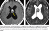
Choroid Plexus Papilloma/Carcinoma
What is it?
Image appearance?
Location in children and in adults?
How to differentiate between papilloma and CA?
- Choroid plexus papilloma is a rare intraventricular tumor. Choroid plexus papilloma is a low-grade (WHO I) neoplasm arising from choroid plexus epithelial cells. It is the most common brain tumor in babies <1 year old, but it may also occur in adults.
- T2-weighted images show a lobulated, heterogeneous or hyperintense mass that avidly enhances on T1-weighted MRI.
- In children, the atrium of the lateral ventricle is the most common location.
- In adults, the fourth ventricle is the most common location.
- Less commonly, choroid plexus papilloma may arise from the third ventricle or cerebellomedullary angle (shown).
- Choroid plexus papilloma and carcinoma (WHO III) are not reliably distinguishable.
Intraventricular Meningioma
Common location? In what population?
How to differentiate this from other intraventricular neoplasms?
- Intraventricular meningioma appears as a solid mass, typically in the trigone of the lateral ventricle. It tends to occur in older patients, similar to other meningiomas.
- Intraventricular meningiomas are typically hypercellular and homogeneously enhance, distinguishing them from other intraventricular neoplasms.

Subependymoma
What is it?
Found in what population?
Most common locations?
- Subependymoma is a non-enhancing low-grade tumor of unclear origin thought to arise from subependymal astrocytes, ependymal cells lining the ventricles, or common precursor cells.
- Subependymoma is a tumor of middle-aged and older adults. It is often found incidentally.
- The most common locations are the obex of the 4th ventricle (inferior 4th ventricle) or at the foramen of Monro in the lateral ventricle. The tumor usually doesn’t enhance.
- Despite their similar names, subependymoma is not related to subependymal giant cell astrocytoma or with ependymoma.
What are the most common tumors to metastasize to the dura?
- Breast (most common)
- Lymphoma
- Melanoma
- Small cell lung cancer
MNEMONIC: BLaMS (kinda goes with interventricular tumors mnemonic lol)

Differential diagnosis for posterior fossa mass in a child
MNEMONIC: Posterior fossa tumors really puts a “HaMpER” on a child’s life. . . and partly in intramedullar spinal tumors!
- Hemangioblastoma (3rd most common intramedullary spinal tumor)
- jPA (note that astrocytoma is most common intramedullary tumor in children)
- Medulloblastoma
- Ependymoma (note that ependymoma is most common intramedullary tumor in adults)
- ATRT

Differential diagnosis for a posterior fossa tumor in an adult
MNEMONIC: I found some “HAMM” in the posterior fossa of this adult.
- Hemangioblastoma
- Astrocytoma
- Medulloblastoma
- Metastasis
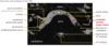
Cerebellopontine Angle (CPA) Masses
Anatomic overview
Important structures
Masses that can develop in CPA
- The cerebellopontine angle (CPA) is the region between the pons and cerebellum and the posterior aspect of the petrous temporal bone.
- Important structures of the CPA include the 5th, 7th, and 8th cranial nerves, and AICA.
- most lesions of the CPA are extra-axial and located in the CPA cistern itself, although some may arise in the internal auditory canal (IAC), temporal bone, or rarely intra-axially from the pons or cerebellum.
- CPA masses are more common in adults.
- Schwannoma, Meningioma, Arachnoid Cyst, Aneurysm, Epidermoid Cyst, Intra-axial Neoplasm.
CPA Meningiomas
Occurrence rate?
Special features?
Contrast to schwannoma?
- Although meningioma is overall the most common extra-axial mass in adults, it is only the second most common mass of the CPA, representing approximately 10-15% of all CPA masses.
- Meningiomas often feature a short segment of dural enhancement and may induce adjacent bony hyperostosis. Approximately 20% calcify, in contrast to schwannomas where calcification is rare.
- In contrast to schwannoma, a CPA meningioma does not enlarge the porus acousticus.

What is the most common tumor that occurs in the CPA?
Imaging appearance?
Characteristic appearance?
What nerves can it affect?
- Schwannoma of the vestibulocochlear nerve, also known as a vestibular schwannoma, is by far the most common cerebellopontine angle mass, representing greater than 75% of all CPA masses.
- Vestibular schwannoma is hyperintense on T2-weighted images and avidly enhances.
- The characteristic ice cream cone appearance describes the “cone” protruding through (and widening) the porus acousticus and the “ice cream” exerting mass effect on the cerebellar-pontine junction. Schwannoma may become cystic, especially when larger.
- Schwannomas of other cranial nerves in the CPA, including the facial or trigeminal nerves, are less common. Trigeminal schwannoma may extend into Meckel’s cave.

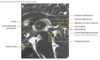
CPA Epidermoid Cyst
What is it?
Characteristic feature? Histopathology?
CT/MR appearance?
How to differentiate with arachnoid cyst?
- An epidermoid cyst is a congenital lesion arising from ectopic ectodermal epithelial tissue.
- Epidermoids progressively enlarge from desquamation of keratinized epithelium lining the cyst. The mass characteristically insinuates in between structures, encasing cranial nerves and vessels. Gross pathology features a characteristic “cauliflower-like” surface.
- On CT, epidermoid cysts may mimic arachnoid cysts and appear as a water-attenuation cystic structure. On MRI, an epidermoid cyst has similar signal characteristics to CSF on T1- and T2-weighted images. Unlike arachnoid cysts, an epidermoid does not usually suppress on FLAIR.
- Diffusion sequences show very bright signal on diffusion-weighted images.
- Postsurgical DWI follow-up is critical to detect any residual focus, which will be DWI bright.
- Rarely, epidermoids may exhibit signal hyperintensity on unenhanced T1-weighted imaging, also known as “white epidermoids.”

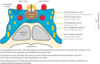
Cavernous Sinus Anatomy Overview
Pituitary Overview - Hormones and Imaging Appearance
- The pituitary gland is formed from Rathke’s pouch, which is a superior invagination from the primitive oral cavity. The pituitary gland sits in the sella turcica, a cup-shaped depression of the basisphenoid bone. The pituitary is composed of an anterior and a posterior lobe.
- Rathke’s pouch closes off to form a vesicle that involutes. Sometimes, the involution is incomplete and a cleft can be left behind, which may give rise to craniopharyngioma or Rathke’s cleft cyst.
- The anterior lobe of the pituitary produces and secretes endocrine hormones, including growth hormone, ACTH, prolactin, TSH, FSH, and LH (FLAT PiG).
- The posterior lobe of the pituitary is derived from neuroectoderm and is composed of axons from the hypothalamus, through which vasopressin and oxytocin are transported.
- The pituitary gland has a wide range of normal morphology, depending on patient age, sex, and hormonal/pregnancy status. The gland may be convex superiorly in adolescent or pregnant females. The normal posterior pituitary is hyperintense on T1-weighted MRI and is called the “posterior pituitary bright spot,” best seen on sagittal images.
Pituitary Macroadenoma
What is it? Typical presentation?
Image findings? Tendencies?
What is pituitary apoplexy?
- A macroadenoma is defined as an adenoma >10 mm in size. Patients usually present with mass effect (e.g., compression of the optic chiasm) rather than endocrine dysfunction.
- The bony sella is often enlarged. Macroadenomas may encase the carotid but tend not to narrow it. In contrast, meningiomas or metastases can narrow the carotid. Pituitary macroadenoma may bleed after medical treatment, producing a complex MRI appearance.
- Intra-tumoral hemorrhage is distinct from pituitary apoplexy. Pituitary apoplexy is a clinical syndrome of a severe headache and endocrine dysfunction caused by hemorrhage into an otherwise normal pituitary.

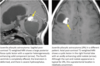
Craniopharyngioma
What is it?
Epidemiology?
Location?
Histological characteristics?
Image characteristic and enhancement pattern?
Contrast to rathke’s cleft cyst!
- Craniopharyngioma is the most common suprasellar lesion of childhood, arising from squamous epithelial remnants of Rathke’s pouch that produce keratin.
- Craniopharyngioma occurs in a bimodal age distribution. The majority of cases are lesions of childhood, but craniopharyngioma may occur uncommonly in late middle age.
- Most involve both the sella and suprasellar regions. Although, craniopharyngioma may rarely involve only the sella, it is almost always separate from the pituitary gland.
- Craniopharyngioma has potential for enamel production and almost always calcifies.
- The characteristic intracystic machine-oil seen on gross examination is composed of desquamated squamous epithelium, keratin, and cholesterol.
- MRI shows a complex cystic mass containing protein or blood products (hyperintense on T1-weighted images).
- There is avid enhancement of the solid elements and cyst walls.
- In contrast to Rathke’s cleft cyst, craniopharyngioma almost always enhances, is almost always calcified, and is almost always separate from the pituitary.

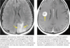
Rathke’s Cleft Cyst
What is it? Epidemiology?
Presentation?
Image appearance?
Contrast to craniopharyngioma!
- Similar to craniopharyngioma, Rathke’s cleft cyst is also a remnant of the embryologic Rathke’s pouch, the precursor of the anterior lobe of the pituitary gland. In contrast to craniopharyngioma, Rathke’s cleft cyst is made of simple columnar or cuboidal epithelium.
- While craniopharyngioma is the most common suprasellar lesion of childhood, Rathke’s cleft cyst is typically seen in middle-aged adults, twice as commonly in females.
- Rathke’s cleft cyst is reportedly very common in autopsy studies (up to 22% incidence), but clinically is usually asymptomatic or discovered incidentally.
- Imaging appearance is dependent on the protein content of the cyst. The intra-cystic fluid may be isointense to CSF if low protein and hyperintense on T1-weighted images if high protein. High protein content may cause incomplete nulling of the intracystic fluid on FLAIR.
- The claw sign represents enhancing pituitary tissue completely wrapped around the cyst.
- It is usually possible to distinguish craniopharyngioma from Rathke’s cleft cyst.
- Unlike craniopharyngioma, Rathke’s cleft cyst does not enhance (although rim enhancement is often seen) and does not calcify. Rathke’s cleft cyst may occasionally be inseparable from the pituitary, but craniopharyngioma is nearly always distinct.

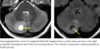
Optic Glioma
Epidemiology?
Prognosis?
Imaging appearance?
- An astrocytoma involving the visual pathway (optic nerve, optic chiasm, and optic tract) is the second most common suprasellar mass in children (craniopharyngioma is most common). A substantial minority of patients with optic pathway glioma have neurofibromatosis type 1.
- In contrast to the low-grade tumor of childhood, optic glioma is an aggressive tumor when it occurs in adults.
- Tumors are isointense on T1-weighted images, hyperintense on T2-weighted images, and usually enhance.

Hypothalamic Hamartoma
What is it?
Epidemiology? Presentation?
Characteristic location and imaging appearance?
- Hypothalamic hamartoma is not a true neoplasm, but represents ectopic hypothalamic neural tissue.
- It is a rare lesion of childhood that classically presents with precocious puberty and gelastic seizures (laughing spells).
- Hypothalamic hamartoma characteristically appears as a sessile mass between the pituitary stalk and the mammillary bodies.
- Hypothalamic hamartoma does not enhance and is isointense to gray matter.

The differential for suprasellar mass in a child:
- Craniopharyngioma
- Hypothalamic Hamartoma
- Langerhans Cell Histiocytosis Hypophysitis
- Optic pathway glioma
- Germ Cell Tumor
MNEMONIC: “CHLOG in the child’s suprasella”

The differential for suprasellar mass in an adult:
- Craniopharyngioma
- Rathke’s cleft cyst
- Aneurysm
- Meningioma
- Pituatary macroadenoma extension
- Lymphocytis or granulomatous hypophysitis
MNEMONIC: “CRAMPL in the suprasellar region of an adult”

Overview and Anatomy of Pineal Region
Where is the pineal gland located? What is located superiorly to the pineal gland?
What is the principle neuronal cell of the pineal gland and what does it produce?
Presence of BBB?
A mass lesion in the pineal region can cause compression of what structures?
Compression of the tectal plate results in what?
What are the steps in evaluating a pineal region mass?
- The pineal gland is located in the midline at the level of the midbrain. It is situated between the thalami at the posterior aspect of the third ventricle. The internal cerebral veins and vein of Galen are located superior and posterior to the pineal gland, respectively.
- The principal neuronal cell of the pineal gland is the pinealocyte, which is a modified retinal neuronal cell that is innervated by the sympathetic plexus originating in the retina. The pineal gland releases melatonin, which modulates the sleep/wake cycle.
- The pineal gland does not have a blood-brain barrier.
- A mass lesion in the pineal region may cause compression of the midbrain, compression of the cerebral aqueduct of Sylvius, or compression of the tectal plate.
- Compression of the tectal plate produces Parinaud syndrome, which is the inability to look up (upward gaze paralysis), pupillary light dissociation, and nystagmus.
- The first step in evaluating a pineal region mass is to determine if the lesion is arising from the pineal gland itself or from an adjacent structure.
- A mass of the pineal gland is extra-axial.
- Tumors of pineal cell origin tend to lift the internal cerebral veins, while tentorial meningiomas tend to depress the internal cerebral veins. The relationship of any pineal region mass to the internal cerebral veins is key for surgical planning and approach.