Maxillo-facial Trauma 01-22 Flashcards
An 18-year-old man presents with right periorbital edema and ecchymosis after an all-terrain vehicle collision. Physical examination shows enophthalmos, diplopia, and pain with eye movements. When asked to look upward from forward gaze, there is upward gaze restriction. A photograph is shown. Which of the following locations is most likely fractured?
A) Greater wing of sphenoid bone
B) Lamina papyracea of ethmoid bone
C) Orbital plate of frontal bone
D) Orbital process of maxillary bone
E) Posterior crest of lacrimal bone

The correct response is Option D.
Limitation with vertical gaze, as described in the vignette, is indicative of extraocular muscle (EOM) entrapment.
The lamina papyracea of the ethmoid bone contributes to the medial orbital wall. Fractures of the medial wall may result in medial rectus entrapment and restriction with lateral gaze. Such injuries can involve the ethmoidal/sphenoidal sinuses.
The greater wing of the sphenoid bone contributes to the lateral orbital wall. Fractures of the lateral wall are less common due to its increased strength, but at the wings of the sphenoid, bone can have serious effects on contents of the superior orbital fissure and may even involve the intraorbital portion of the optic (II) nerve.
The orbital plate of the frontal bone contributes to the superior orbital roof. Fractures of the superior roof may result in superior rectus and/or oblique entrapment. Such injuries can involve the frontal sinus, frontal lobe, supraorbital nerve, and/or supratrochlear nerve (resulting in loss of sensation of the forehead and upper eyelid).
The orbital process of the maxillary bone contributes to the inferior orbital floor. Fractures of the inferior floor may result in inferior rectus/oblique entrapment and restriction with upward gaze. Such injuries can involve the maxillary sinus and/or infraorbital nerve (resulting in malar and superior alveolar numbness).
The posterior crest of the lacrimal bone contributes to the medial orbital wall. Fractures of the medial wall may result in medial rectus entrapment and restriction with lateral gaze. Such injuries can involve the ethmoidal/sphenoidal sinuses.
An otherwise healthy 74-year-old man presents with traumatic brain injury from an open comminuted anterior and posterior table frontal sinus fracture sustained in a motor vehicle collision. Persistent clear fluid rhinorrhea is observed when the patient is upright. The patient is initially medically unstable, and surgical intervention is delayed by 5 days. Which of the following factors represents the greatest increase in risk for a central nervous system infection?
A) Cerebrospinal fluid leak
B) More than 48 hours before operative repair
C) Open fracture injury
D) Patient age
E) Presence of traumatic brain injury
The correct response is Option B.
There are many algorithms for treating frontal sinus fractures. Unfortunately, many of these patients have other injuries that may limit their surgical options. Conservative management has been shown to be successful at managing comminuted, displaced fractures, pre-injury comorbidities, or those with cerebrospinal fluid (CSF) leak. Factors that are not associated with an increased risk for serious infection include: preoperative CSF leak, persistent CSF leak, surgical procedure performed, age, gender, and penetrating or open injury. Factors that do impact the risk for serious infection include: greater than 48 hours from injury to operating room intervention, use of CSF catheter diversion, and soft-tissue infection.
A 20-year-old man has severe hypotension and bradycardia after sustaining multiple facial fractures in a motor vehicle collision. He has no other injuries. Repairing which of the following structures will most likely improve the patient’s symptoms?
A) Mandible
B) Mental nerve
C) Nasal bone
D) Orbital floor
E) Zygomatic arch
The correct response is Option D.
The oculocardiac reflex can precipitate marked bradycardia and hypotension in the setting of trauma with significant orbital and maxillofacial injury. Prompt identification and management with vagolytic agents or definitive surgical intervention may prevent morbidity or mortality. Patients who sustain maxillofacial trauma involving the orbit, most commonly the orbital floor, are at risk for developing the oculocardiac reflex. These patients tend to be young, and common symptoms include nausea and vomiting. The oculocardiac reflex is not static and may evolve during a patient’s clinical course.
Elderly patients are more likely than younger patients to have which of the following facial fractures?
A) Nonoperative Le Fort fracture from a bicycle accident
B) Nonoperative mandible fracture from a motor vehicle collision
C) Nonoperative maxillary fracture from a fall
D) Operative nasal fracture from interpersonal violence
E) Operative orbital fracture from a workplace injury
The correct response is Option C.
Elderly patients are more likely to sustain injury from falls than younger patients and are less likely to be in fights. Nonoperative maxillary fractures and nasal bone fractures are more common in elderly patients, and mandible fractures are more common in younger patients.
A 50-year-old man receives preoperative radiation therapy for a large calvarial osteosarcoma that will require a 9-cm2 craniectomy. Which of the following is the most appropriate material to use for reconstruction?
A) Bone allograft with bone morphogenic protein (BMP)
B) Calcium phosphate
C) Polyether ether ketone (PEEK) implant
D) Titanium mesh
The correct response is Option C.
Any calvarial defect greater than 6 cm2 should be reconstructed. Titanium mesh can be used for craniectomy reconstruction; however, radiologic surveillance of disease recurrence can be challenging with the degree of artifact seen on imaging. Polyether ether ketone (PEEK) implants are radiolucent and therefore do not produce artifact on surveillance imaging and can be customized based on the size of the planned resection. Bone allograft with bone morphogenic protein (BMP) is contraindicated in cases of malignancy since BMP is a growth factor that induces bone formation and is involved in tumorigenesis. Calcium phosphate can be used for smaller calvarial defects, but it is associated with a high risk for infection and would be contraindicated in a patient who received preoperative radiation therapy.
A 50-year-old man sustained multiple visceral injuries, prolonged loss of consciousness, and a fracture of the orbital floor in an accident 3 months ago. He deferred repair of the orbital floor fracture at the time of injury, but is now seeking help for symptoms related to the fracture. The patient is alert and oriented. Orbital floor fracture repair is most likely to achieve correction of which of the following findings in this patient?
A) Blindness
B) Ectropion
C) Enophthalmos
D) Globe volume
E) Vertical restriction
The correct response is Option C.
Orbital floor fractures risk the pathoanatomy of changing the volume of the orbit, which can affect the position of the globe, as well as entrapping the inferior rectus muscle, leading to restriction of globe movement. Therefore, indications for surgery include: vertical globe dystopia (vertical change in globe position from inferior displacement of floor), enophthalmos (retro-positioning of globe from increased orbit volume), and globe entrapment (inability for globe to look up).
For this patient 3 months after unrepaired orbital floor fracture, enophthalmos would be the most likely correctable by delayed orbital floor repair, which reduces the orbital volume closer to its pre-injury state, thereby restoring the globe to its pre-injury position.
Blindness would be from optic nerve injury, which would not be improved by orbital floor fracture repair.
Ectropion would usually be from lower eyelid contracture, which would not be improved by orbital floor fracture repair.
Globe volume refers to volume of the globe itself (not the orbit volume), which would not be improved by orbital floor fracture repair.
Vertical restriction could be due to muscle entrapment, which needs to be repaired urgently otherwise permanent damage to the muscle could result. This patient is presenting 3 months after injury for delayed repair, therefore orbital floor fracture repair would no longer be able to repair a permanently damaged muscle entrapment scenario.
A 30-year-old woman presents with the dental findings shown in the diagram. Which of the following best describes the dental relationship?
A) Angle class I
B) Angle class II
C) Angle class III
D) Negative overjet
E) Overbite

The correct response is Option B.
The images show an Angle class II relationship. The Angle classification system describes the relative positions between the mesial buccal cusp of the maxillary first molar and the buccal groove of the mandibular first molar. Angle class I molar relationship implies that the mesiobuccal cusp is in line with the buccal groove. In an Angle class II molar relationship, the maxillary mesiobuccal cusp is anterior to the mandibular buccal groove. Class II is subdivided into two divisions. In class II, division 1, patients have minimal crowding of the maxillary teeth and proclination of the upper central incisors, and a significantly increased overjet. In a class II, division 2 relationship, the central incisors are retroclined. An Angle class III molar relationship exists when the maxillary mesiobuccal cusp lies posterior to the mandibular buccal groove.
A 23-year-old man sustains a severe right orbital floor fracture in a physical altercation. Reconstruction with a pre-bent orbital floor plate and intraoperative CT scanning is planned. Which of the following is the most likely to be optimized using this imaging modality intraoperatively?
A) Operative time
B) Plate positioning
C) Rate of corneal abrasion
D) Rate of plate extrusion
E) Risk for lid malposition
The correct response is Option B.
Use of intraoperative computed tomography has been gaining traction in maxillofacial trauma. Stated benefits include decreased re-operation rate, improved accuracy of plate positioning, and decreased postoperative enophthalmos. Although the use of intraoperative CT scans may increase time in the operating room, it has no effect on rates of corneal abrasion, lid malposition, or plate extrusion.
Which of the following best represents the likelihood that a patient with a frontal sinus fracture would have a concurrent intracranial injury?
A) 1%
B) 15%
C) 30%
D) 55%
E) 90%
The correct response is Option D.
In an acute trauma setting, the recognition of mild traumatic brain injury (mTBI) is a diagnostic challenge as there are often competing diagnoses that take immediate priority. Furthermore, within this cohort, patients with craniofacial fractures have been shown to be at risk for delayed or missed diagnosis for all degrees of TBI, although with a higher likelihood of missed or delayed diagnosis for mTBI compared with moderate to severe TBI. Previously, it was hypothesized that facial fractures buffered the forces transmitted during blunt head trauma, thereby protecting intracranial structures. This conceptual framework has since been questioned as evidence has mounted that individuals with facial fractures are at increased risk for head injury. The biomechanics resulting in different types of facial fractures and the amount of force required to fracture the different components of the facial bony structure have been well described. The nasal bone has the lowest tolerance for fracture at 25 to 75 lbs, while the frontal bone has the highest tolerance at 800 to 1600 lbs. Recent studies have proposed that craniofacial fractures can serve as clinical markers for brain injury and Mulligan et al. suggest that the prevalence of overall head and cervical spine injuries in the setting of facial fractures is high enough to warrant a change in current protocols.
In this context, the prevalence of mTBI and moderate to severe TBI in patients with isolated facial fractures in the National Trauma Databank (NTDB) was evaluated, and further characterized the association of isolated facial fractures with different degrees of TBI in patients with mild, moderate, and severe TBI. Facial fractures can serve as objective clinical markers for the potential presence of mTBI and moderate to severe TBI in trauma patients. As mTBI patients have been shown to benefit from simple, easy-to-administer educational interventions, trauma patients with facial fractures may benefit from automatically receiving education about mTBI and TBI recovery, given the clinically meaningful prevalence of mTBI and TBI in this population. As one moves up the craniofacial skeleton, the forces are transmitted more reliably to the intracranial space. Therefore, a frontal sinus fracture is at extremely high risk (usually a 45 to 65% chance) of having an associated intracranial injury.
A 27-year-old man sustained multiple facial fractures when he was involved in a motorcycle collision. On arrival to the emergency department, blood pressure is 80/50 mmHg and heart rate is 150 bpm. Significant retropharyngeal bleeding is noted. Trauma workup reveals no other injuries. CT angiography shows active bleeding from the right maxillary artery. Angioembolization is planned and massive transfusion protocol is initiated. Which of the following is the most appropriate intravenous resuscitation in this patient?
A) Fresh frozen plasma (FFP) and packed red blood cells (pRBC) in a 1:1 ratio; discontinuation of crystalloids
B) FFP and pRBC in a 1:1 ratio; crystalloids via rapid transfuser (max rate)
C) FFP and pRBC in a 1:4 ratio; crystalloids at 125 cc/h
D) FFP and pRBC in a 1:4 ratio; discontinuation of crystalloids
E) FFP and pRBC in a 4:1 ratio; crystalloids via rapid transfuser (max rate)
The correct response is Option A.
For initiation of a massive transfusion protocol, transfusing fresh frozen plasma (FFP) and packed red blood cells (pRBC) at a 1:1 ratio and discontinuing intravenous crystalloids is the most appropriate next step in patient management.
Massive Transfusion Protocol guidelines have been set forth by the American College of Surgeons through its Trauma Quality Improvement Program (TQIP). Recommendations for initiating a massive transfusion protocol include:
Beginning universal blood product infusion rather than crystalloid or colloid solutions,
Transfusing universal pRBC and FFP in a ratio between 1:1 and 1:2 (FFP:pRBC),
Transfusing one single donor apheresis or random donor platelet pool for each six units of pRBC.
It is also suggested to deliver PRBC and FFP by a rapid transfuser and through a blood warmer, and that the initial rate of transfusion should restore perfusion while allowing for “permissive hypotension” until the operation or angioembolization to stop the bleeding begins.
A 65-year-old man develops a hemorrhagic stroke requiring decompressive craniotomy. The bone is found to be unusable and a customized polyetheretherketone prosthesis is planned. Which of the following is the most common complication of using this material?
A) Cerebrospinal fluid leak
B) Contour deformity
C) Dehiscence
D) Hematoma
E) Infection
The correct response is Option E.
Reports on using polyetheretherketone (PEEK) as an alloplast for cranial reconstruction vary in terms of outcomes and complications. The larger studies conclude that it is a reliable material compared with other alloplastic alternatives and has the advantage of being custom made for a variety of craniofacial defects. However, infection remains the most common complication, and choosing this material should be weighed against the risk for microorganism seeding through, wound dehiscence, hematogenous spread, or indolent colonization of the wound bed.
A 32-year-old man comes to the emergency department after being hit in the right eye. Examination shows enophthalmos, hyphema, and numbness over the cheek. There is no diplopia. CT scan shows a large orbital floor fracture with herniation of contents into the maxillary sinus. Which of the following findings requires urgent management?
A) Cheek numbness
B) Enophthalmos
C) Hyphema
D) Maxillary sinusitis
E) Orbital floor fracture
The correct response is Option C.
Hyphema is marked by presence of blood in the anterior chamber and is an emergent concern. It can lead to permanent damage to the vision. All the other options are urgent concerns but can be addressed after the hyphema is treated.
A 65-year-old man who wears glasses sustained a massive injury to the left side of the face causing a ruptured globe with total loss of the upper and lower eyelids. Which of the following is the best aesthetic option to recommend?
A) Eye patch
B) Hemifacial prosthesis
C) Ocular prosthesis
D) Orbital prosthesis
The correct response is Option D.
In this case the patient has had severe orbital trauma with loss of lids and globe. Natural-looking and functional total-lid reconstruction is challenging. Lids would be needed to support an ocular prosthesis. An orbital prosthesis would likely provide this patient a comfortable and aesthetically satisfactory prosthesis. Eyeglasses can help mask the seam of the prosthesis. The hemifacial prosthesis is larger than necessary for this patient and has unnatural seams. An eye patch would not improve symmetry or be reconstructive.
In a patient undergoing reconstructive cranioplasty, an increased rate of complications is most likely if which of the following is present?
A) Frontal location
B) Occipital location
C) Parietal location
D) Sphenoidal location
E) Temporal location
The correct response is Option A.
Early decompressive craniectomy is a life-saving maneuver for certain traumatic brain injuries and can be performed far forward in the theater of war. Patients treated with decompressive craniectomy for combat injuries are a unique understudied population. Outcome of treatment of this patient cohort has been previously reported using a standardized cranial defect treatment protocol using custom alloplast implants. Two subgroups of patients (large endocranial dead space and frontal orbital bar injuries) were identified as often having higher rates of complications than other cranial reconstruction cohorts.
A 28-year-old man is brought to the emergency department after sustaining injury during a motor vehicle collision. Cranialization of the frontal sinus is planned. Which of the following best describes the components of cranialization?
A) Removal of the anterior table, reconstruction of the posterior table with a titanium plate, and closure of the dura
B) Removal of the posterior table, sinus mucosa, and closure of the sinonasal tract
C) Repair of both the posterior and anterior tables with bioabsorbable plates, and obliteration of the frontal sinus
D) Repair of the anterior table and obliteration of the frontal sinus
E) Repair of the posterior table with bioabsorbable plates, removal of the sinus mucosa, and closure of the dura
The correct response is Option B.
Cranialization involves removal of the posterior table (not repair), closure of the dura, sinonasal tract, and obliteration of the sinus mucosa. Management of the anterior table is as indicated.
Surgical repair of the anterior table is indicated if there is nasofrontal duct involvement, or, in the absence of nasofrontal duct involvement (such as a minimally displaced anterior table), patient desire for a better aesthetic outcome. If there is nasofrontal duct involvement, the nasofrontal duct and frontal sinus can be obliterated (repair of the anterior table and obliteration of the frontal sinus).
Bioabsorbable or titanium plates can be used to fixate the fractured anterior table. It is not used for the posterior table.
A 30-year-old man sustains significant mid face injuries following a motor vehicle collision, and has a large laceration in the vicinity of the medial canthal region. Canalicular injury is confirmed intra-operatively. Which of the following is the most appropriate method for repairing this patient’s canalicular injury?
A) Delayed dacryocystorhinostomy
B) Direct microsurgical suture repair
C) Healing by secondary intention
D) Immediate dacryocystorhinostomy
E) Placement of silicone canalicular stents
The correct response is Option E.
When canalicular injury is suspected, the lacrimal system should be investigated for patency. Typically, this involves performing a Jones I and II test to determine if fluorescein navigates from the lower lid fornix into the nose. If canalicular interruption is suspected and identified, the proximal and distal stumps of the canaliculus are joined by placing a silicone stent and leaving this in place for 3 to 6 months to allow for healing.
Direct microsurgical suturing is not preferred because of the high likelihood of cicatricial obstruction.
Dacryocystorhinostomy is generally reserved as a “salvage” procedure for patients who have lacrimal obstruction after being treated with a stent. Healing by secondary intention is incorrect since it would likely result in canalicular obstruction.
A 40-year-old man and his 80-year-old father are assaulted. They both have facial fractures. The older victim is more likely to have which of the following?
A) Decreased chance of noncraniofacial injuries
B) Higher mortality
C) Less severe injuries
D) Mandibular body fracture
E) Shorter hospital stay
The correct response is Option B.
In recent years many publications focused on craniofacial injury in the elderly as not only the mode of trauma differs compared with the younger population, but also the associated injuries and morbidities. In general, most related comorbidities in patients older than 60 to 65 (depending on the study) versus those younger are worse, including: longer hospital stays, need for assistance upon discharge, more severe injuries, likely to have noncraniofacial injuries like limb and spine fractures, and, of greatest concern, a much higher death rate. In a recent article though, Mundinger et al, showed that panfacial and mandible fractures were more common in the nongeriatric population, whereas mid face, orbital, and condylar fractures were more common in those older than 60 years of age.
A 20-year-old man desires correction of a depressed, retracted, post-tracheostomy scar. Which of the following is the best recommendation for improving the scar?
A) Perform autologous fat grafting and laser resurfacing
B) Reconstruction tracheal ring and detach adhesions
C) Scar excision and interposition of acellular dermal matrix
D) Scar excision and reapproximation of strap muscles
E) Scar revision
The correct response is Option D.
After decannulation, the tracheostomy site heals by secondary intention. Often the patient is left with a soft, small asymptomatic scar. On occasion, the scar is painful and the skin has adhesions to tissue deep to the strap muscles. This may lead to pulling and retraction with swallowing as well as a scar that is not aesthetically pleasing to the patient. The depressed retracted tracheostomy scar requires reapproximation of platysma and approximation of the sternothyroid and sternohyoid for correction. Fat grafting is unlikely to address retraction or fully correct the depression. Laser resurfacing and fat grafting will have minimal improvement of retraction. Several studies support use of cadaver materials or fascia to support the coverage of the strap muscles when tissue is missing or heavily damaged. The tracheal ring does not need to be reconstructed for routine tracheostomy scar revision. Care must be taken when working around the trachea. Communication with anesthesia about oxygen content and fire risk is important for surgical safety.
A patient underwent open reduction and internal fixation of naso-orbital-ethmoid fractures 12 months ago, and epiphora was noted on follow-up examination. After 6 months of observation and persistent epiphora, which of the following is the most appropriate next step to evaluate the function of the patient’s nasolacrimal system?
A) Conjunctivorhinostomy tube placement
B) Continued observation, as function is likely to return
C) Jones tests
D) Lacrimal system flushing
E) Schirmer tests
The correct response is Option C.
The Jones test is used to evaluate lacrimal drainage. Divided into two parts, the Jones I test investigates lacrimal outflow under normal physiologic conditions. A drop of sterile 2% fluorescein solution or a moistened fluorescein strip is placed into the conjunctival fornix and a cotton-tipped wire applicator is placed into the inferior nasal meatus in the region of the ostium of the nasolacrimal duct at 2 and 5 minutes to check for fluorescein. As this test occasionally yields abnormal results in normal patients, it is not uniformly performed. The Jones II test determines the presence or absence of fluorescein when the residual fluorescein is flushed from the conjunctival sac with clear saline to determine whether there is reflux of fluorescein.
Naso-orbital-ethmoid (NOE) fractures can be challenging fractures, and either through direct instrumentation with transcanthal wiring or from the fractures themselves, the lacrimal drainage system can be affected. Postoperative epiphora can be very common and is present in at least 50% of patients who have undergone open reduction and internal fixation (ORIF) of an NOE fracture. After 3 to 6 months approximately half of this epiphora resolves, with the other half of patients (25%) requiring consideration for other investigations to evaluate lacrimal drainage. Schirmer test is used to evaluate dry eyes and is not appropriate in this patient.
A 22-year-old man is evaluated for multiple facial fractures after he was assaulted. Which of the following fractures is most likely associated with an increased risk of temporomandibular joint dysfunction?
A) Bilateral parasymphyseal mandible
B) Comminuted unilateral condylar mandible
C) Complete Le Fort I maxillary
D) Displaced unilateral subcondylar mandible
E) Unilateral zygomaticomaxillary
The correct response is Option B.
Temporomandibular joint (TMJ) dysfunction symptoms are serious, often overlooked complications of facial fractures and their treatments. They can range from clicking and pain to locking, malocclusion, and trismus. Overt ankylosis can occur in rare circumstances. Fractures that result in significant disruption of the condylar/glenoid apparatus are more likely to result in TMJ dysfunction symptoms than more anatomically remote fractures. Condylar fractures are most susceptible to post-fracture TMJ dysfunction. This is especially true in comminuted condylar head fractures. One recent study demonstrated an increase in TMJ dysfunction symptoms in patients with condylar fractures and concomitant contralateral mandibular body/angle fractures. Le Fort I and zygomaticomaxillary complex (ZMC) fractures are unlikely to be associated with TMJ symptoms.
A 7-year-old boy is brought to the emergency department after being injured in a domestic violence incident. Physical examination shows bruising around the right eye. The patient reports pain and nausea when looking upward. A CT scan shows an entrapped inferior rectus muscle. Three weeks later, the floor of the orbit is repaired with an orbital floor implant. One year later, he continues to have diplopia. Which of the following is the most likely reason for the persistent diplopia?
A) Exophthalmos
B) Location of prosthesis
C) Nerve damage
D) Persistent swelling
E) Timing of surgery
The correct response is Option E.
Timing of pediatric orbital floor fractures is well studied. Unlike adult fractures, significant delays for surgery in children, especially more than 7 days after injury, is associated with varying degrees of diplopia. Many consider this pathology an emergency and should be treated within 24 hours. Assuming a typical, standard of care approach is performed well from a technical standpoint, only delays in time to treat were shown to predict such a poor outcome.
Proper reduction of a zygomaxillary complex (ZMC) fracture requires reduction and realignment of which of the following?
A) Zygomatic arch, infraorbital rim, alveolus
B) Zygomaticofrontal suture, infraorbital rim, alveolus
C) Zygomaticofrontal suture, zygomaticomaxillary buttress, infraorbital rim
D) Zygomaticofrontal suture, zygomaticomaxillary buttress, orbital floor
E) Zygomaticomaxillary buttress, infraorbital rim, alveolus
The correct response is Option C.
A zygoma fracture involves displacement of the zygoma that articulates with the frontal bone, maxilla, and sphenoid. In order to stabilize the fracture after adequate reduction, the zygomaticofrontal, zygomaticomaxillary buttress, and infraorbital rim need to be fixated. If there is a large (>2 cm2) defect in the orbital floor after reduction, reconstruction of the orbital floor is also necessary to prevent enophthalmos.
While the nasomaxillary buttress is one of the vertical buttresses of the face, the zygoma does not articulate with the nasal bones.
A 10-year-old boy is brought to the physician after sustaining a nondisplaced fracture of the mandibular body in a fall. Soft diet is recommended. Two days later, he is brought back to the office and reports pain in the right mandibular lateral incisor when drinking cold liquid. The base of the defect appears yellow and is tender when probed. Examination shows a lingual fracture of the tooth crown. On the basis of these findings, which of the following is the deepest layer of exposed tooth?
A) Cementum
B) Dentin
C) Enamel
D) Pulp cavity
E) Root canal
The correct response is Option B.
This patient has a fracture of the tooth crown that extends through the dental enamel into the deeper parts of the tooth. This is evidenced by the sensitivity to touch and cold, a finding not characteristic of a fracture limited to the enamel. The yellow color to the base of the fracture indicates exposed dentin, which resides just under the hard outer enamel layer of the tooth. If the fracture had extended deeper into the pulp cavity, the area where the vessels and nerves reside, the base of the fracture would appear as a blood-filled cavity. These injuries often challenge the viability of the tooth and often require drilling and packing of the pulp space (root canal). The fracture described is of the crown and there is no indication that it involves the root of the tooth or the surrounding structures. Cementum is a bone-like covering of the tooth root and would not be affected by this injury.
The Ellis classification provides a useful system of categorizing these injuries. There are 9 categories:
Ellis I: enamel fracture. The tooth is non tender and treatment is smoothing of the rough surfaces and, possibly, application of a filling or amalgam.
Ellis II: fracture of the enamel and dentin. Tooth is tender to air, cold, and probing and the base of the defect often appears yellow.
Ellis III: involves the enamel, dentin, and the pulp space. The tooth is sensitive as in Ellis II, but the base of the defect appears red or bloody.
Ellis IV: a nonviable tooth.
Ellis V: luxation of the tooth.
Ellis VI: tooth avulsion.
Ellis VII: displacement without fracture.
Ellis VIII: fracture of entire crown.
Ellis IX: fracture of deciduous teeth.
Which of the following is the most common complication of a fracture to the temporal bone?
A) Cerebrospinal fluid leak
B) Facial nerve injury
C) Hearing loss
D) Meningitis
E) Temporomandibular joint ankylosis
The correct response is Option A.
Cerebrospinal fluid leak is the most common complication of a temporal bone injury. The majority of these will resolve spontaneously within a week. If they persist longer, then there is higher risk for meningitis, but this is not common. Facial nerve injury is the second most common injury and prognosis is dependent on the severity and delay of onset. Incomplete nerve loss or delayed onset is associated with a better prognosis for recovery. Hearing loss is the third most common complication seen with this fracture. Temporomandibular joint ankylosis is an unlikely sequela of this type of injury.
In adults, which of the following bones is most commonly fractured in isolated orbital floor fractures?
A) Ethmoid
B) Frontal
C) Lacrimal
D) Maxillary
E) Zygomatic
The correct response is Option D.
Most isolated orbital fractures involve the orbital floor made up of the maxillary bone. The maxillary bone is quite thin behind the infraorbital rim and is perforated by the infraorbital nerve passing in a canal below it. Most pure blow-out fractures involve the orbital floor with the maxillary bone making the majority of the orbital floor.
A retrospective study by Hwang et al. evaluated 391 patients with orbital bone fractures from a variety of accidents that were treated at the department of Plastic and Reconstructive Surgery, Inha University Hospital, Incheon, South Korea, between February 1996 and April 2008. The medical records of these patients were reviewed and analyzed to determine the clinical characteristics and treatment of the orbital bone fractures. The following results were obtained. The mean age of the patients was 31.1 years, and the age range was 4 to 78 years. The most common age group was the third decade of life (32.5%). There was a significant male predominance in all age groups, with a ratio of 4.43:1. The most common etiology was violent (assault) or nonviolent traumatic injury (57.5%) followed by traffic accidents (15.6%) and sports injuries (10.7%). The most common isolated orbital bone fracture site was the orbital floor (26.9%). The largest group of complex fractures included the inferior region of the orbital floor and zygomaticomaxilla (18.9%). Open reduction was performed in 63.2% of the cases, and the most common fracture reconstruction material was MEDPOR (56.4%) followed by a resorbable sheet (41.1%). The postoperative complication rate was 17.9%, and there were no statistically significant differences among the reconstruction materials with regard to complications. During follow-up, diplopia, hypoesthesia, and enophthalmos occurred as complications; however, there was no significant difference between porous polyethylene sheet (MEDPOR) and resorbable sheet groups.
Long-term epidemiologic data regarding the natural history of orbital bone fractures are important for the evaluation of existing preventative measures and for the development of new methods of injury prevention and treatment.
A patient underwent open reduction and internal fixation of naso-orbital-ethmoid fractures 12 months ago, and epiphora was noted on follow-up examination. After 6 months of observation and persistent epiphora, which of the following is the most appropriate next step in management of the patient’s nasolacrimal system?
A) Conjunctivorhinostomy tube placement
B) Continued observation
C) Dacryocystorhinostomy
D) Jones tests
E) Lacrimal system flushing
The correct response is Option D.
Naso-orbital-ethmoid (NOE) fractures can be challenging fractures, and either through direct instrumentation with transcanthal wiring or from the fractures themselves, the lacrimal drainage system can be affected. Postoperative epiphora can be very common and is present in at least 50% of patients who have undergone open reduction and internal fixation (ORIF) of an NOE fracture. After 3 to 6 months approximately half of this epiphora resolves, with the other half of patients (25%) requiring consideration for other investigations to evaluate lacrimal drainage.
A 22-year-old man comes to the office because of a history of nasal trauma with resultant nasal deformity, C-shaped septal fracture, and nasal obstruction. Two weeks after injury, he undergoes closed reduction of the nasal fractures, but significant nasal obstruction persists. Which of the following is the most likely reason for his residual nasal deformity and nasal obstruction?
A) Inadequate time of nasal casting
B) Nonunion of the nasal bones
C) Presence of a septal fracture
D) Turbinate hypertrophy
E) Unidentified septal hematoma
The correct response is Option C.
One of the most important causes of failure of closed reduction to address the nasal fracture is simultaneous nasoseptal fracture. Murray, et al. found 30 to 40% residual nasal deformity after closed reduction. The cadaver study showed failure consistently associated with a C-shaped nasoseptal deviation and fracture when the external nose deviated at least 1/2 of the nasal bridge width. The theory is that the interlocking of the septal fracture creates tension causing the nasal bone to displace.
Untreated septal hematoma results in thickening of the cartilage and nasal obstruction, but not with inadequate reduction. Nasal casting for 7 to 10 days is sufficient to allow the reduction to set. Nonunion is a rare cause of inadequate reduction, usually in comminuted or open nasal fractures. Turbinate hypertrophy can cause nasal obstruction but does not interfere with nasal bone reduction.
A 26-year-old woman who is at 32 weeks’ gestation sustains a traumatic head injury during a boating collision. CT scan shows subarachnoid hemorrhage and pan-facial fractures. The patient is cleared by the neurosurgeon for facial fracture repair. In the ICU, blood pressure is 112/70 mmHg and heart rate is 95 bpm. Fetal monitoring shows no distress. The patient is taken to the operating room and placed in supine position. On the operating table, blood pressure is 80/50 mmHg and heart rate is 130 bpm. Which of the following is the most appropriate next step in management?
A) Administer fluid bolus intravenously
B) Logroll the patient to the left
C) Obtain immediate chest x-ray study
D) Prepare and drape the patient for the planned procedure
E) Start vasopressors
The correct response is Option B.
The most appropriate next step in this scenario is to logroll the patient 4 to 6 inches (or 15 degrees) to the left, decompressing the inferior vena cava (IVC). Women in the second half of pregnancy may become hypotensive when placed in the supine position, caused by compression of the inferior vena cava by the enlarged uterus, reducing venous return to the heart by up to 30%. Spinal precautions should be maintained for any patient whose spine has not been appropriately cleared.
Vasopressors should be used as a last resort in restoring the blood pressure of pregnant trauma patients, as these drugs further reduce uterine blood flow, resulting in fetal hypoxia. The placental vasculature is exquisitely sensitive to catecholamine stimulation.
Crystalloid fluid resuscitation would be indicated if the patient’s vital signs did not return to baseline after repositioning and IVC decompression. Similarly, a chest X-ray could be obtained as part of the workup for unresponsive hypotension.
Ignoring this patient’s hemodynamic changes and proceeding with surgery would be a mistake, as the placenta would most likely be deprived of vital perfusion, resulting in fetal distress.
A 25-year-old man is brought to the emergency department after sustaining injury during a roll-over motor vehicle collision. CT scan shows multiple facial fractures and systemic injuries. Which of the following is a CONTRAINDICATION to nasotracheal intubation in a trauma patient?
A) Base of skull fracture
B) Bilateral mandibular condyle fractures
C) Cervical spine fracture
D) Depressed mental status
E) Fracture of the maxilla
The correct response is Option A.
Of the options listed, base of skull fracture is the only contraindication to nasal intubation. In these patients, there is a risk of the nasotracheal tube passing through the cribriform plate into the frontal lobes of the brain. Other absolute contraindications include mid face instability, suspected epiglottitis, coagulopathy, or apnea/impending respiratory arrest. Relative contraindications to nasotracheal intubation include nasal polyps, suspected nasal foreign bodies, recent nasal surgery, upper-neck hematoma or infection, and a history of frequent epistaxis. Depressed mental status, suspected cervical spine fracture, hypotension, and bilateral mandibular condyle fractures are not contraindications to nasotracheal intubation.
The options for intubating a trauma patient include nasal, oral, and surgical airways. The indications for nasotracheal intubation include intraoral and oropharyngeal surgery, complex intraoral procedures involving the mandible (e.g., segmental mandibulectomy, osteotomy, mandibular reconstructive procedures), and dental surgery.
The advantages of nasotracheal intubation include uninhibited access to the mouth as well as an enlarged surgical field. Despite these advantages, one must be cognizant of the contraindications and when in doubt (e.g., during initial advanced trauma life support management), the airway should be secured using either oral intubation or tracheostomy. Other drawbacks to nasotracheal intubation are that it often requires multiple attempts, there can be an abrupt rise in intracranial pressure, and it is difficult to attempt in an uncooperative patient.
In the presence of an isolated maxillary fracture nasal intubation may be preferred so that premorbid occlusion can be established.
The method of insertion of a nasotracheal tube includes using a well-lubricated tube with the cuff fully deflated. The tube should be inserted into either naris at a right angle to the face. Once the tube is beyond the nasopharynx, a laryngoscope is placed in the oral cavity and the tube is advanced under direct vision.
A 21-year-old man undergoes repair of the parotid gland and Stensen duct after sustaining an injury while being assaulted with a glass bottle. Ten days later, salivary leakage from the wound is noted. Which of the following is the most appropriate next step in management?
A) Pressure dressings and limited oral intake
B) Radiation therapy
C) Re-exploration of the wound and repair of the duct injury
D) Tympanic neurectomy
E) Observation only
The correct response is Option A.
Sialocele and cutaneous fistula may occur as a complication of a major salivary gland injury or after attempted parotid gland or duct repair. Prior to intervention, the surgeon should confirm the presence of salivary fluid in the aspirate by evaluating for amylase (>1000 U/L). This helps to rule out hematoma vs. infection. At this time, the accepted initial treatment of sialocele and cutaneous fistula is conservative management. This includes pressure dressings, repeated aspirations, limited intake by mouth, and the use of antisialagogues to decrease salivary flow. Most studies have shown that most sialoceles and fistulas will resolve within 2 to 3 weeks with this approach.
Secondary repair of the duct is generally difficult because of granulation and scar tissue. This runs the risk for delayed or poor wound healing as well as facial nerve injury. Radiation therapy has been used in the past but has been found to have a high failure rate in ductal injuries. More than 6 weeks is needed for gland atrophy to occur after radiation. There is also the risk of inducing malignancy, which has led to the abandonment of radiation for the treatment of sialoceles and cutaneous fistulas. Antisialagogues primarily have a role as an adjunct therapy to decrease salivary flow. Alone, they have had mixed reviews in the literature and have been found generally ineffective in major salivary duct injuries. Tympanic neurectomy has a high failure rate and does not speed recovery. Internalization of salivary flow could be considered for failures of conservative management. It has lower surgical risk than wound exploration and duct repair.
A 40-year-old man is evaluated for orbital fractures from a direct blow to the right eye. A swinging flashlight test is performed to evaluate a suspected afferent pupillary defect. Which of the following is the most appropriate description of this procedure?
A) The flashlight shone into the left eye causes both eyes to constrict; then when the light is shone in the right eye, the right eye constricts but the left eye does not
B) The flashlight shone into the left eye causes consensual constriction, and then both eyes dilate when the light is shone in the right eye
C) The flashlight shone into the left eye causes consensual constriction; then when the light is shone in the right eye, the right eye dilates while the left eye constricts
D) The flashlight shone into the left eye causes consensual dilation followed by paradoxical constriction when swung to the right eye
E) The flashlight shone into the left eye causes the left eye to constrict while the right eye remains dilated; both eyes are constricted when the light is shone in the right eye
The correct response is Option B.
A relative afferent pupillary defect, or Marcus Gunn pupil, results from an optic nerve dysfunction. The physical examination maneuver used to evaluate the optic nerve for a relative afferent pupillary defect is the swinging flashlight test described above. The most common cause of Marcus Gunn pupil is a lesion on the optic nerve, optic neuritis, or severe retinal disease but can also be caused by trauma. The normal response to light in one eye is constriction of both pupils and when the light is taken away both eyes dilate (consensual light reflex). In an abnormal exam the normal eye/optic nerve will cause a normal constriction of both pupils but the light shone in the affected eye will not have normal light perception (minimal pupillary constriction). The afferent pathway refers to the nerve impulse sent from the pupil to the brain via the optic nerve of one eye. The efferent pathway is the impulse sent back to both pupils via CN III and ciliary ganglia, causing both pupils to constrict.
A 21-year-old man is evaluated because of pain and double vision 2 weeks after being punched in the face. An orbital fracture is suspected. Which of the following characteristics of an orbital floor fracture is the most common indication for surgical repair?
A) Concomitant naso-orbito-ethmoid fracture
B) Contralateral zygomaticomaxillary complex fracture
C) Herniation of orbital contents with diplopia
D) Medial wall fracture without additional symptoms
E) Nondisplacement
The correct response is Option C.
The facial skeleton acts as a buffer and barrier against injury to the brain and deep structures. Reconstruction of the buttress system is integral to reforming the form and function of the facial skeleton.
Zygomaticomaxillary complex (ZMC) fractures, sometimes referred to as tripod fractures, may involve fractures of the following: zygomaticotemporal suture, maxillary sinus, and occasionally sphenoid bone. Correction involves reduction of the fracture, commonly with miniplate fixation or with 2-point wire fixation. These fractures can also involve the orbital floor and maxillary wall.
Symptoms may include numbness in the maxillary nerve (V2 of trigeminal (V) nerve), subconjunctival hemorrhage, diplopia, and enophthalmos. Orbital floor fractures can be isolated, as in a blowout, or involve other structures (e.g., orbital rim, orbital medial/lateral walls, orbital apex). Additional fractures are typically associated with higher-force injury and may require more extensive surgical repair.
Diagnostic study typically includes CT scan of the facial bones to assess the fractures and to delineate the extent of the facial trauma.
There are three main indications to treat isolated orbital fractures:
Entrapment of the extraocular muscles: this may cause injury and permanent dysfunction of the globe if not reduced.
Prevention of globe malposition: malposition of the globe may occur leading to diplopia or enophthalmos. This may not be apparent immediately after the injury because of soft-tissue edema. Reduction is indicated if there are signs of early enophthalmos, displacement of more than 50% of the orbital floor, or significant soft-tissue displacement.
Lateral orbital wall displacement: when the injury is more severe, the lateral orbital bone (sphenoid bone) may impact the orbital apex or even the mid-cranial fossa.
The CT scan should be examined for muscle entrapment, herniation of orbital contents, and associated fractures (naso-orbito-ethmoid, zygomaticomaxillary, Le Fort). Open reduction and internal fixation can be performed emergently or after edema has subsided, and usually after the orbit has been examined by an ophthalmologist for ocular injury.
Repair of the orbital floor can be done using several different types of materials—alloplastic and autologous. This is usually dictated by the surgeon’s preference and the extent of the injury.
A 50-year-old man is evaluated for facial trauma sustained after falling off a bicycle 5 days ago. The patient reports mid-facial pain and resolved swelling. Physical examination of the face shows no signs of trauma. Intraoral examination shows traumatic loss of tooth No. 8. He has no cervical spine tenderness or pain to active range motion. Which of the following is the most appropriate next step in management?
A) Obtain cervical spine MRI
B) Obtain maxillofacial CT scan to evaluate for facial fracture
C) Recommend elevation of the head of the bed and ice pack to the face
D) Recommend soft diet and referral to a dentist
E) No further management is necessary
The correct response is Option B.
The patient described most likely has a facial fracture and, thus, the most appropriate next step in management is to obtain a maxillofacial CT scan. A retrospective analysis of patients with facial trauma who had maxillofacial CT scan for the evaluation of facial fractures identified five independent criteria highly predictive of facial fracture. Any of the following five physical examination criteria – the Wisconsin criteria – were predictive of facial fracture: bony step-off or instability, periorbital swelling or contusion, Glasgow Coma Scale score less than 14, malocclusion, or tooth absence (sensitivity, 98.2%). Because the patient has a missing tooth, he should be evaluated with a maxillofacial CT scan. Although soft diet and eventual referral to a dentist as well as elevation of the head of bed with cool compress are reasonable recommendations, the first important step is obtaining a diagnosis.
Because the patient does not have tenderness of the cervical spine, there is no indication for obtaining cervical spine x-ray studies.
Systematic and efficient decision instruments in the management of multisystem trauma patients are critical in identifying all injuries including those of the maxillofacial skeleton and the cervical spine. The accurate use of systematic decision instruments could potentially result in a dramatic decrease in the number of CT scans and x-ray studies performed annually in the United States.
An 18-year-old woman is evaluated for persistent fullness of the left cheek 3 months after undergoing open reduction and internal fixation of a left zygomaticomaxillary complex fracture. She has no pain. The fracture was fixated at the inferior orbital rim (1.3-mm-thick titanium plate), the frontozygomatic suture (1.3-mm-thick titanium plate), and the lateral buttress (2.4-mm-thick titanium plate). On physical examination, the incisions are well healed. Which of the following is the most likely cause of this patient’s cheek fullness?
A) Excessive profile of the lateral buttress and orbital rim plate fixation
B) Malreduction of the fracture
C) Mucocele from the maxillary sinus
D) Residual soft-tissue edema
E) Subclinical hardware infection
The correct response is Option D.
Fullness of the soft-tissue envelope is expected after any severe trauma, regardless of location. This is further compounded by operative dissection to effect open reduction and internal fixation. Although most soft-tissue swelling around a fracture site dissipates in several weeks, there will be persistent thickening of the soft tissue in the area for many months after the trauma.
It is nearly impossible to “over-reduce” a zygomaticomaxillary (ZMC) fracture in the sagittal plane. Malreduction of these injuries characteristically results in flattening of the cheek (not fullness), vertical dystopia, or enophthalmos. Prominent hardware can create contour irregularities, but this would be localized and would not lead to global cheek fullness. Furthermore, the plate choices in areas where they could potentially be visible (i.e., the orbital rims) here are quite low profile and would be very unlikely to create excessive soft-tissue fullness. Hardware infection can rarely occur and lead to fullness of the cheek. Nevertheless, the absence of pain and the presence of healed wounds make this improbable. Mucocele from the maxillary sinus after repair of ZMC fracture is a rare but described entity and can result in localized soft-tissue fullness. These often occur in the context of a concurrent sinus infection and present many months to years after the injury. Soft-tissue swelling is usually accompanied by pressure and/or pain, and the symptoms can wax and wane. External drainage is not uncommon.
A 23-year-old man is brought to the emergency department 30 minutes after sustaining a self-inflicted shotgun wound to the face. Physical examination shows loss of soft tissue of the mid face with exposed mandible and maxilla, sonorous respiration, and periods of apnea. Heart rate is 100 bpm and blood pressure is 65/30 mmHg. Which of the following is the most appropriate course of management?
A) Assess and establish airway, control bleeding, perform secondary survey, stabilize cervical spine
B) Assess and establish airway, stabilize cervical spine, control bleeding, perform secondary survey
C) Assess and establish airway, stabilize cervical spine, perform secondary survey, control bleeding
D) Stabilize cervical spine, control bleeding, assess and establish airway, perform secondary survey
E) Take the patient to the operating room for debridement of facial wound with reconstruction
The correct response is Option B.
The appropriate management sequence in this patient is to assess and establish the airway, stabilize the cervical spine, control bleeding, and perform a secondary survey. Only after the patient’s condition has been stabilized using standard Advanced Trauma Life Support (ATLS) protocol would he be taken to the operating room for debridement and eventual facial reconstruction. While the deformity is obvious, and will require near-immediate attention in the operating theater, it can easily distract the evaluator from executing the ATLS management protocol.
The ATLS protocol was developed by the American College of Surgeons (ACS) Committee on Trauma (COT) in 1980 and is the standard of care for all trauma patients. The steps of the primary survey are remembered by the mnemonic ABCDE (Airway, Breathing, Circulation, Disability, Environment/exposure). The airway is the first priority and is assessed by determining the ability of air to pass unobstructed into the lungs. Treatment may require endotracheal intubation or establishment of a surgical airway. Breathing should then be evaluated to determine the patient’s ability to ventilate and oxygenate. Circulation is evaluated by identifying hypovolemia and external sources of hemorrhage. Disability is determined by performing gross mental status and motor examinations. The final step includes patient exposure and control of the immediate environment. The secondary survey should only be completed after following the fundamental steps of the ATLS protocol.
Cervical spine injury should be assumed in all trauma patients and should be managed as such until it can be definitively excluded. This patient with a gunshot wound to the face should have the cervical spine stabilized immediately and strict cervical spine precautions should be maintained during the assessment of the patient’s airway and breathing. During assessment of the airway, the cervical spine should not be flexed, extended, or rotated. If the external neck support must be removed, a member of the trauma team should maintain control of the head and neck using the in-line immobilization technique.
A 21-year-old man is evaluated after being struck in the chin with a lead pipe during a bar fight. Physical examination shows a 6-mm displaced bicortical right parasymphyseal fracture and left nondisplaced subcondylar fracture with open crossbite. A full trauma evaluation is performed; no injury to the cervical spine or intracranial injury is noted. Which of the following is the most appropriate treatment in this patient?
A) Liquid diet for 3 weeks, followed by mechanical soft diet for 3 weeks
B) Maxillomandibular fixation followed by open reduction and internal fixation (ORIF) in 4 weeks
C) Maxillomandibular fixation alone for 4 to 6 weeks
D) ORIF of the parasymphyseal fracture followed by immediate maxillomandibular fixation
E) Maxillomandibular fixation followed by immediate ORIF of the parasymphyseal fracture
The correct response is Option E.
The proper surgical repair is rigid maxillomandibular fixation, followed by open reduction and internal fixation (ORIF) of the parasymphyseal fracture. Patients with mandibular fractures sustain significant blunt force trauma and require a full trauma assessment. Closed head injury and cervical spine trauma must be excluded. Additionally, a thorough assessment of the facial skeleton for other fractures is necessary.
Mandibular fracture repair is based on the paramount principle of restoring proper dental occlusion. As such it is necessary to place the patient into maxillomandibular fixation (MMF), rigidly with arch bar technique prior to addressing the parasymphyseal fracture. MMF will effectively reduce the subcondylar fracture and realign the bicortical anterior fracture, allowing plate and screw fixation to be done anatomically. Rigid fixation is needed in this particular case due to the need for full immobilization of several weeks while the subcondylar fracture sets and heals.
Mandibular fracture rigid fixation with plates and screws without proper restoration of dental occlusion can potentially lead to severe consequences. Patients will have temporomandibular joint pain due to the malalignment of the maxilla and mandible. They can develop associated painful trismus. Nonunion with or without osteomyelitis can also occur due to the force of the muscles of mastication and the abnormal mandibular forces created by the malocclusion.
Starting the surgical repair with ORIF of the parasymphyseal fracture followed by MMF is inappropriate since proper dental occlusion is not assured prior to the rigid fixation. With the presence of the subcondylar fracture, the MMF placed after plating the parasymphyseal fracture can still achieve dental occlusion by further displacement of the subcondylar fracture, rather than reducing it.
MMF with elastics followed by open repair of the parasymphyseal fracture in 4 weeks is inappropriate. Because of displacement of both fractures, rigid MMF is needed. Furthermore, waiting 4 weeks to reduce and fixate the parasymphyseal fracture will likely lead to fibrous non-union or malunion. Facial fractures in adults should be repaired within 10 to 14 days so as to readily manipulate the fracture elements for anatomical reduction.
MMF with elastics as the only treatment is also inappropriate for the treatment as indicated above. Similarly liquid and soft mechanical diets without fixation is inappropriate and will lead to nonunion or malunion with subsequent malocclusion and pain.
A 52-year-old man is evaluated 7 months after sustaining an isolated left-sided orbital blowout fracture for which no surgical intervention was performed. Physical examination shows extraocular movements are intact. He reports gradual and progressive episodes of double vision over the past 3 months. Which of the following physical findings is most likely associated with this condition?
A) Ectropion
B) Muscle entrapment
C) Exophthalmos
D) Vertical dystopia
E) Orbital proptosis
The correct response is Option D.
The physical finding that would most likely be associated with the finding of double vision 7 months after having sustained an orbital blowout fracture is enophthalmos. The position of the globe is altered in enophthalmos. Because of increased orbital volume, the globe is situated deeper or more inferiorly, such that the direct line of vision of the affected eye no longer matches that of the unaffected eye. The two eyes no longer track appropriately, causing double vision or diplopia by virtue of not having the same visual field.
Orbital fractures frequently occur in conjunction with other facial fractures, most commonly the zygomaticomaxillary complex fracture. Often the orbital floor or sidewall can be fractured without concomitant fractures. This occurs from a direct blow to the orbit/eye, most often by a punch or elbow/ball in sports. The absolute indication for operative repair acutely is the presence of entrapment, most often of the inferior rectus muscle. The belly of the muscle herniates into and remains within the fracture site in the orbital floor. The finding of diplopia on upward, lateral gaze with the affected eye having reduced movement in that upward direction indicates entrapment. The forced duction test further documents this condition. Surgical repair is indicated acutely to avoid permanent damage and fibrosis to the inferior rectus muscle and consequent visual difficulties.
In the absence of entrapment or orbital hematoma, surgical repair is a judgment call based on the location and extent of the fractures. If the fracture is deemed sizable enough to increase the orbital volume, then the risk of subsequent enophthalmos and diplopia is considered high and surgical repair is indicated. The repair involves reduction of orbital contents and restoration of the orbital floor, most often with titanium mesh. Large fractures may require primary bone grafting.
The conventional means of correcting post-traumatic enophthalmos is bone grafting. However, it is often difficult to accurately judge the correction needed due to multimodal increase in orbital volume from the combination of increase orbital volume and potential reduction in periorbital fat from traumatic atrophy. New techniques of alloplastic floor restoration and periorbital fat grafting have shown encouraging results.
Exophthalmos, ectropion, and proptosis are not conditions typically associated with isolated orbital blowout fractures. Furthermore, these conditions frequently occur without diplopia.
Entrapment of the periorbital muscles, most usually the inferior oblique, can cause entrapment by limiting proper movement of the involved globe. However, this is usually an acute symptom and would not present initially several months after the trauma.
In patients without loss of posterior vertical height, which of the following is the most appropriate duration of maxillomandibular fixation (MMF) for treatment of minimally displaced bilateral subcondylar fractures of the mandible?
A) 1 week
B) 4 weeks
C) 8 weeks
D) None; stabilize mandible with gunning splint for 8 weeks
E) None; treat patient with soft diet
The correct response is Option B.
Closed reduction has historically been the standard treatment for subcondylar fractures of the mandible. Its widespread use is attributed to the idea that closed reduction results in fewer complications with similar functional and aesthetic outcomes compared with open reduction and internal fixation (ORIF). For instance, complications such as facial nerve damage and excessive scarring are significantly decreased because of the noninvasive nature of this approach. However, as highlighted by ongoing debate, a consensus regarding outcomes between open and closed reduction is not evident in the literature. In short, some studies conclude that both approaches produce roughly similar results, while other studies have associated an array of unfavorable outcomes with closed reduction. These outcomes include facial asymmetry, deviation upon mouth opening, skeletal malocclusion, and chronic pain of the temporomandibular joint (TMJ). The fact that many of these parameters lack standardization in the duration of the treatment further obscures the debate. Larger studies with consistent parameters are needed to reassess outcomes with the surgical techniques and technology present today. However, it is unlikely that a large enough trial will deliver granular evidence to conclusively quell this debate.
Another controversial point regarding closed reduction is the length of time a patient should spend in maxillomandibular fixation (MMF). Many surgeons choose to apply fixation for a very short period (i.e., 2 weeks) to avoid ankylosis of the TMJ secondary to forced immobilization during MMF. While the etiology of ankylosis is not completely understood, it is hypothesized that trauma leading to intracapsular hematoma results in fibrosis and excessive bone formation, ultimately causing hypomobility of the affected side.
Given the current hypothesis, ankylosis of the TMJ is most likely a manifestation of direct injury within the joint capsule or condylar head itself. It is imperative to point out that as a result, there should be a lower risk of ankylosis in subcondylar fractures compared with fractures of the condylar head. Therefore, the position of the fracture line relative to the joint capsule should be closely examined, and a longer period of MMF should be employed if there is no involvement of the condylar head, disc, or capsule. A longer period of MMF commonly results in better union of the fractured segments with no increase in the incidence of ankylosis. In a nondisplaced fracture or minimally displaced fracture with a functional occlusion, 4 to 6 weeks of MMF is recommended, followed by 2 to 3 weeks of guiding elastics. The same treatment applies in the case of a nondisplaced bilateral fracture. However, this scenario is less common because the force parameters to cause the bilateral fracture are often greater and tend to displace the fracture fragments significantly, necessitating ORIF. The patient will most likely develop shortening of the masseter muscle and stiffness with prolonged immobilization during 8 to 10 weeks. With bilateral instability, the fracture pattern is unstable and soft diet alone would likely lead to malocclusion. A gunning splint is often used in edentulous mandible only and is not functional MMF.
Which of the following concomitant fractures is most likely to affect the perceived reduction when performing open reduction and internal fixation of a zygomaticomaxillary complex fracture?
A) Anterior maxillary wall comminution with loss of bone
B) Articular tubercle of the zygomatic arch
C) Le Fort I fracture
D) Naso-orbito-ethmoid fracture
E) Orbital floor blowout fracture
The correct response is Option D.
When surgically reducing a zygomaticomaxillary complex (ZMC) fracture (tripod fracture), the surgeon uses multiple landmarks to confirm adequate reduction. The lateral orbital sidewall is the most reliable landmark as it is a three-dimensional landmark (junction of frontal bone, sphenoid, and zygoma). Other landmarks include the zygomaticofrontal suture, the zygomaticomaxillary lateral buttress, the orbital rim, and the zygomatic arch. Because the orbital rim is easily visualized, surgeons may rely heavily on this landmark for alignment. Sometimes due to orbital swelling or poor visualization, the lateral orbital wall is not easy to assess for adequate reduction of the fracture. If the orbital rim appears to be reduced (well aligned) but the ZMC fracture is not well reduced, then an ipsilateral unreduced naso-orbito-ethmoid (NOE) fracture is the most likely reason. It is important to reduce the NOE fracture before aligning the orbital rim for the ZMC fracture reduction. Postoperative deformity will ensue if the NOE fracture is not reduced and the ZMC is plated in position based on the orbital rim alignment. The deformity will lead to enlarged orbital volume and facial widening, loss of malar projection, and enophthalmos. Proper reduction and initial management are important, as secondary corrections are more difficult and have more morbidity.
Isolated orbital fractures most commonly occur in which of the following bones?
A) Ethmoid
B) Frontal
C) Lacrimal
D) Maxillary
E) Zygomatic
The correct response is Option D.
Most isolated orbital fractures involve the orbital floor, a majority of which is made up of the maxillary bone. The maxillary bone is quite thin behind the infraorbital rim, and is perforated by the infraorbital nerve passing in a canal below it. Most pure blowout fractures involve the orbital floor.
Long-term epidemiologic data regarding the natural history of orbital bone fractures are important for the evaluation of existing preventive measures and for the development of new methods of injury prevention and treatment.
A 24-year-old man is brought to the emergency department after being ejected from a vehicle at high speed. Physical examination shows massive oronasal bleeding and an unstable maxilla. He is hemodynamically unstable, and other sources of marked bleeding have been excluded. Endotracheal intubation is performed. Which of the following is the most appropriate next step in management?
A) Establishment of mandibulo-maxillary fixation
B) Nasendoscopy with bipolar coagulation
C) Operative ligation of the external carotid arteries
D) Placement of anterior and posterior nasal packing
E) Transcatheter embolization
The correct response is Option D.
After establishing an airway, the best first step to controlling massive oronasal hemorrhage is nasal packing. This can be quickly accomplished by inflating Foley catheters in the posterior choanae, followed by anterior packing with either a nasal tampon or ribbon gauze. Nasal packing has been shown to control bleeding in 29% of such patients and decrease it in another 44% of patients.
Establishment of mandibulo-maxillary fixation (MMF) may also control oronasal hemorrhage. However, achieving MMF in the emergency department is often limited by the availability of fixation devices and is complicated by the presence of an endotracheal tube.
Emergent transcatheter arterial embolization, when available, is highly effective in identifying and controlling oronasal hemorrhage when packing has failed to do so. The internal maxillary and superficial temporal arteries are most often responsible for such bleeding.
Operative ligation of the external carotid arteries is rarely effective to control oronasal hemorrhage due to rich collateral blood flow in the head and neck.
The utility of nasendoscopy is limited in the presence of marked bleeding as visualization is poor and the bleeding vessel may not be readily visible.
A 38-year-old truck driver is examined in the emergency department following a motor vehicle collision. He is sedated and intubated on a spine board. On examination, ecchymoses over the cheeks, swelling over the left jaw, and pulsation of the left globe are noted. Which of the following is the potential fracture of most concern?
A) Mandibular body
B) Medial maxillary buttress
C) Nasal bone
D) Orbital roof
E) Zygomatic-malar complex
The correct response is Option D.
Fractures of the orbital roof that enter the middle cranial fossa may allow communication between the cavernous sinus and the carotid artery. Other findings include associated bruit and ipsilateral blindness, which would not be appreciated in the obtunded patient. The remaining fractures are important but are not as critical as a carotid-cavernous fistula.
Proper reduction of an isolated zygoma fracture requires reduction and realignment of which of the following?
A) Zygomaticofrontal suture, zygomaticomaxillary buttress, and infraorbital rim
B) Zygomaticofrontal suture, zygomaticomaxillary buttress, and orbital floor
C) Zygomaticofrontal suture, zygomaticonasal suture, and infraorbital rim
D) Zygomaticomaxillary buttress, infraorbital rim, and nasomaxillary buttress
E) Zygomaticomaxillary buttress, orbital floor, and alveolus
The correct response is Option A.
A zygoma fracture involves displacement of the zygoma that articulates with the frontal bone, maxilla, and sphenoid. In order to stabilize the fracture after adequate reduction, the zygomaticofrontal, zygomaticomaxillary buttress, and infraorbital rim need to be stabilized. If there is a large (>2 cm2) defect in the orbital floor after reduction, reconstruction of the orbital floor is also necessary to prevent enophthalmos.
Although the nasomaxillary buttress is one of the vertical buttresses of the face, the zygoma does not articulate with the nasal bones.
A 23-year-old man sustains multiple fractures to the middle and upper face during a motor vehicle collision. The patient reports disturbances of smell immediately afterwards. Injury to which of the following bones is the most likely reason for this patient’s anosmia?
A) Ethmoid
B) Frontal
C) Nasal
D) Sphenoid
E) Vomer
The correct response is Option A.
The cribriform plate is the horizontal component of the ethmoid bone which supports the olfactory bulb and creates a passageway for the olfactory nerves.
The cribriform plates (right and left) have many foramina that act as passageways for olfactory nerves and are in intimate contact with the meninges. In addition to anosmia (loss of smell), injury to the cribriform plate may cause tearing of the meninges with cerebrospinal fluid leakage. The crista galli is a midline prominence of the ethmoid, immediately above the cribriform plates, which serves as a point of attachment for intracranial soft tissue. Other components of the ethmoid bone are the vertical portion and the lateral masses. The vertical portion is called the perpendicular plate, which forms part of the nasal septum. The lateral masses of the ethmoid bone contain a plate of bone called the lamina papyracea, which forms part of the medial orbital wall and the ethmoid air cells.
While anosmia and taste abnormalities can occur following a variety of facial fracture patterns, including frontal bone, naso-orbital-ethmoid, nasal, Le Fort, and zygoma, the close anatomic relationship of the ethmoid cribriform plate to the olfactory nerve makes it most likely.
A 21-year-old woman comes to the emergency department because she has had ocular pain and decreased vision since she was struck in the face with a bottle 2 hours ago. On examination in a dark room, both pupils constrict when a light is shone in the right or left sides directly. When the light is shifted from the right to the left, the pupils dilate. Which of the following is the most likely pathologic condition in this patient?
A) Central retinal artery occlusion
B) Oculomotor (III) nerve injury
C) Preexisting cataract
D) Symmetrical acute glaucoma
E) Vitreous hemorrhage
The correct response is Option A.
A Marcus Gunn pupil is a relative afferent pupillary defect caused by a lesion of the optic nerve (between the retina and the optic chiasm) or severe retinal disease. It is observed during the swinging-flashlight test whereupon the patient’s pupils constrict less (therefore appearing to dilate) when a bright light is swung in front of the unaffected eye to the affected eye. The affected eye still senses the light and produces pupillary sphincter constriction to some degree, albeit reduced. Conditions that do not cause a Marcus Gunn pupil include cataracts, vitreous hemorrhage, injury to the oculomotor nerve (cranial nerve III), or symmetrical acute glaucoma.
A 23-year-old man is brought to the emergency department unconscious after a head-on motor vehicle collision. Physical examination shows left periorbital bruising. When a light is shined into the left eye, no pupillary constriction is noted, and thereafter, the light is shined into the right eye, and both pupils constrict. When the light is subsequently shined again into the left eye, the right pupil constricts and the left pupil dilates. Which of the following best explains this finding?
A) Extraocular muscle entrapment
B) Hyphema
C) Inadequate brain perfusion
D) Optic nerve injury
E) Tentorial herniation
The correct response is Option D.
The vignette illustrates findings consistent with a Marcus Gunn pupil. A lesion at the level of the posterior globe (retina) and optic nerve anterior to the chiasm alters the afferent response normally expected from shining a light in the eye. The test is done by shining a light in the pupil of the affected side first, and this will cause little or no constriction on that side, yet there will be a consensual response on the normal side. Then, the light is shined in the normal pupil, resulting in constriction of both pupils (normal ipsilateral and consensual response). The third step is to swing the light back to the injured side, and this typically will cause paradoxical dilatation of the affected pupil. A fracture in the posterior orbit may cause pressure on the optic nerve directly by impingement of a fracture fragment or from hematoma.
Extraocular muscles control the directionality of the globe for the purpose of binocular vision. There is no direct distal anatomical connection to the pupillary sphincter muscle (parasympathetic) or the iris dilator (sympathetic), both of which control pupillary size. However, there is a proximal commonality in that some extraocular muscles (superior/medial/inferior recti, inferior oblique) and the pupillary muscles are supplied by the oculomotor nerve. Most commonly, it is the inferior oblique with or without the inferior rectus muscle that is entrapped in an orbital floor fracture, and thus has no direct bearing on pupillary size. Therefore, pupils should react normally in an isolated orbital floor fracture with muscle entrapment.
Hyphema is the presence of blood in the anterior chamber. It does not cause a Marcus Gunn pupil, or any other abnormal pupillary reaction.
The pupils in a patient with inadequate brain perfusion are usually dilated and fixed, or react sluggishly to light, but always symmetrical.
In tentorial herniation, due to a laterally expanding intracranial hematoma, the oculomotor nerve is susceptible to compression as it exits the mid brain. The pupillary examination will show one ipsilateral dilated pupil that is fixed or sluggish, and the globe will be laterally displaced due to the unopposed tone generated by cranial nerve VI on the lateral rectus muscle over a paralyzed medial rectus muscle.
Normal pupillary constriction as a reaction to light begins with a light stimulus on the retina that triggers a signal via afferent fibers in the optic (II) nerve. These fibers connect to the pretectal nucleus and both Edinger-Westphal nuclei. A response is generated from there through parasympathetic fibers via the oculomotor (III) nerve, which supplies the sphincter pupillae.
A 35-year-old man is evaluated because of pain in one of the right lower molar teeth 6 months after he underwent open reduction and internal fixation of a right mandibular angle fracture. Examination shows grayish discoloration and tenderness to axial percussion in the mandibular second molar. A panoramic x-ray study (Panorex) shows an intact mandibular second molar with 1.5-cm radiolucency at its apex. Which of the following is the most likely cause of these findings?
A) Dentigerous cystnsufficiency
B) Mucous retention cyst
C) Odontogenic keratocyst
D) Periapical cyst
E) Traumatic bone cyst
The periapical cyst is the most common odontogenic cyst. The usual etiology is a nonviable tooth that becomes infected, leading to necrosis of the pulp. Toxins exit the apex of the tooth, leading to periapical inflammation leading to a radiolucency seen on x-ray study.
The dentigerous cyst is the second most common odontogenic cyst which develops within the normal dental follicle surrounding an unerupted tooth such as a mandibular or maxillary third molar or maxillary canine. Most are asymptomatic and found incidentally on x-ray study.
The odontogenic keratocyst or OKC is the third most common odontogenic cyst. It is a great mimic and can have a wide variety of clinical presentations. These cysts are rapid growing and aggressive and can be difficult to remove; recurrence rates are high. They are also a component of the basal cell nevus syndrome.
Mucous retention cyst, or mucocele, is a pseudocyst that arises from trauma to the minor salivary glands in the lips.
Traumatic bone cyst is a self-limiting radiolucent lesion of uncertain etiology that forms an empty or fluid-filled cavity most commonly within the mandible but also elsewhere in the body.
A 25-year-old man comes to the office for evaluation of unilateral proptosis, visual impairment, and limitation of ocular movements. History includes basilar skull fractures and repair of panfacial fractures 4 weeks ago. On physical examination, the left eye appears to be pulsating. Which of the following is the most appropriate next step?
A) Beta-2 transferrin assay
B) Carotid duplex
C) Cerebral angiography
D) Craniotomy
E) Noncontrast CT scan
The correct response is Option C.
This case described represents the classic presentation of a posttraumatic carotid-cavernous fistula (CCF). Symptoms include pulsatile proptosis, ocular and orbital erythema, chemosis, diplopia, headaches, and visual loss. This pathology occurs through abnormal connections between the internal carotid artery and the cavernous sinus, and, on rare occasions, may appear between the internal carotid or branches of the external carotid artery and the venous plexus of the skull. Traumatic CCF can lead to blindness as the disorder progresses and, in rare cases, can result in paralysis, unconsciousness, and even death. It is important that clinicians operating on the craniofacial region, and especially those who manage craniofacial trauma, have a thorough understanding of this potentially lethal entity. Although CCF occurs rarely after craniofacial trauma, this disorder is thought to occur relatively frequently in patients with basilar skull fractures. Once there is a suspicion, a prompt evaluation of the arterial vasculature around the cavernous sinus is required. Most commonly, a cerebral angiogram is used to make the diagnosis and, using related techniques, a variety of curative measures (e.g., embolization) can then be enacted. A craniotomy-type modality would be relegated to only the most refractory of cases and would be for cure and not diagnosis. A noncontrasted CT scan would only show posttraumatic bony derangements. Beta-2 transferrin assay could be used if there is an accompaniment of clear rhinorrhea to rule out a craniospinal fluid leak. CCF can be mistaken for other pathologies such as an orbital apex syndrome or even stroke. The latter suspicion may lead to the ordering of a carotid duplex, which would be expected to be normal.
An 18-month-old girl is brought to the office because of a soft spot with pulsations on the right parietal region of her skull. At age 6 months, the patient sustained a linear skull fracture due to head trauma, which was managed nonoperatively. CT scan shows a growing skull fracture. Which of the following is the most likely cause of this patient’s growing fracture?
A) Dural injury at the time of the fracture
B) Genetic bone healing disorder
C) Isolated right unicoronal craniosynostosis
D) Patent anterior fontanelle
E) Trauma-related infection
The correct response is Option A.
Growing skull fractures occur in children. If a child sustains a fracture that appears linear but has an underlying dural injury, then the fracture may fail to heal. As the skull continues to grow, so does the cranial defect.
Trauma alone, open fontanelles, genetic bone disorders, and craniosynostosis are not associated with growing skull fractures.
Children under 2 years of age have a tremendous ability to regenerate bone and heal fractures or even large defects. Having normal, healthy dura is very important to bone healing.
A 14-year-old boy is brought to the emergency department after a fall. Physical examination shows ocular entrapment. A CT scan is shown. Surgical correction is planned. Which of the following is the most appropriate location for incision in this patient?
A) Bicoronal
B) Lateral brow
C) Orbital rim
D) Subciliary
E) Transcaruncular

The correct response is Option E.
The CT scan shows an isolated medial wall fracture with entrapment of the medial rectus muscle. The transcaruncular approach can be used for isolated medial wall surgery or combined with a retroseptal transconjunctival approach to the orbital floor (with or without a lateral canthotomy).
Although a bicoronal incision will give the necessary exposure to reduce the entrapment and repair the fracture, it has a higher morbidity associated with it, primarily in regards to blood loss and scarring. In this case, it would be excessive in regards to what is needed to expose and treat an isolated medial wall fracture. The bicoronal incision is more appropriate in complex cases where multiple fractures are involved or if access to a naso-orbital-ethmoid or frontal sinus fracture is needed.
In this case, subciliary, orbital rim, or lateral brow incisions would not give adequate exposure to reduce the entrapped muscle and repair the fracture.
A 27-year-old man is brought to the emergency department after he sustained a small-caliber gunshot wound through the cheek, obliterating the intraoral orifice of the parotid duct. Which of the following is the most appropriate management?
A) Ligation
B) Primary repair
C) Secondary intention healing
D) Stenting
E) Superficial parotidectomy
The correct response is Option D.
It is recommended that injury to the orifice of the parotid duct be managed with a stent or a drain to maintain patency during healing and guide the flow of saliva into the mouth.
Allowing the ductal injury to heal secondarily will likely result in blockage of the orifice with scar tissue, which could lead to a sialocele or cutaneous fistula.
Ligation of the proximal duct is recommended for extensive injuries of the glandular/duct system in which neither end of the duct is amenable to repair. This will result in eventual atrophy and loss of function of the parotid gland.
Primary repair alone would not be recommended given the degree of soft-tissue loss and contusion with this mechanism of injury.
Superficial parotidectomy would be considered for management of chronic glandular/duct system fistulas.
A 33-year-old man is brought to the emergency department after he is struck in the forehead with a baseball bat. The patient is awake and alert. Examination shows an obvious deformity in the frontal region. Emergency CT scan shows a displaced and comminuted fracture of the anterior wall of the frontal sinus and a nondisplaced fracture of the posterior wall. The frontonasal duct is patent. Which of the following is the most appropriate treatment?
A) Cranialization of the sinus
B) Craniotomy
C) Obliteration of the sinus and fat grafting
D) Observation and intravenous administration of antibiotics
E) Open reduction and internal fixation of the anterior wall of the frontal sinus
The correct response is Option E.
Frontal sinus fractures can be assessed by patency of the nasofrontal duct and by whether the fracture involves the anterior wall of the sinus, the posterior wall, or both. In this case, CT scanning shows significant damage to the anterior wall and a minimal fracture in the posterior wall. The normal nasofrontal duct would allow salvage of the sinus with minimal risk of a mucocele or an infection. The underlying brain is uninvolved, so craniotomy is unnecessary. The obvious deformity indicates open reduction and fixation, and because the posterior wall is minimally involved, repair of the anterior wall without craniotomy could be performed.
A 5-year-old boy is evaluated because of downward and lateral displacement of the left globe and diplopia that has increased steadily for the past 18 months. History includes open reduction and internal fixation of a frontal bone fracture at 2 years of age. An image is shown. This deformity was most likely caused by which of the following at the time of repair?
A ) Failure to obliterate the frontal sinus
B ) Failure to recognize dural injury and insufficiency
C ) Failure to repair periorbital lacerations
D ) Poorly aligned bony fixation
E ) Use of absorbable plates

The correct response is Option B.
Head injuries in infants and young children accompanied with skull fracture often contain dural laceration. Although rare in occurrence, when these dural tears are not recognized (especially when extensive), a growing skull fracture (GSF) can occur. Repair of these dural tears acutely prevents the development of GSF and the potential for secondary cortical injury. The frontal sinus, unlike the maxillary and ethmoid sinuses, is not present at birth and only starts to develop at age 2 years, which, in this case, was when the original repair was performed. Thus, obliteration of the sinus was not warranted. Lacerations of the periorbita, poor bone fixation, and the use of absorbable plates are not linked to GSF.
A 25-year-old man comes to the office 12 hours after sustaining a maxillofacial injury during a bicycle collision. Physical examination shows that the injured tooth is stable but sensitive to cold liquids, hot liquids, and air. No other abnormalities are noted. The patient?s pain suggests a fracture extending into which of the following?
A ) Alveolar bone
B ) Cementum
C ) Dentin
D ) Enamel
E ) Pulp
The correct response is Option C.
Dental trauma can result from a variety of mechanisms. Significant trauma should be evaluated completely in the emergency department, but injury that is isolated to the teeth can be managed in an outpatient setting. Tooth fractures can be minor, involving only the outer enamel layer, or more significant. Beneath the enamel is the dentin. This yellowish substance overlays and protects the inner tooth pulp. The visible and exposed one third of the tooth is the crown; it consists of outer enamel, inner dentin, and a small amount of the pulp. The two thirds of the tooth in the alveolar bone is called the root and consists of outer cementum, inner dentin, and the pulp chamber.
X-ray studies are useful to diagnose the extent of a dental facture. The sensitivity to cold and air that the patient is experiencing suggests a deeper fracture extending into the dentin. Prolonged exposure of the dentin is not only painful but also risks infection that can progress into the pulp and destroy the tooth. The tooth should be capped soon, and follow-up x-ray studies should be performed in 3 to 4 months to make sure that the pulp has not died.
Treatment of an injury to the pulp itself would require extracting the dying tooth and replacing it with a prosthetic. Alternatively, a root canal — removing all the dying pulp and filling the tooth — can be performed to avoid extraction. Fractures of the alveolar bone cause the teeth to be very unstable. An arch wire is applied while the bone heals. The tooth is then assessed with x-ray study to make sure it is viable. A root canal may be required in the future.
A 23-year-old male pitcher is struck in the face with a baseball during practice and sustains an isolated blowout fracture of the medial orbital wall. A transcaruncular approach is chosen for exposure. After incising the medical conjunctiva, dissection is best carried out between which of the following two structures to reach the posterior lacrimal crest?
A ) Inferior rectus muscle and capsulopalpebral fascia
B ) Lockwood ligament and inferior oblique muscle
C ) Medial canthal ligament and lacrimal duct
D ) Medial orbital septum and Horner muscle
E ) Whitnall ligament and medial rectus muscle
The correct response is Option D.
The appropriate plane of dissection is between the medial orbital septum and Horner muscle to expose the periosteum immediately posterior to the posterior lacrimal crest. This approach attempts to minimize injury to the Lockwood ligament which is more inferior and the lacrimal sac which is more anterior. Preservation of the septum will minimize spillage of fat into the surgical field.
A 45-year-old woman is evaluated because of enophthalmos caused by trauma to the left orbit sustained 6 months ago. Which of the following is the minimum increase of orbital volume that can result in post-traumatic enophthalmos?
A ) 1%
B ) 5%
C ) 10%
D ) 25%
E ) 50%
The correct response is Option B.
If the anatomical volume of the bony orbit is increased as a result of fracture displacement following trauma or inadequate restoration of the normal anatomy, patients may develop enophthalmos. This may be seen with an increase in volume in excess of 5% and may be commonly encountered following fracture of the orbital floor or the medial wall of the orbit.
A 27-year-old man is brought to the emergency department 1 hour after sustaining a knife wound to the left cheek. Physical examination shows a wound just anterior to the left ear that extends intraorally. A photograph is shown. He is able to elevate the brow, close the eyes, smile, and evert the lower lip. The laceration is irrigated thoroughly. Which of the following is the most appropriate next step in management?
A) Application of wet-to-dry dressings
B) Cannulation of Stensen duct
C) Closure of the facial wound and administration of sialogogues
D) Starch-iodine test
E) Testing of the distal branches of the facial nerve with a stimulator

The correct response is Option B.
After ruling out facial nerve injury, the next priority in management of a cheek laceration is to rule out injury to the parotid (Stensen) duct. Failure to repair a parotid duct laceration can result in a salivary fistula or sialocele. A small amount of methylene blue dye injected via an intravenous catheter, introduced through the ductal papilla in the mouth opposite the maxillary second molar, may be effective in identifying lacerations. Whenever possible, it is best to repair lacerations primarily using fine suture (eg, 8-0 nylon) over a stent. If the duct cannot be repaired because of extensive damage, ligation of the duct can be considered.
Wet-to-dry dressing changes and healing by secondary intention are not indicated in an early, uninfected facial wound. If a ductal injury can be ruled out, the wound should be closed primarily. It is not necessary to explore the facial nerve when the patient has clinically intact facial motor function. Such exploration risks inadvertent injury to the nerve. Anticholinergic medications, such as glycopyrrolate, can be administered to limit salivary secretion during healing and to help prevent salivary fistula or sialocele formation from an unrecognized ductal injury or glandular laceration. Sialogogues increase salivary secretion and are not effective in limiting complications. The starch-iodine test is used to assess gustatory sweating thought to occur because of inappropriate sympathetic reinnervation of the facial sweat glands after parotid surgery and is not indicated in the initial management of the patient described.
A 41-year-old man was punched in the eye two days ago and now has numbness in his cheek and double vision. Physical examination shows paresthesias in the V2 distribution, edema of the eyelids, and proptosis. Diplopia occurs at 40 degrees of upward gaze, but there are no definite signs of entrapment. A coronal CT is shown. Which of the following is the absolute indication for repair of the orbital floor fracture in this patient?
A ) Diplopia on upward gaze
B ) Extent of orbital floor loss
C ) Medial maxillary sinus wall fracture
D ) Medial orbital wall fracture
E ) Paresthesia between the lower eyelid and upper lip

The correct response is Option B.
The absolute indication for repair of the orbital floor fracture in the patient described is the CT finding of loss of greater than 50% of the orbital floor. Without repair, this patient is prone to enophthalmos and long-term diplopia.
Diplopia, without evidence of entrapment, is not an absolute indication for operative repair of orbital floor fractures, especially when not within 20 to 30 degrees of primary gaze. Diplopia in extreme gazes is not particularly dysfunctional; therefore, it is only a relative indication for surgery. Definite entrapment noted on examination would be an indication for surgery, but it was not demonstrated in the patient described. Note on the CT the round shape (relative to the left side) of the inferior rectus muscle in coronal section. This shape is indicative of edema in the muscle, which is the likely cause of the diplopia in extreme upward gaze.
Concomitant medial maxillary sinus wall fracture is not an indication for operative repair of the orbital floor. Further, the patient described does not demonstrate such a fracture on the CT.
A fracture of the medial orbital wall is not present on the CT image. When present, a fracture of the medial orbital wall may make enophthalmos more likely. Enophthalmos would be an indication for surgery but also was not demonstrated.
Numbness between the lower eyelid and upper lip indicates injury to the infraorbital nerve, which is present in nearly all orbital floor fractures. It is usually a neurapraxic injury, which improves to some degree with time. It is not improved by surgery, and therefore this finding is not an indication for surgery.
A 42-year-old man comes to the office for consultation regarding the appearance of the right eye 10 months after sustaining a fracture of the right orbitozygomatic complex and multiple lacerations in a motor vehicle collision. He was unable to undergo treatment of the fractures because of other injuries. A photograph is shown. Which of the following is the most likely cause of the enophthalmos?
A ) Atrophy of globe fat
B ) Cicatrix of adnexal tissue
C ) Eyelid lacerations
D ) Increased orbital volume
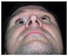
The correct response is Option D.
Posttraumatic enophthalmos is primarily the result of increased bony orbital volume. Atrophy of globe fat and cicatrix of adnexal tissue may also contribute to enophthalmos, but minimally. Eyelid lacerations do not lead to changes in orbital volume. Clinical features of enophthalmos include narrowing of the palpebral fissure and deepening of the supratarsal fold. Enophthalmos is best assessed on submental view to evaluate globe projection. Posttraumatic enophthalmos results from displaced zygoma fractures, medial wall or floor blow-out fractures, and nasoorbito-ethmoidal fractures with disruption of the medial wall resulting in increased orbital volume.
A 12-year-old girl is brought to the emergency department after she sustained injuries in a motor vehicle collision. Physical examination shows extensive lacerations of the right medial orbit and forehead (shown) with complete transection of the medial canthal tendon (MCT). For effective reattachment of the MCT with transnasal wiring, which of the following is the most appropriate direction of resuspension of the tendon in relation to the anterior lacrimal crest?
(A) Anterior and inferior
(B) Anterior and superior
(C) Directly horizontal
(D) Posterior and inferior
(E) Posterior and superior

The correct response is Option E.
The medial canthal tendon (MCT) consists of three limbs: 1) a prominent anterior limb that inserts medially on the anterior lacrimal crest, 2) a thinner posterior limb that attaches to the posterior lacrimal crest, and 3) a vertical limb of fascia that inserts on the medial orbital rim inferior to the nasal frontal suture. The resultant vectors of these attachments suggest that resuspension of the entire complex of the MCT following disruption should be posterior and superior to the anterior lacrimal crest.
The 27 -year -old woman shown above comes to the office for evaluation because she has had diplopia for the past 18 months. Physical examination shows orbital dystopia, enophthalmos, and malar flattening on the right. On the basis of the current findings, this patient €™s prior injury was most likely which of the following fractures?
(A) Le Fort II fracture
(B) Le Fort III fracture
(C) Orbital floor blow €‘out fracture
(D) Orbital roof fracture
(E) Orbitozygomatic complex fracture

The correct response is Option E.
The clinical findings of orbital dystopia, enophthalmos, and malar flattening on the patient €™s right side are typical for orbitozygomatic fractures. With the downward pull of the muscles, the zygoma rotates and the orbital volume is increased. This results in enophthalmos and possible dystopia, depending on the degree of displacement. The rotation of the zygoma causes loss of malar projection. This patient had only her orbital floor repaired with synthetic mesh. This repair was inadequate for her type of fracture, and stresses the importance of correct diagnosis and treatment in order to prevent secondary post €‘traumatic deformities.
Orbital floor or roof fractures would not result in malar flattening. Le Fort II and III fractures would cause a malocclusion, which is not present in this patient.
A 1-month-old infant is brought to the office by her parents for evaluation of a skull deformity. CT scan of the head is shown above. Which of the following cranial dysmorphologies is the most likely diagnosis?
(A) Dolichocephaly
(B) Plagiocephaly
(C) Scaphocephaly
(D) Trigonocephaly
(E) Turricephaly

The correct response is Option D.
The CT scan demonstrates an infant with a narrowed forehead and decreased bitemporal distance. There is also evidence of orbital hypotelorism and an obliterated or fused metopic suture. These findings are consistent with trigonocephaly.
Scaphocephaly is a cranial shape excessive in the anterior-posterior dimension and narrow in the bitemporal dimension. This results in a long and narrow (boat-like) shape of the head. This results from premature fusion of the sagittal suture.
Dolichocephaly is another term for scaphocephaly.
Plagiocephaly or “twisted head” is used to describe anterior or posterior deformities. Plagiocephaly is classified as anterior or posterior as well as synostotic and nonsynostotic or deformational. Anterior synostotic plagiocephaly refers to coronal craniosynostosis and posterior synostotic plagiocephaly refers to lambdoid craniosynostosis. Anterior or posterior nonsynostotic plagiocephaly refers to deformational plagiocephaly or skull molding from a persistent sleep position.
Turricephaly or oxycephaly is used to describe vertically tall head shapes that are usually associated with the brachycephaly (short in the anterior-posterior dimension) of bicoronal syndromic craniosynostosis.
The CT scan shown demonstrates a fracture of which of the following bones?
(A) Ethmoid
(B) Maxillary
(C) Palatine
(D) Parietal
(E) Sphenoid

The correct response is Option E.
In the CT scan, the lateral orbital wall is fractured. The lateral orbital wall is made up of the zygoma and the greater wing of the sphenoid. The medial orbital wall is made up of the ethmoid and palatine bones, and no fracture of these is demonstrated. The parietal bone largely is a cranial bone, which is not fractured in this CT scan. The maxillary bone, making up the buttresses and walls of the maxillary sinus, a portion of the orbital floor, and the medial aspect of the inferior orbital rim, is not fractured in this CT scan.
A 35-year-old man has swelling and tenderness of the nose and deviation of the nose to the left after being accidentally struck in the face while playing squash. Intranasal examination shows a localized purple mass on the left side of the septum. A CT scan is shown on page 4. Which of the following is the most appropriate initial management?
(A) Immediate closed reduction and placement of an external splint
(B) Immediate open reduction of the nasal fractures
(C) Immediate incision and drainage of the septal mass
(D) Immediate submucous resection of the septal fracture

The correct response is Option C.
The most appropriate initial step in the management of this patient is immediate incision and drainage of the septal hematoma. If left untreated, septal hematomas cause fibrosis and narrowing of the nasal passages, distortion of the septum, and/or formation of an abscess. They can also cause pressure necrosis of the septum, leading to septal perforation and eventually to complete necrosis with formation of a saddle-nose deformity.
Fracture management is undertaken after the hematoma has been evacuated. In patients who have significant swelling obscuring the nasal structure, fracture reduction is delayed until the swelling resolves, typically within five to 10 days. If swelling is minimal, then reduction can be performed immediately after evacuation.
Although closed reduction is appropriate in most patients with nasal fractures, open reduction may be necessary to obtain anatomic reduction. If lacerations are present, they may be used as incisions for surgical reduction. However, it should be noted that open techniques have been associated with a small incidence of necrosis of the traumatized nasal mucosa. In addition, unsatisfactory results following surgery are likely to lead to severe injury of the nasal septum that may be difficult to correct. Formal rhinoplasty may be performed at a later date to resolve breathing difficulties or improve the aesthetic appearance of the nose.
A 27-year-old man has malocclusion and tenderness around the orbits and bridge of the nose after sustaining facial injuries in a motor vehicle collision. A photograph and CT scan are shown above. The patient is to undergo open reduction and internal fixation of the fractures.
Which of the following is the most appropriate management of the lacrimal system?
(A) Observation
(B) Exploration of the lacrimal duct
(C) Placement of a silicone stent
(D) Immediate dacryocystorhinostomy
(E) Primary repair followed by dacryocystorhinostomy in three months

The correct response is Option A.
In this patient who has sustained a naso-orbitoethmoid fracture, the most appropriate management of the lacrimal system is observation. The incidence of injury to the nasolacrimal duct in patients who undergo open reduction and internal fixation of naso-orbitoethmoid fractures but have no overlying lacerations is fairly low. Although swelling or fracture may contribute to blockage of the duct at the time of the initial injury, approximately 90% of patients will experience improvement of symptoms with resolution of the swelling and reduction of the fracture.
Exploration and/or manipulation of the duct are not recommended if there is no obvious injury to the duct. These procedures may only result in further damage because of the edema and friability of the tissues. Immediate dacryocystorhinostomy is not warranted for the same reason.
Patients who have persistent epiphora after resolution of swelling should undergo further evaluation. Dacryocystography can be performed for assessment of possible nasolacrimal duct occlusion. If occlusion is present, dacryocystorhinostomy is indicated.
The CT scans shown above are from a 25-year-old man who sustained facial injuries in a motor vehicle collision. In this patient, rigid fixation at which of the following points is most likely to result in stable reduction of the fractures?
(A) Nasomaxillary buttress, inferior orbital rim, and zygomaticomaxillary buttress
(B) Zygomatic arch, central fragment, and nasomaxillary buttress
(C) Zygomatic arch, inferior orbital rim, and zygomaticomaxillary buttress
(D) Zygomaticofrontal suture, inferior orbital rim, and zygomaticomaxillary buttress
(E) Zygomaticofrontal suture, orbital floor, and zygomaticomaxillary buttress

The correct response is Option D.
This patient has sustained a fracture of the zygomaticomaxillary complex, also known as a zygoma fracture. To ensure stable reduction of the fracture, rigid fixation is applied at the zygomaticofrontal suture, inferior orbital rim, and zygomaticomaxillary buttress. Partial relapse may occur without this three-point rigid fixation.
Central fragments are associated with naso-orbitoethmoid fractures, not zygomaticomaxillary complex fractures. This fragment is comprised of the ascending frontal process of the maxilla and the descending internal angular process of the frontal bone. It provides the bony support for the medial canthus.
The nasomaxillary buttress is comprised of the piriform rim and is osteotomized routinely during elective Le Fort I maxillary advancement procedures. This structure is typically involved in midface fractures and not in fractures of the zygomaticomaxillary complex.
The orbital floor is often involved in zygomaticomaxillary complex fractures but not in simple fractures of the zygoma. Therefore, open reduction and internal fixation of the orbital floor may not be necessary to ensure correction of the zygomaticomaxillary fracture.
The zygomatic arch is contained within a periosteal sleeve and is often reduced and fixed adequately with adequate reduction and fixation of the zygomaticomaxillary complex. Open reduction and internal fixation through a coronal approach may be required if the zygomatic arch is comminuted significantly, but this is rare.