Lower Extremity 01-22 Flashcards
A 46-year-old woman presents with new-onset pain following below knee amputation. Medical history includes a Gustilo IIIB left leg injury and failed limb salvage 3 years ago. The patient reports phantom sensation, pain in her great toe, burning that ascends the limb, and several points along the distal stump that are inappropriately tender.On examination, a well - healed amputation stump without evidence of unstable skin or skin changes consistent with pressure - related trauma is noted.Targeted muscle reinnervation surgery is planned. Which of the following is the most likely evolution of neuropathic pain at 4 weeks and 6 months postoperatively in this patient?

The correct response is Option C.
Targeted muscle reinnervation (TMR), as originally described by Dumanian and Kuiken, is a procedure in which sensory and/or mixed nerves are transferred or coapted to motor nerve branches, in an effort to promote organized nerve growth and also to improve prosthetic control. While initially explored for improvement of prosthetic functionality, researchers observed a concomitant reduction in neuropathic and residual limb pain.
Though more research is needed, targeted muscle reinnervation may be most successful in decreasing or preventing pain when performed at the time of the amputation. The natural history of pain following TMR performed secondarily (not at the time amputation) includes a period of immediate relief (nerves are cut proximal to the neuroma), followed by activation of the nerves and increased pain (3 to 6 weeks), and plateau and reduction of pain (6 weeks to 6 months).
Nearly all patients report a decrease in pain and improvement in quality of life. The degree with which the pain is decreased and life is improved may be related to the timing of the operation. Data suggest that the earlier the operation is performed, the better the results, perhaps because of the centralization of the somatic pain response, though more work is needed to elucidate this mechanism.
A 25-year-old man presents with a comminuted tibia plateau fracture sustained during a self-inflicted gunshot wound. A CT scan is shown. During open reduction and internal fixation of the fracture, a 5-cm gap in the common peroneal nerve is noted. Tendon transfer, nerve repair with grafting, and nerve transfer are planned. Which of the following fascicular nerve transfers is most likely to aid in ankle dorsiflexion?
A) Flexor digitorum longus to soleus
B) Flexor digitorum longus to tibialis posterior
C) Flexor hallucis longus to tibialis anterior
D) Peroneal longus to extensor digitorum longus
E) Soleus to extensor digitorum brevis

The correct response is Option C.
Common peroneal nerve repairs tend to have less favorable results than their upper extremity counterparts. Traction injuries showed only good outcomes in 42% of patients, whereas sharp injuries showed good results in 61% of patients. Gunshot wounds, on the other hand, showed only 49% good outcomes due to the blast injury. This patient had damage of his nerve from penetrating trauma from the fracture fragments as well as damage from the blast injury. Because of the size of the defect, primary nerve repair would not be feasible. Nerve grafts less than 6 cm have been found to have a more favorable result than those greater than 6 cm. When combined with blast injury, a favorable result has only been found in 31% of patients.
In addition to nerve repairs, tendon transfers of the posterior tibial tendon have been useful for more immediate dorsiflexion. Fascicular nerve transfers have found some success in dorsiflexion. Of the options given, only transfer from the flexor hallucis longus (tibial nerve) to the tibialis anterior (deep peroneal nerve) would result in dorsiflexion. Although flexor digitorum longus is a common donor from the tibial nerve, innervating the soleus would result in plantar flexion and the soleus is also innervated by the tibial nerve, so it was not injured in the gunshot. Transfer from the flexor digitorum longus to the tibialis posterior (tibial nerve) would plantarflex the foot and not dorsiflex it. Although transfer to the extensor digitorum longus can lead to dorsiflexion, transfer from the peroneal longus (superficial peroneal nerve) to extensor digitorum longus would not result in a functional nerve repair, since the peroneal longus is innervated by the damaged nerve. Finally, transfer from the soleus to the extensor digitorum brevis (EDB, deep peroneal nerve) would not be a nearby transfer, nor would the EDB dorsiflex the ankle.
A 36-year-old man is evaluated for coverage of a 4 × 3-cm middle-third lower extremity soft-tissue defect with an exposed fracture of the mid tibia. Reconstruction with a proximally based medial hemisoleus muscle flap is planned. Which of the following arteries provides the major contribution to the blood supply of this flap?
A) Anterior tibial
B) Dorsalis pedis
C) Medial sural
D) Peroneal
E) Posterior tibial
The correct response is Option E.
The soleus muscle is a bipennate muscle that is located in the superficial posterior compartment of the lower extremity, deep to the gastrocnemius muscle. The soleus muscle has a medial head that originates from the posterior aspect of the tibia and a lateral head that originates from the posterior surface of the fibula. The soleus functions in plantar flexion of the foot in conjunction with the gastrocnemius as they unite to form the Achilles tendon. Branches from the popliteal artery contribute to the blood supply of the proximal soleus muscle.
The soleus muscle flap is a reliable local flap for coverage of moderate-sized soft tissue defects of the middle-third of the lower extremity. It is customarily elevated as a muscle-only flap and covered with a split-thickness skin graft. Branches from the posterior tibial artery contribute to the blood supply of the medial hemisoleus muscle flap, which is more reliable when based proximally.
Branches from the peroneal artery contribute to the blood supply of the lateral hemisoleus muscle.
The medial sural artery is the dominant blood supply to the medial gastrocnemius muscle flap.
The anterior tibial artery and the dorsalis pedis artery do not contribute to the blood supply of the medial hemisoleus flap
A 51-year-old man presents for evaluation of nerve injury following varicose vein stripping of the left leg. Which of the following findings would be expected with saphenous nerve injury in this patient?
A) Anesthesia around the left medial malleolus
B) Hypersensitivity along the dorsum of the left foot
C) Inability to dorsiflex the left foot
D) Increased insertional activity in the tibialis anterior muscle
E) Numbness near the left lateral heel
The correct response is Option A.
Nerve injury is a relatively rare but significant complication of varicose vein stripping. Knowledge of anatomy can help identify which nerve is involved in most injuries. Injury to the saphenous nerve would cause anesthesia over the medial calf and medial malleolus. Injury to the deep peroneal nerve would cause weakness in dorsiflexion and would result in increased insertional activity on electromyography of the tibialis anterior. Hypersensitivity on the dorsal foot or numbness over the lateral heel would come from an injury to the superficial peroneal nerve and the sural nerve, respectively.
A 20-year-old man presents to the emergency department 6 hours after a bicycle accident with an open tibial fracture. The patient was traveling at 10 miles per hour at the time of the accident. The wound is 5 cm in length, and there is moderate contamination. The fracture is a mid-shaft tibial fracture with moderate comminution, with an associated closed fibula fracture. Which of the following Gustilo classifications is most appropriate for this injury?
A) I
B) II
C) IIIA
D) IIIB
E) IIIC
The correct response is Option B.
Though it was never designed to predict treatment, the Gustilo classification has stood the test of time as a highly utilized grading system for lower extremity trauma. It is often used to predict the need for flap coverage, to estimate the risk for osteomyelitis, and to guide antibiotic use.

A 34-year-old woman presents with a 1-year history of progressive ankle and dorsal foot pain and paresthesias in the first dorsal web space. Electrodiagnostic study is significant for changes in the extensor digitorum brevis muscle. Which of the following nerves is the most likely source of this patient’s symptoms?
A) Deep peroneal
B) Saphenous
C) Superficial peroneal
D) Sural
E) Tibial
The correct response is Option A.
Anterior tarsal tunnel syndrome, also known as deep peroneal nerve (DPN) entrapment, is the result of compression of the DPN at the superior border of the inferior extensor retinaculum at the ankle joint and beneath the extensor hallucis longus tendon. Entrapment can occur as a result of wearing tight-fitting shoes or boots. It is important to rule out exertional anterior compartment syndrome or common peroneal nerve entrapment as the cause of symptoms. The nerve can also experience traction injury caused by chronic ankle instability due to ankle sprains.
The DPN travels in the leg between the extensor digitorum longus (EDL) and tibialis anterior and distally between the EDL and extensor hallucis longus just proximal to the ankle before dividing into the lateral and medial branches, 1.3 cm proximal to the ankle joint. The lateral branch innervates the extensor digitorum brevis (EDB) and the tarsometatarsal (TMT) and metatarsophalangeal joints. The medial branch, a sensory branch, travels to the first dorsal web space and has a dorsomedial cutaneous branch to the second toe and a dorsolateral cutaneous branch to the great toe. Entrapment of the medial branch can occur from the extensor hallucis brevis (EHB) tendon, as it travels over the nerve at the first and second TMT joints. Patients typically present with pain along the dorsum of the foot with intermittent numbness radiating to the first dorsal web space. A Tinel sign may be elicited over the superior and inferior retinaculum along the DPN, resulting in tingling over the first dorsal web space of the foot. The EDB can be weak or atrophied. Patients may also report aching and tightness along the ankle joint or numbness at the first dorsal web space when the ankle is placed in plantar flexion with the toe extended. Electrodiagnostics should be ordered to confirm the diagnosis and location of the entrapment.
A 53-year-old man underwent excision and primary closure with extensive wide subcutaneous undermining for squamous cell carcinoma of the leg 7 days ago. Examination shows margins are positive, and reexcision resulting in a wound with exposed tendon with no peritenon is noted. Which of the following is the most appropriate reconstructive option for this patient?
A) Bipedicled advancement flap
B) Keystone flap
C) Mustardé rotational flap
D) Split-thickness skin graft
E) V-Y advancement flap
The correct response is Option A.
The appropriate reconstructive option from the given choices for this patient would be bipedicled advancement flap. It is important to understand the blood supply to local skin flaps. The bipedicled advancement flap is a random pattern flap with perfusion from the dermal/subdermal plexus. Unlike a standard, single-pedicle random pattern flap, the bipedicled flap maintains skin connections in two directions allowing for a 2:1 length-to-width ratio. The donor defect of the bipedicled flap is typically skin grafted to minimize opposing tension on the flap and allow for greater advancement. The wide subcutaneous undermining 7 days prior would have allowed for the “delay phenomenon,” potentially making the flap more reliable.
The extensive wide subcutaneous undermining during initial primary closure likely injured perforators to the skin. The V-Y advancement flap is typically islandized during closure and would be compromised by adjacent wide undermining. Similarly, a keystone flap would be compromised if the skin was delaminated from the underlying tissues. An adjacent perforator flap is perfused by a cutaneous perforator (septocutaneous or muscular) which likely would have been injured during wide undermining. Split-thickness skin graft is not appropriate because there is no peritenon on the tendon. A Mustardé rotational flap is used for cheek advancement.
A 25-year-old man is brought to the emergency department with a large degloving injury with exposed tibia and ankle joint (Gustilo Type IIIB) sustained during a motorcycle collision. After multiple debridements, the plastic surgeon is consulted 6 days later. Routine laboratory testing shows a platelet count of 1.5 million/mL, increased from normal range on admission. Free flap reconstruction is planned. Which of the following is the most appropriate next step in management in this patient?
A) Consult hematology for bone marrow biopsy
B) Delay reconstruction until platelet count is within normal range
C) Initiate antiplatelet therapy
D) Proceed with free tissue transfer
The correct response is Option C.
Antiplatelet therapy should be initiated to minimize the risk for flap loss in this patient. This patient has a reactive thrombocytosis which can commonly occur following trauma, infection, inflammation, or major surgery (e.g., post-splenectomy). This temporary elevation in platelet count is usually transient and peaks at approximately 2 weeks after injury. The traditional pharmacologic agents for microvascular thrombosis have minimal (e.g., aspirin) or no anti-platelet effects (e.g., heparin, hirudin, and thrombolytic agents). There are no guidelines or standards of care, but Hollenbeck et al. advocate for administration of glycoprotein IIb/IIIa antagonists (e.g., abciximab, tirofiban, and eptifibatide), which block the final common pathway for platelet aggregation. Thienopyridines (e.g., clopidogrel) often reach steady state after 4 to 7 days, which would further delay surgery. Platelet apheresis has also been described to reduce platelet count and secondary flap salvage.
Acute trauma patients with elevated preoperative platelet counts are at higher risk for both intraoperative and postoperative microvascular complications (especially arterial thrombosis). Proceeding with free tissue transfer without any precautions is high risk for failure.
A hematology consult and bone marrow biopsy is not necessary since the hypercoagulable state is usually self-limited but can take several weeks. This is not a chronic medical condition requiring further workup or treatment.
Multiple studies suggest that patients with complex open fractures requiring soft-tissue coverage have improved outcomes with early free flap coverage, so it is not appropriate to wait until after this condition resolves. The platelet elevations can peak at approximately 2 weeks, which would delay reconstruction and increase morbidity. Delaying reconstruction increases the risks for infection, nonunion, and ultimately failure of limb salvage.
A patient presents with a traumatic heel loss. The surgeon decides on an innervated medial plantar artery flap. The medial plantar nerve is a terminal branch of which of the following other nerves?
A) Common peroneal
B) Lateral plantar
C) Saphenous
D) Sural
E) Tibial
The correct response is Option E.
The answer is tibial nerve. The sciatic nerve comes off the sacral plexus, then it branches into sural nerve, common peroneal nerve, and tibial nerve. The tibial nerve terminal branches are the medial sural cutaneous nerve, medial plantar nerve, and lateral plantar nerve. The medial plantar nerve innervates the abductor hallucis, flexor digitorum brevis, flexor hallucis brevis, and first lumbrical muscle. The cutaneous branches innervate the skin of the medial 2/3 of the sole of the foot as well as plantar digital toes. There are also terminal branches to the intertarsal and tarso-metatarsal joints. The flap is supplied by the medial plantar artery off the posterior tibial artery. The artery is found between the abductor hallucis and flexor digitorum brevis. It travels along the medial border of the foot and anastomosis with the first plantar metatarsal artery. Next, the perforators run between the abductor hallucis muscle and the plantar aponeurosis to the skin of the instep.
A 50-year-old man undergoes a total glossectomy for tongue cancer. A microvascular free flap reconstruction is planned with an anterolateral thigh free flap. After a vertical thigh incision is made, no perforators are found between the rectus femoris and vastus lateralis muscles. Exploration of the medial thigh demonstrates a large perforator between the rectus femoris and vastus medialis muscles. Which of the following is the most likely origin of this perforator?
A) Ascending branch of the lateral circumflex femoral artery
B) Common femoral artery
C) Descending branch of the lateral circumflex femoral artery
D) Medial circumflex femoral artery
E) Profunda femoris artery
The correct response is Option C.
An adequate perforator is not found on dissection of the anterolateral thigh (ALT) free flap in up to 5% of cases. When an ALT flap perforator traveling between the rectus femoris and vastus lateralis or through the vastus lateralis to supply the anterolateral thigh skin is not found, there is a higher chance of finding a perforator that supplies the anteromedial thigh skin. This perforator, present in about 50% of thighs, most commonly arises from the descending branch of the lateral circumflex femoral artery, via a branch that supplies the rectus femoris muscle. This perforator can take a course through the rectus femoris or between the rectus femoris and vastus medialis. Alternately, one or more anteromedial thigh perforators may arise directly from the superficial femoral artery.
When an ALT perforator cannot be located, the surgeon may salvage the situation by changing to an anteromedial thigh (AMT) free flap rather than exploring another donor site. Another alternative is to harvest a tensor fascial lata free flap, based on the ascending branch of the lateral circumflex femoral artery. The medial circumflex femoral artery, common femoral artery, and profunda femoris artery do not give rise to previously described cutaneous perforator flaps.
A 63-year-old man presents to the office with a 4 × 4-cm heel defect. A local podiatrist has debrided the calcaneus, and there is healthy granulation tissue present. After extensive discussion with the patient, it is decided to proceed with a distally based sural fasciocutaneous flap for coverage. Which of the following risk factors is associated with the highest rate of flap-related complications?
A) Coronary artery disease
B) Diabetes mellitus
C) Hypertension
D) Obesity
E) Venous insufficiency
The correct response is Option E.
A recent meta-analysis of 61 papers showed that venous insufficiency is associated with a nine-fold increase in risk of developing a complication in a distally based sural flap. Other risk factors, such as peripheral vascular disease, diabetes, obesity, and hypertension, have been associated with increased rates of complications previously in the literature, but venous insufficiency is associated with the highest rate of complications. The pooled data from this series showed a 26% complication rate, a 3.2% flap loss rate, and a 15.3% partial flap loss rate. As the design of the sural flap is distally based, the physiology of the flap requires reversed flow through the venous system.
Advanced age is also associated with an increased complication rate and there is literature to suggest a delay procedure in this patient population. Other papers have found smoking to have a higher risk of complications as well. There is still much debate over which patients should undergo a delay procedure or venous supercharging, but in general, high-risk patients with multiple comorbidities should be considered for this additional surgery. Coronary artery disease has not been found to be associated with increased risk of flap-related complications.
A 56-year-old woman with a traumatic defect of the upper third of the tibia undergoes open reduction and internal fixation with tibial nail. Soft tissue coverage with a gastrocnemius flap is planned. Which of the following arteries provides the dominant blood supply for this flap?
A) Anterior tibial
B) Peroneal
C) Popliteal
D) Posterior tibial
E) Sural
The correct response is Option E.
Each head of the gastrocnemius muscle is supplied by the sural artery: either the medial sural or lateral sural artery for medial and lateral gastrocnemius, respectively. The arteries arise from the popliteal artery about 3-4 cm above the head of the fibula and enter the medial and lateral heads of the gastrocnemius at about the level of the head of the fibula. The flap can be rotated to cover soft-tissue defects of the anterior distal aspect of the knee. The flap ranges from 5 to 9 cm in width and from 13 to 20 cm in length. It provides a vascular bed for a skin graft and improves the delivery of oxygen and systemic antibiotics. The other listed arteries do not supply the gastrocnemius muscles.
A 40-year-old man presents to the emergency department because of severe pain after sustaining a crush injury to the left lower extremity from a forklift. On physical examination, the lower leg is tense and swollen circumferentially. Sensation to the foot is diminished. Distal pulses are palpable. X-ray study does not show any fractures. Which of the following is the most appropriate next step in management?
A) Ace wrap compression
B) CT angiography
C) Emergent fasciotomy
D) MRI
E) Observation and leg elevation
The correct response is Option C.
The patient displays the signs and symptoms of acute compartment syndrome, a surgical emergency requiring emergent fasciotomy. Acute compartment syndrome requires prompt diagnosis and expeditious treatment in order to minimize morbidity. Compartment syndrome can occur following a substantial soft tissue crush injury, even in the absence of a fracture, such as in this clinical scenario. Severe pain is usually the presenting complaint. It may be out of proportion to the injury and unresponsive to analgesics. The presence of paresthesias can signify nerve hypoxia from elevated compartment pressures. Pallor, paralysis, and pulselessness are very late signs. Nerve and muscle do not tolerate long periods of ischemia and may undergo irreversible damage if surgical decompression is delayed.
Compartment syndrome is primarily a clinical diagnosis, but measurement of compartment pressures can provide additional information especially if the diagnosis of compartment syndrome is less obvious. If compartment pressures are greater than 30 mmHg or if the differential pressure (difference between diastolic blood pressure and compartment pressure) is less than 30 mmHg, then fasciotomy is recommended.
Observation and leg elevation would not be appropriate management in the setting of acute compartment syndrome. CT angiography would not be indicated in this case, where there is a low suspicion of vascular injury. MRI has been used in the diagnosis of chronic exertional compartment syndrome but has little value in the setting of acute trauma.
A 35-year-old man presents for evaluation of a laceration to the lateral aspect of the right lower leg 5 cm distal to the knee that he sustained when he fell from a bicycle 2 months ago. Findings on electromyography and nerve conduction studies are consistent with an isolated complete injury of the common peroneal nerve. Which of the following deficits is most likely on physical examination?
A) Dorsiflexion of ankle
B) Plantarflexion of great toe
C) Sensation of lateral foot
D) Sensation of medial foot
E) Sensation of plantar foot
The correct response is Option A.
The common peroneal nerve forms as the sciatic nerve bifurcates at the apex of the popliteal fossa. It then follows the medial border of the biceps femoris muscle and tendon. The nerve then passes over the posterior aspect of the fibular head and winds around the neck of the fibula. The common peroneal then divides into the deep and superficial peroneal nerve branches. The deep branch supplies the anterior muscles of the leg, the dorsum of the foot, and the skin of the first web space. The superficial branch supplies the peroneus longus and brevis muscles and the skin on the distal third of the lower leg and dorsum of the foot. Because of its relatively superficial position, the common peroneal nerve is the most commonly injured nerve of the lower extremity. Transection of the common peroneal nerve results in paralysis of all muscles in the anterior and lateral compartments of the leg (dorsiflexors and ankle evertors). This pattern of injury results in the classic picture of a foot drop. The distribution of sensory loss would include the anterolateral leg and dorsum of the foot.
Sensation of the medial foot is from the saphenous nerve and branches of the medial plantar nerve. Lateral foot sensation is provided by the sural nerve. Sensation of the plantar aspect of the foot is from the terminal branches of the tibial nerve (medial and lateral plantar nerves). All of the muscles of plantar flexion of the ankle and toes (i.e. gastrocnemius, soleus, plantaris, and tibialis posterior, flexor hallucis longus, flexor digitorum longus, and the intrinsic plantar foot muscles) are innervated by the tibial nerve.
When a pedicled sural flap is raised to the heel, which of the following is the origin of the arterial blood supply?
A) Descending genicular artery
B) Lateral sural artery
C) Medial femoral circumflex artery
D) Medial plantar artery
E) Peroneal artery
The correct response is Option E.
The reverse sural flap is a fasciocutaneous flap often used for ankle or heel wounds. The blood supply of the flap can be from a median superficial artery or the arterial plexus that travels with the sural nerve; the origin is a lower peroneal perforator located approximately 5 cm proximal to the lateral malleolus.
The lateral sural artery would be the appropriate blood supply for perfusion of a pedicled lateral gastrocnemius flap. The gracilis flap blood supply derives from the medial circumflex artery. The descending genicular artery provides the blood supply of the medial femoral condyle flap. The medial plantar artery is the blood supply for the medial plantar artery flap.
A 19-year-old man is brought to the emergency department because of an injury to the right heel sustained during a lawn mower accident. After serial debridement is performed, there is a 2 x 2-cm soft tissue defect with exposed calcaneus. Which of the following innervated flaps is most appropriate for coverage of this defect?
A) Anterior lateral thigh flap with anterior femoral cutaneous nerve
B) Medial plantar artery flap with division of posterior tibial nerve
C) Radial forearm flap with superficial branch of the radial nerve
D) Reverse sural artery flap with saphenous nerve
E) Ulnar forearm flap with deep branch of ulnar nerve
The correct response is Option B.
Heel reconstruction is a difficult surgical problem with limited local options, relatively poor vascularity in the region, and weight-bearing requirements. Flap options include a variety of local flaps including transposition or rotation flaps, fasciocutaneous flaps (e.g., medial plantar); local muscle flaps (e.g., abductor hallucis, flexor digitorum brevis, and abductor digiti minimi); reversed fasciocutaneous flaps (e.g., sural); and free flaps. Although innervated (and therefore potentially sensate) free flaps can be performed, these are less predictable than local options. From the above answer choices, the best option is a local flap based on the medial plantar artery, which had sensation from the medial plantar nerve, a branch of a division of the posterior tibial nerve. Another advantage of the medial plantar artery flap is that it covers the heel with glabrous skin, which may better be able to withstand weight-bearing. The other options are not correctly matched with flaps and nerves. Correct pairings of flap and cutaneous innervation are:
Anterior lateral thigh flap - lateral femoral cutaneous nerve
Ulnar forearm flap - medial antebrachial cutaneous nerve
Radial forearm flap - lateral antebrachial cutaneous nerve
Reverse sural artery flap - sural nerve
16-year-old boy with a Gustilo Type IIIB open tibial fracture underwent wound coverage with an anterolateral thigh (ALT) flap including muscle. Which of the following is the most likely muscular complication in this patient?
A) Weakness of knee extension
B) Weakness of knee flexion
C) Weakness of thigh abduction
D) Weakness of thigh adduction
E) Weakness of thigh flexion
The correct response is Option A.
An understanding of the actions of donor muscles is necessary when using muscle flaps for reconstruction. A knowledge of the specific muscles, which are included as part of specific flaps also helps one understand what donor deficits may be produced when using a particular flap.
The anterolateral thigh (ALT) flap perforators often traverse part of the vastus lateralis muscle, and the muscle may need to be dissected and therefore can be injured during flap harvest. It is rare that any long-term sequelae, such as weakness, are noted without muscle harvest. If the muscle is taken, as is often necessary for the filling of a dead space, weakness of knee extension may be noted as the vastus lateralis is a large part of the quadriceps muscle of the thigh, which is primarily responsible for knee extension.
Thigh abduction is accomplished by the tensor fascia lata and sartorius muscles. The pectineus, adductor longus and brevis, gracilis, and adductor magnus are responsible for thigh adduction. Thigh flexion is achieved by the pectineus, adductor brevis, and adductor magnus. Knee flexion is done primarily by the hamstrings-semitendinosus, semimembranosus, and biceps femoris muscles.
A 48-year-old man presents to the emergency department because of spontaneous progressive pain, swelling, cyanosis, and edema of the left lower extremity for the past 24 hours. A photograph is shown. Medical history includes prophylactic inferior vena cava (IVC) filter placement in the setting of prolonged immobilization secondary to traumatic closed head injury sustained 2 years ago. Physical examination shows no dyspnea. Oxygen saturation is 98% on room air. Venous ultrasonography and CT scan show total left deep femoral thrombosis extending into the lower IVC at the indwelling filter. Which of the following is the most appropriate next step in management?
A) Catheter-directed thrombolysis
B) Femoral vein to IVC vascular bypass
C) Isolated extracorporeal membrane oxygenation (ECMO) support to the affected extremity
D) Open thrombectomy
E) Oral anticoagulation

The correct response is Option A.
The patient is presenting with extensive acute thrombotic occlusion resulting in clinically evident symptomatic venous insufficiency of the extremity. If the occlusion is left untreated, progressive cyanosis and secondary ischemia followed by gangrene develop. Locally delivered thrombolytic agents via catheter-directed thrombolysis with or without percutaneous transluminal angioplasty is an effective first line of treatment in this scenario where the patient presents within a few days of symptom onset (ie, prior to clot fibrosis) and is not high-risk for bleeding. In patients who are high-risk for bleeding (eg, acute intracerebral hemorrhage, gastrointestinal bleeding), alternative methods of restoring venous outflow include clot retrieval through other percutaneous or open techniques (eg, transluminal aspiration thrombectomy, open inferior vena cava (IVC) thrombectomy with or without temporizing groin arteriovenous fistula creation). Systemic thrombolysis can be considered when other first line therapies are not available but has been associated with high frequency of major bleeding complications in several randomized trials (14% for streptokinase).
Systemic anticoagulation infusion helps prevent progression but does not restore acute compromised ischemic limb secondary to venous outflow obstruction. Oral anticoagulation is not indicated for acute management of a limb-threatening thrombosis. Femoral vein to IVC vascular bypass is not a described procedure for venous insufficiency. Limb-threatening thrombo-occlusive venous insufficiency resulting in a painful swollen blue leg, such as that pictured (also known as “phlegmasia cerulea dolens,” literally “painful blue edema”) was first described with heparin-induced thrombocytopenia. It has also been associated with cancer or life-threatening critical illness. More recently, a growing population of patients are at risk due to unretrieved IVC filters. While IVC filter placement may protect the pulmonary vascular bed, it does not lessen thrombotic predisposition or incidence in the lower extremities, and IVC thrombosis with or without phlegmasia cerulea dolens has been reported to occur in 3 to 30% of patients following IVC filter placement. Filter retrieval following its initial indicated need can lessen secondary thrombotic complications, but data suggest that only a fraction of retrievable filters are later removed. In a systemic review, overall retrieval was 34% with a high percentage of nonretrieval occurring for a variety of reasons, including loss to follow up (particularly in trauma centers), limited life expectancy, and/or unresolved underlying conditions.
A 62-year-old woman with non-insulin-dependent diabetes mellitus is undergoing lower extremity angiogram to determine her suitability for forefoot reconstruction. Which of the following is the most appropriate therapy for the prevention of contrast-induced nephropathy in this patient?
A) Ascorbic acid
B) Intravenous saline
C) N-acetylcysteine
D) Simvastatin
E) Sodium bicarbonate
The correct response is Option B.
Contrast-induced nephropathy (CIN) is a significant problem in patients undergoing procedures that require contrast administration. The mechanism is believed to be an ischemic injury to the renal medulla. It is the third most common cause of hospital-acquired renal failure. Independent of renal failure, the development of even mild CIN is associated with increased rates of morbidity and mortality. The major risk factor for developing CIN is pre-existing renal dysfunction. This is particularly associated with patients with diabetes and those who have a creatinine clearance less than 60. The best method of prevention is appropriate risk stratification, intravenous hydration with normal saline and withholding of nephrotoxic medications. Intravenous fluid hydration with normal saline is the mainstay of practice in the prevention of CIN. It is low-risk, carries few side effects, and is cost-effective. Randomized trials have found intravenous hydration with normal saline to be consistently effective. The administration of intravenous fluids increases intravascular volume, promotes diuresis, diminishes the overall intravascular contrast load and supports vasodilation. Although intravenous administration of sodium bicarbonate has also gained popularity in the prevention of CIN, recent publications have demonstrated mixed results. The use of N-acetylcysteine, statin drugs and ascorbic acid are not recommended for the prevention of CIN.
A 40-year-old man is diagnosed with a posterior thigh sarcoma. He undergoes resection of the tumor as well as some of the surrounding muscle. Partial sacrifice of the sciatic nerve is required, leaving a 40% circumferential defect and an 11-cm gap between proximal and distal ends. A photograph is shown. Which of the following is the most appropriate method of nerve reconstruction?
A) Mobilization and primary coaptation
B) Polyglycolic acid nerve tube
C) Processed human allograft conduit
D) Saphenous vein graft
E) Sural nerve cable graft

The correct response is Option E.
Fundamentals of nerve repair include coaptation in a tension-free manner. If there is any tension, nerve grafts or conduits are indicated. In this clinical scenario, there is a large nerve gap that precludes tension-free primary coaptation, even with extensive proximal and distal mobilization. Therefore, a nerve graft is indicated. Common choices include sural, lateral, or medial antebrachial cutaneous. For the size and length of the defect and the fact that multiple cable grafts would be needed, the sural is the most appropriate choice.
Nerve conduits such as PGA tubes and processed human allograft conduits serve as scaffolds to promote nerve regeneration, although these are typically used for gaps less than 3 cm. Given the distance involved, a sural nerve graft using a grouped fascicular or epineurial repair is the most appropriate choice, although a gap this large is almost certain to leave permanent deficits. Appropriate levels of expectation must be set with the patient.

An 18-year-old woman comes to the office because of a large osteosarcoma of the distal shaft of the right femur. A 15-cm bone resection is planned, with a resulting large intercalary segmental defect. The overlying skin and soft-tissue is not involved. The patient is very motivated to proceed with limb preservation. Which of the following is the most appropriate option for reconstruction of this defect?
A) Bone allograft
B) Contralateral vascularized fibula free flap
C) Contralateral vascularized fibula free flap with bone allograft
D) Ilizarov bone transportation
E) Ipsilateral pedicled vascularized fibula flap
The correct response is Option C.
In a young patient who desires limb preservation after sarcoma resection, a contralateral vascularized fibula free flap with bone allograft (Capanna technique) is the most appropriate option for a large intercalary segmental defect. This involves placing the fibula flap within an allograft construct and bridging both osteotomy sites. There are advantages to using the allograft with the fibula flap, as a fibula flap alone may have difficulty with weight-bearing and potential fracture. In select cases a double barrel configuration can be used; however, in this patient the defect is too large. An ipsilateral pedicled flap would have difficulty reaching this large defect and would still have issues with fractures from weight-bearing. Ilizarov bone transportation can be performed for smaller defects (4 to 6 cm), but not in a defect this large. Finally, bone allograft alone is an option; however, this has a high rate of nonunion (34% versus 8 to 10%).
A 45-year-old woman who underwent Achilles tendon repair through a posterior midline incision 3 weeks ago develops a postoperative wound infection and subsequent skin necrosis. Physical examination shows a 3 x 3-cm wound directly overlying the Achilles tendon in the absence of peritenon. A fasciocutaneous propeller flap from the medial leg is designed to cover this defect. The septal perforators to this flap run between which of the following structures?
A) Flexor hallucis longus and gastrocnemius
B) Gastrocnemius and soleus
C) Peroneus longus and peroneus brevis
D) Soleus and flexor digitorum longus
E) Tibialis anterior and extensor digitorum longus
The correct response is Option D.
This defect may be reconstructed with a posterior tibial artery perforator propeller flap. These vessels emerge between the flexor digitorum longus and the soleus muscle. In one anatomic study, there were three clusters of perforators: 4 to 9 cm, 13 to 18 cm, and 21 to 26 cm from the intermalleolar line. The peroneal artery perforators often arise through the posterior peroneal septum, and the anterior tibial artery perforators are often found between the extensor digitorum longus and the peroneus longus or between the tibialis anterior and the extensor digitorum longus.
A 27-year-old woman sustains a Grade IIIB degloving injury of the left lower extremity in a motor vehicle collision. Latissimus dorsi free flap placement is planned. Which of the following is the most likely outcome in this patient in terms of donor site morbidity?
A) Decreased seroma formation but increased hematoma formation
B) Inability to maintain sitting-up position when back is not supported
C) Initial decreased shoulder range of motion that improves by one year
D) Permanent loss of external rotation of the shoulder and inability to reach forward
The correct response is Option C.
Most studies that demonstrate shoulder weakness and loss of motion show that the loss of function is greatest in the early postoperative period and returns to baseline, or close to baseline, at 1 year or more after surgery.
All studies comparing types of latissimus flaps demonstrate less morbidity with perforator or muscle-sparing flaps as compared to traditional or extended latissimus dorsi (LD) flaps. Lower functional morbidity is observed with more native muscle preserved as is other flaps. This assumes that the muscular branches of the motor nerve to the latissimus are spared.
A recent meta-analysis does show higher functional impairment than expected after latissimus flap transfer. The number of patients who required a change in occupation was less than 10%. This was likely because of difficulty with activities such as ladder climbing, painting overhead, and sustained reach overhead.
The function of the latissimus dorsi muscle is shoulder adduction, extension and internal rotation. Other muscles of the rotator cuff perform similar functions and will assist in compensation for the loss of the latissimus. Patients who do develop weakness report it in activities involving shoulder adduction and internal rotation. Paradoxically, limitations in range of motion are mostly in shoulder flexion and abduction possibly related to tight skin closure and internal scarring.
Donor site seroma formation is particularly problematic, with published rates ranging from 3.9 to 79%.
Core muscles such as rectus abdominis, external oblique, gluteus maximus, medius, and minimus, and erector spinae all contribute to rotation, balance, and stabilization during sitting and standing.
A 21-year-old man undergoes reconstruction with a free flap. Photographs are shown. This procedure places the patient at risk for claw toe with loss of active flexion of the great toe. The muscle responsible for this functional loss is located in which of the following compartments in the lower leg?
A) Anterior
B) Deep posterior
C) Lateral
D) Superficial posterior

The correct response is Option B.
Claw toe or loss of active flexion of the great toe interphalangeal joint can result from harvest of the flexor hallucis longus for free fibula flaps. The flexor hallucis longus is present within the deep posterior compartment of the lower leg and should be resuspended to the interosseus membrane and posterior tibial muscles as needed to maintain proper tension. Physical therapy is initiated after adequate wound healing to maintain the mobility of the great toe and ankle. The deep posterior compartment musculature is composed of the tibialis posterior, the flexor digitorum longus, the flexor hallucis longus, and the popliteus.
The superficial posterior compartment musculature is composed of the gastrocnemius, soleus, and plantaris.
The anterior compartment musculature is composed of the tibialis anterior, the extensor digitorum longus, extensor hallucis longus, and the peroneus tertius.
The lateral compartment musculature is composed of the peroneus longus and brevis muscles.
A 67-year-old man comes to the office because of nerve deficit of the left lower extremity which occurred after undergoing a femoral-distal bypass 5 days ago. Physical examination shows numbness of the plantar surface of the foot and weakness in plantarflexion. Which of the following nerves is most likely injured in this patient?
A) Femoral
B) Obturator
C) Peroneal
D) Sural
E) Tibial
The correct response is Option E.
This patient appears to demonstrate symptoms of a tibial nerve injury. The tibial nerve is a branch of the sciatic nerve. It travels through the popliteal fossa and gives off branches to gastrocnemius, soleus, plantaris, and popliteus muscles. The tibial nerve travels in proximity to the posterior tibial artery. In the leg, it gives off branches to the flexor digitorum longus, tibialis posterior, and flexor hallucis longus. Distally in the foot, it branches to give rise to the medial and lateral plantar nerves, which provide sensation to the plantar surface of the foot. Injury to the tibial nerve results in deficits of plantarflexion, as well as anesthesia to the plantar surface of the foot.
The femoral nerve innervates muscles of the anterior thigh, including the quadriceps group, iliacus, and sartorius. Injury to the femoral nerve results in weakness of leg extension.
The obturator nerve provides innervation to the medial thigh muscles (adductor group), including adductor brevis, longus, and magnus, as well as the gracilis and obturator externus. The cutaneous branch provides sensation of the medial thigh. Injury to the obturator nerve results in weakness in thigh adduction, and sensory deficits in the medial thigh.
The peroneal nerve is divided into superficial and deep branches at the area of the fibular neck. The superficial peroneal nerve supplies the lateral compartment of the leg, giving motor branches to peroneus longus and brevis, as well as sensory contributions to the lateral aspect of the leg. Injury to the superficial peroneal nerve results in anesthesia of the lateral aspect of the leg and weakness in eversion and plantarflexion of the foot. The deep peroneal nerve travels in the anterior compartment of the leg, and gives branches to the tibialis anterior, extensor hallucis longus, and extensor digitorum longus and brevis, as well as peroneus tertius. The sensory distribution of the deep peroneal nerve is in the area of the first web space. Injury to the deep peroneal nerve causes weakness in dorsiflexion of the foot.
The sural nerve travels on the posterior aspect of the leg between the lateral malleolus and calcaneus. It provides sensation to the lateral aspect of the foot, and does not have a motor component. It is commonly sampled in nerve biopsy and used as a source of nerve graft. Injury or sacrifice of the sural nerve would result in numbness of the lateral foot.
A 52-year-old man presents with a chronic soft-tissue ulcer of the plantar surface of the first metatarsal head. Medical history includes type 2 diabetes mellitus. Examination shows the wound is not infected, and there is no evidence of peripheral vascular disease. Which of the following is the most appropriate initial treatment?
A) Achilles tendon lengthening
B) Creation of a custom-molded shoe insert
C) Hyperbaric oxygen therapy
D) Knee-high total contact casting
E) Metatarsal head resection
The correct response is Option D.
For the noninfected, nonischemic, neuropathic diabetic foot ulcer, pressure reduction through offloading measures is of critical importance. The International Working Group on the Diabetic Foot strongly recommends the use of a non-removable knee-high offloading device as first line treatment. This is supported by high-level quality evidence and multiple studies. These non-removable knee-high offloading devices include the use of total contact casts and non-removable knee-high walker devices. Removable offloading devices, such as custom molded and other therapeutic shoe inserts, as well as multiple therapeutic shoe designs, have consistently been shown to be less effective in healing chronic wounds than non-removable devices. This may be largely because of patient non-compliance. Surgical interventions that may decrease plantar pressures, such as Achilles tendon lengthening, metatarsal head resection, and metatarsal-phalangeal joint arthroplasty, may be of less utility and should only be considered when nonsurgical methods have failed. Hyperbaric oxygen therapy may play a significant role in Wagner grade III (bone involvement) or greater diabetic foot wounds in terms of increased healing and decreased amputation rate; however, evidence is lacking to suggest its routine use in soft-tissue-only diabetic foot ulcers.
A 54-year-old woman sustains an open fracture of the right ankle in a motorcycle collision. Flap coverage of the associated distal-third leg wound is planned. Which of the following is the most significant advantage of using a fasciocutaneous flap instead of a muscle flap?
A) Better fill of dead space
B) Higher flap survival rate
C) Improved clearance of osteomyelitis
D) Less donor site morbidity
E) Quicker dissection
The correct response is Option D.
Muscle flaps were “workhorses” for lower extremity reconstruction for years, but harvest of muscle always leaves some donor site functional morbidity because of loss of the muscle function. Survival rates, speed of dissection, and treatment of osteomyelitis are not significantly different between the flap types. Muscle flaps tend to fill dead space easier than fasciocutaneous flaps.
A 42-year-old man presents with an open tibia fracture sustained during a motor vehicle collision 4 hours ago. Physical examination shows a 3-cm puncture wound at the fracture site, no dirt or debris in the wound, and no exposed bone. X-ray studies show a transverse fracture of the tibia and fibula without comminution. Which of the following is the appropriate initial antibiotic coverage?
A) First generation cephalosporin
B) First generation cephalosporin, aminoglycoside, and penicillin
C) First generation cephalosporin and aminoglycoside
D) Third generation cephalosporin
E) Third generation cephalosporin, aminoglycoside, and penicillin
The correct response is Option A.
The Gustillo-Anderson classification system is used to grade open fractures based on the extent of bone and soft tissue injury, and the extent and nature of wound contamination. Aggressive debridement, administration of prophylactic antibiotics, application negative pressure dressing while the wound is open, and early definitive wound coverage (less than 5 days) reduces the infection risk. The open fracture described is a grade II injury and a first-generation cephalosporin alone provides appropriate antibiotic coverage. A concurrent vascular or neural injury or gross contamination could escalate this into a grade III injury, but there is no mention of these factors in the clinical scenario described.
A 12-year-old boy is referred to a multidisciplinary sarcoma treatment center because of a deep localized rhabdomyosarcoma of the right thigh. After neoadjuvant radiotherapy, radical resection with curative intent, including a 20-cm segmental intercalary resection of involved distal femoral diaphysis, is performed. Skin and major neurovascular structures will be spared. Postoperative chemotherapy is planned. Which of the following is the most appropriate method for management of the bony defect in this patient?
A) Distraction osteogenesis
B) Free fibula transfer with femoral allograft (Capanna technique)
C) Induced membrane (Masquelet) technique
D) Lower extremity rotationplasty (Van Ness procedure)
E) Pedicled medial femoral condyle flap
The correct response is Option B.
Rhabdomyosarcomas represent the most common soft-tissue sarcoma of childhood and are responsible for approximately half of all soft-tissue sarcomas in this age group. They are thought to originate from immature cells that are destined to form striated skeletal muscle, although they can arise anywhere in the body. With modern multimodal management, the cure rates for localized disease are generally greater than 70% overall. The primary goal of local tumor control in extremity rhabdomyosarcoma is limb-sparing complete resection where possible.
Vascularized bone grafting represents the gold standard for reconstructing segmental bone loss greater than 6 cm associated with a compromised local soft-tissue environment that occurs with radiotherapy and chemotherapy. For large weight-bearing intercalary reconstruction, significant literature supports the combination of a large structural allograft combined with vascularized fibula as described by Capanna in 1980. With this combination, the neoosteogenic properties of the free fibula are supplemented by the immediate structural support of the bulk allograft and provide a durable single-stage biological reconstruction.
Distraction osteogenesis is a technique of de novo bone formation that capitalizes on normal bone healing with gradual, surgically controlled distraction of adjacent osteotomy defects and has the advantage of simultaneously expanding surrounding soft-tissue envelopes. The technique requires viable bone in proximity to one another following a latency phase and is useful in limb lengthening and craniofacial procedures but has limited utility in long segmental tumor reconstruction.
The induced membrane technique proposed by Masquelet is a two-step procedure where a segment of bone loss is first filled with an acrylic spacer and later replaced by cancellous bone graft in the so-called self-induced reactive “periosteal” membrane. The technique requires two stages and is less favored in the setting of planned radiation or chemotherapy where experience has shown that vascularized flaps or supplemented vascularized allografts are beneficial. The medial femoral condyle flap has been used for small osteoperiosteal, corticoperiosteal, and osteocartilaginous flaps based off either the articular descending genicular or superomedial genicular arteries. It would be insufficient in size for a 20-cm-long bone defect.
The Van Ness rotationplasty is a type of autograft where functional limb below the knee is used to reconstruct more proximal defects. It can be a useful “spare part” reconstructive option in composite proximal extremity resections by repurposing a functional ankle joint more proximally in a rotated configuration for preserved gait advantage at the repurposed knee. A rotationplasty would not be indicated for intercalary resections sparing joint and metaphysis.
An otherwise healthy 62-year-old woman presents with mild edema, some hemosiderin deposition, and a clean, shallow, painful ulcer about 2 cm in size above the left medial malleolus. Medical history includes a left ankle fracture 15 years ago. She does not smoke cigarettes. She has a job which requires that she stand for 8-hour shifts. Distal pulses are present and ankle brachial index is .94. Which of the following is the most appropriate initial management?
A) Debride the wound and apply a split-thickness skin graft
B) Elevate and apply serial compression dressings (Unna boot)
C) Hyperbaric oxygen therapy (HBOT)
D) Optimize the wound bed with bilaminate neodermis (Integra)
E) Strip the greater saphenous vein and ligate the perforators
The correct response is Option B.
Venous insufficiency is staged using the CEAP (clinical, etiologic, anatomical, and pathophysiologic) classification. The patient presented in this scenario meets the criteria for C6 (Clinical 6) criteria with the presence of an active ulcer. Compression and keeping the wound clean are the initial, primary, and mainstay therapies for healing venous ulcers. The only option listed that provides compression and wound care is to clean the wound, elevate, and apply serial compression dressings (Unna boot). After a trial of compression and wound bed optimization, closure can be considered. The literature does not provide conclusive evidence that skin grafting is a superior or desired closure. There are studies that demonstrate the superiority of Apligraf in achieving wound closure. If the perforators are found to be the source of the issue, ligation may reduce the recurrence of ulcers in the area but studies comparing ligation and wound care do not show earlier closure of ulcers present. Hyperbaric oxygen therapy (HBOT) is not indicated in this situation.
Which of the following conditions is a relative CONTRAINDICATION for use of the flap in the image shown for reconstruction of an 8 x 10-cm anterior ankle wound?
A) Diabetes mellitus
B) Hypertension
C) Joint exposure with loss of the joint capsule
D) Occlusion of the peroneal artery
E) Underlying osteomyelitis

The correct response is Option D.
Hypertension does not preclude the use of any fasciocutaneous flaps in the lower extremity.
Diabetes mellitus can be associated with peripheral vascular disease, but by itself, would not prevent successful use of the reverse sural artery flap for foot or ankle reconstruction. Appropriate preoperative workup would include noninvasive ultrasound study of the lower extremity vasculature to prove the peroneal artery was patent.
Vascularized flaps, including the reverse sural artery flap, provide excellent coverage for foot/ankle wounds, including those with underlying osteomyelitis. Effective treatment would necessitate adequate debridement and antibiotic therapy as part of the reconstructive paradigm.
The distally based sural artery flap receives its blood supply from a few sources, the most robust of which are perforators from the peroneal artery. The most distal of these perforators arise between 4 and 7 cm proximal to the lateral malleolus. Additional perfusion arises from neurocutaneous perforators from the sural nerve and venocutaneous perforators from the lesser saphenous vein.
A 61-year-old man comes to the office for evaluation of a 3 x 3-cm calcaneal defect with exposed bone. Medial plantar flap reconstruction is planned. The principal blood supply to this flap arises from which of the following arteries?
A) Arcuate
B) Dorsalis pedis
C) Peroneal
D) Plantar arch
E) Posterior tibial
The correct response is Option E.
The primary blood supply to the medial plantar flap is the medial plantar artery, a terminal branch of the posterior tibial artery. The dorsalis pedis is the continuation of the anterior tibial artery and does not contribute to this flap. The peroneal artery is a proximal branch of the posterior tibial artery and descends in the deep posterior compartment posterior to the tibialis posterior and anterior to the flexor hallucis longus; it does not contribute to this flap. The arcuate artery is the terminal branch of the anterior tibial artery. The plantar arch runs on the plantar aspect of the foot at the level of the metatarsals; it is formed from a confluence of the lateral plantar artery and the deep plantar artery from the dorsalis pedis.
A 40-year-old man sustains an avulsion of the weight-bearing portion of the medial heel. Coverage with an instep flap is planned. Sensation to this flap is provided by which of the following?
A) Lateral plantar nerve from the deep peroneal nerve
B) Lateral plantar nerve from the superficial peroneal nerve
C) Lateral plantar nerve from the sural nerve
D) Medial plantar nerve from the deep peroneal nerve
E) Medial plantar nerve from the tibial nerve
The correct response is Option E.
The medial plantar artery flap, or instep flap, provides sensate, full-thickness glabrous skin and subcutaneous tissue that can be transferred as a pedicled or free flap. The tissue is well suited for weight-bearing areas of the foot but has also been used as a free tissue transfer for palmar defects. Because the instep donor site is non–weight-bearing, the donor site can be covered with a skin graft. The innervation of the medial instep flap comes from the medial plantar nerve, a branch of the tibial nerve.
A 65-year-old man is referred for evaluation of a 3 x 4-cm wound with exposed tendon over the distal anterior tibia after sustaining fracture to the lateral malleolus, which was successfully treated with cast immobilization. The wound had been managed with local wound care for the past several weeks. Physical examination shows a clean wound with some fibrinous exudate. Periosteum and peritenon are intact. Pulses cannot be palpated. Pencil Doppler signals in dorsalis pedis and posterior tibialis are noted. Which of the following studies is the most appropriate next step in management?
A) Ankle brachial index
B) CT angiography
C) MRA
D) MRI
E) Percutaneous angiography
The correct response is Option A.
This patient has a pressure sore from cast immobilization. He also has asymptomatic peripheral vascular disease, as is evidenced from his clinical examination. For the lower extremity to heal, adequate blood flow is required and this can be most effectively quantified with an ankle brachial index measurement. Ankle brachial index less than or equal to 0.9 establishes the presence of peripheral artery disease. Ankle brachial index between 0.5 and 0.79 yields wound healing issues and less than 0.5 results in rest pain and arterial insufficiency.
CT angiography, MRI, MRA, and percutaneous angiography can assist in delineating anatomy but they do not yield clinically helpful information about perfusion, prognosis, or stratification of peripheral artery disease.
A 50-year-old man comes to the office because of a persistent nonhealing wound 6 months after he underwent open reduction and internal fixation of an open ankle fracture. Examination shows palpable pedal pulse with retained protective sensation of the foot. Which of the following is the most appropriate initial step in management of this patient?
A) Application of collagenase ointment
B) Core needle bone culture
C) Coverage with a free flap
D) Operative debridement
E) Referral for hyperbaric oxygen therapy
The correct response is Option D.
The patient is at high risk for fracture nonunion and osteomyelitis. The best next course of management is operative debridement ideally along with the treating orthopedist to make judgments about bone viability and debridement and the risks and benefits of hardware removal. Enzymatic wound debridement would not address the concerns about the deeper wound issues. The role for hyperbaric oxygen in the scenario presented is not well established. Bone cultures at the time of operative debridement should be obtained; but, percutaneous core needle cultures alone would not likely be adequate to obtain best healing. Free flap coverage may be required but is not indicated at this time.
A 40-year-old man sustains an avulsion of the weight-bearing portion of the medial heel. Coverage with an instep flap is planned. Sensation to this flap is provided by which of the following?
A) Lateral plantar nerve from the deep peroneal nerve
B) Lateral plantar nerve from the superficial peroneal nerve
C) Lateral plantar nerve from the sural nerve
D) Medial plantar nerve from the deep peroneal nerve
E) Medial plantar nerve from the tibial nerve
The correct response is Option E.
The medial plantar artery flap, or instep flap, provides sensate, full-thickness glabrous skin and subcutaneous tissue that can be transferred as a pedicled or free flap. The tissue is well suited for weight-bearing areas of the foot but has also been used as a free tissue transfer for palmar defects. Because the instep donor site is non–weight-bearing, the donor site can be covered with a skin graft. The innervation of the medial instep flap comes from the medial plantar nerve, a branch of the tibial nerve.
A 65-year-old man is referred for evaluation of a 3 x 4-cm wound with exposed tendon over the distal anterior tibia after sustaining fracture to the lateral malleolus, which was successfully treated with cast immobilization. The wound had been managed with local wound care for the past several weeks. Physical examination shows a clean wound with some fibrinous exudate. Periosteum and peritenon are intact. Pulses cannot be palpated. Pencil Doppler signals in dorsalis pedis and posterior tibialis are noted. Which of the following studies is the most appropriate next step in management?
A) Ankle brachial index
B) CT angiography
C) MRA
D) MRI
E) Percutaneous angiography
The correct response is Option A.
This patient has a pressure sore from cast immobilization. He also has asymptomatic peripheral vascular disease, as is evidenced from his clinical examination. For the lower extremity to heal, adequate blood flow is required and this can be most effectively quantified with an ankle brachial index measurement. Ankle brachial index less than or equal to 0.9 establishes the presence of peripheral artery disease. Ankle brachial index between 0.5 and 0.79 yields wound healing issues and less than 0.5 results in rest pain and arterial insufficiency.
CT angiography, MRI, MRA, and percutaneous angiography can assist in delineating anatomy but they do not yield clinically helpful information about perfusion, prognosis, or stratification of peripheral artery disease.
A 60-year-old man sustains a Gustilo type IIIB open fracture of the distal left tibia during a boating accident. There is severe contamination of the wound, and the patient undergoes multiple formal washouts in the operating room. There is no neurovascular compromise of the extremity. He undergoes external fixation to stabilize the limb. Which of the following is the most appropriate next step in treatment?
A) Coverage with a free tissue transfer
B) Negative pressure wound therapy until secondary healing is achieved
C) Pedicled gastrocnemius muscle and skin grafting
D) Primary bone allografting
E) Split-thickness skin grafting
The correct response is Option A.
The Gustilo classification describes open fractures of the tibia by the severity of the soft tissue injury overlying the fracture. In patients with IIIB injuries, there is extensive soft-tissue loss and periosteal stripping, but no vascular compromise requiring repair.
Gustilo classification:
Type I: The wound is less than 1 cm long. There is little soft-tissue damage and no sign of crush injury. There is no or minimal comminution of the fracture.
Type II: The laceration is more than 1 cm long but there is no extensive soft tissue damage, flap, or avulsion. There is slight to moderate crushing injury, moderate comminution of the fracture.
Type III: Extensive damage to the soft tissues, including muscle, skin, and neurovascular structures, and a high degree of contamination.
Type IIIA: Soft tissue coverage of the bone is adequate.
Type IIIB: Extensive injury to or loss of soft tissue, with periosteal stripping and exposure of bone, massive contamination, and severe comminution of the fracture from high-velocity trauma
Type IIIC: Any open fracture with a vascular injury requiring repair.
Free tissue transfer will bring healthy, nontraumatized tissue into the area to cover the exposed broken bone. Multiple recent studies have shown equivalence of muscle versus skin/fat/fascia flaps for coverage of the open fracture even in patients with osteomyelitis. Negative pressure wound therapy has proven to be an excellent adjunct in the management of patients with these injuries. Between washouts, negative pressure devices can help decrease edema and isolate the wound and bone from the outside world. In a patient with a IIIB injury, there is insufficient tissue available to cover the wound. Therefore, secondary intention would not close the wound.
Split-thickness skin grafting provides an epithelial barrier to help seal off a wound from outside contamination. Grafts require a viable wound bed to survive. There must be a pliable bed to help grafts resist minor trauma in the future. With the periosteal stripping in this type of injury, a graft would not survive. In addition, graft placed directly on bone with periosteum would be very vulnerable to breakdown from minor trauma.
Bone allografting can be used to bridge defects in many circumstances. In the patient described, the severe contamination of the initial injury would make bone allografting much less appealing than autografting. Because of contamination, any type of bone grafting may need to be delayed until after achieving stable soft tissue coverage of the fracture.
A pedicled gastrocnemius muscle flap provides excellent coverage for defects about the knee, including the proximal tibia. Although the free gastrocnemius muscle flap could be transferred to any location, the pedicled flap would not be able to reach the distal tibia.
A 26-year-old man comes to the office for evaluation after sustaining an open injury to the right knee during a motorcycle collision 2 weeks ago. Physical examination shows a 2-cm defect over the patella. A medial gastrocnemius flap is planned to close the defect. Which of the following is the dominant vascular supply to this muscle?
A) Anterior tibial
B) Inferior geniculate
C) Medial sural
D) Posterior tibial
E) Superior geniculate
The correct response is Option C.
The gastrocnemius flap is the primary flap used to cover soft-tissue defects of the upper third of the tibia and knee. The gastrocnemius muscle is a bipennate muscle located on the posterior surface of the lower leg. The muscle originates from the medial and lateral condyles of the femur and inserts into the Achilles tendon. The dominant blood supply of the muscle is the medial and lateral sural arteries, which are branches of the popliteal artery. Generally only one head of the gastrocnemius flap is harvested to cover soft-tissue defects. The muscle alone is generally taken and is covered with a split-thickness skin graft for lower extremity reconstructions. The geniculate arteries primarily supply the bone around the knee joint.
A 50-year-old woman with systemic lupus erythematosus is evaluated because of a nonhealing ulcer of the right lower extremity. It started as a small pustule 3 months ago and steadily worsened to an ulcerative lesion. Examination of a biopsy specimen ruled out malignancy. Cultures have been negative for more than 4 weeks. Debridement of the wound and skin grafting are attempted but result in loss of the graft and development of similar ulcerative areas at the donor site. Which of the following is the most appropriate next step in management?
A) Bilayer skin substitute
B) Fasciocutaneous flap
C) Hyperbaric oxygen therapy
D) Long-term antibiotic therapy
E) Systemic corticosteroid therapy
The correct response is Option E.
The most appropriate next therapy option for this patient is systemic corticosteroids. These ulcerative lesions are most likely pyoderma gangrenosum (PG), an ulcerative cutaneous condition of unknown etiology. This condition is most likely associated with other systemic diseases like inflammatory bowel disease, or immunologic diseases. This diagnosis is usually one of exclusion, and one must have a high index of suspicion for ulcerative wounds that are persistent despite adequate workup and treatment. One must be especially aware of PG’s association with a condition known as pathergy. This is a phenomenon in which surgical manipulation of the area or distant sites may trigger worsening of the ulcerative condition and/or development of the condition in an area of skin trauma. First-line therapy for PG involves the use of prednisone. Other anti-inflammatory agents, including immunosuppressive agents, and biologic agents have also been used. The prognosis is generally good; however, the disease can recur and residual scarring is common. Because of these factors, the other options are not the most appropriate next steps in the treatment of this patient.
A 55-year-old man comes to the office because of an exposed knee prosthesis. Repair with a gastrocnemius flap is planned, using the entire muscle for reconstruction of the anterior knee defect and hardware coverage. The biomechanical consequence of using this flap is most likely to be observed by which of the following motions?
A) Dorsiflexion
B) Foot eversion
C) Foot inversion
D) Leg extension
E) Plantar flexion
The correct response is Option E.
The most appropriate answer is plantar flexion. The gastrocnemius muscle originates as two heads off the femur. The medial head comes off the medial condyle of the femur, just above the condyle and the lateral head comes off just above the lateral condyle. The muscle inserts onto the posterior calcaneus via the calcaneal tendon. This is the common tendon shared with the soleus muscle. Because of this fact, both heads can be harvested and the patient still maintains 75% of plantar flexion strength. The function of the gastrocnemius muscle is to plantar flex the foot and also flex the leg at the knee. Plantar flexion is the only biomechanical consequence listed above, although minimal. The blood supplies to the gastrocnemius muscle are from the sural branches of the popliteal artery and are independent. The medial head is the larger of the two and will have a larger arc of rotation. The innervation is via separate branches to each head off the tibial nerve.
A 68-year-old man presents 3 months after undergoing reconstruction of a large mandibular defect following tumor resection with a right fibula osteocutaneous flap. Postoperatively, immediate footdrop is noted. Which of the following is the most appropriate next step in management?
A) Clinical observation, conservative management, and re-evaluation in 3 months
B) Exploration of the peroneal nerve, neurolysis, and primary repair if transected
C) Exploration of the sural nerve, neurolysis, and primary repair if transected
D) Exploration of the tibial nerve, neurolysis, and primary repair if transected
The correct response is Option B.
It is recommended that the proximal 4 to 8 cm of the fibula be preserved in order to prevent knee instability and to avoid injury to the peroneal nerve. In large resections, more fibula length is required and the fibular head is often useful in the reconstruction. The common peroneal nerve is formed by the lateral division of the sciatic nerve. The peroneal nerve wraps around the lateral surface of the biceps femoris tendon and fibular head and courses into the anterolateral portion of the leg. The common peroneal nerve trifurcates into the superficial peroneal nerve, the deep peroneal nerve, and the recurrent articular branch. The deep peroneal nerve innervates the anterior compartment muscles of the leg, and provides ankle dorsiflexion. Injury to the common or deep peroneal nerve can result in footdrop or weakened dorsiflexion. Given that this patient had a large resection, the footdrop is indicative of a peroneal nerve injury. Exploration is warranted. The tibial nerve is a branch of the sciatic nerve and runs in the popliteal fossa to pass below the arch of the soleus muscle. The sural nerve is a sensory nerve in the calf. Injury to the tibial or sural nerve would not cause a footdrop.
A 40-year-old man is brought to the emergency department because of a grade IIIB open fracture of the right distal aspect of the tibia and fibula sustained during a motorcycle collision. The plastic surgeon is consulted after initial debridement and external fixation of the fracture. Examination shows a 10-cm open wound of the right medial ankle with complete transection of the tibial nerve. The tibia fracture is comminuted but without marked bone loss. The foot is well perfused, but single-vessel run-off through the anterior tibial artery is noted. Which of the following is the most appropriate management of this patient’s condition?
A) Debridement and free muscle flap without nerve repair
B) Debridement, tibial nerve repair, and coverage with a bilaminar acellular dermal regeneration template
C) Debridement, tibial nerve repair, and coverage with a free fasciocutaneous flap
D) Debridement, tibial nerve repair, and coverage with a pedicled reverse sural fasciocutaneous flap
E) Primary below-knee amputation
The correct response is Option C.
The answer is debridement with repair of the tibial nerve and coverage with an anterolateral thigh (ALT) free flap. This patient is presenting with a grade IIIB open fracture of the distal tibia and fibula with an open medial wound. Traditionally, lower extremity injuries with an insensate plantar foot were considered unsalvageable. However, more recent data have demonstrated that an insensate foot by itself should not be considered a contraindication to limb salvage if repair is otherwise possible. Studies have shown equivalent long-term outcomes with limb salvage and primary amputation and that half of the patients with tibial nerve injuries will regain plantar sensation within two years post-injury. This vignette describes a situation in which one could reasonably expect a successful outcome.
The size and location of the wound are most amenable to coverage with a microvascular free flap. In this instance, a fasciocutaneous flap will provide adequate soft-tissue coverage with minimal donor morbidity and potentially better long-term outcome if secondary bone grafting or hardware revision is required.
The reverse sural fasciocutaneous is a versatile flap that can be used for reconstruction of many different wounds of the distal lower extremity. The flap is a neurocutaneous flap supplied by the vascular axis of the sural nerve as well as distal peroneal artery perforators. In this case, the peroneal artery was damaged from the trauma and the pedicle of the flap is in the zone of injury, making it an inappropriate choice for reconstruction when better options are available.
The use of biologic materials such as bilaminar acellular dermal regeneration templates have been used successfully to cover wounds with exposed vital structures such as bone, tendon, and nerve. However, its use is best suited for smaller wounds or in patients in whom microvascular reconstruction is contraindicated or not desired. This patient required soft-tissue coverage over a major nerve repair and a comminuted fracture with internal hardware. A microvascular free flap would be the method of choice in this patient when feasible.
As stated previously, up to fifty percent of patients with tibial nerve injuries will recover plantar sensation after nerve repair. Nerve repair should be attempted to improve the overall outcome.
A 49-year-old man comes to the office because of dull, aching pain over the dorsum of the foot. Nerve study shows no abnormalities of the nerves around the knee but chronic denervation in the extensor digitorum brevis at the dorsum of the foot. Which of the following nerves is most likely to be entrapped?
A) Anterior tibial
B) Deep peroneal
C) Lesser saphenous
D) Medial plantar
E) Medial sural
The correct response is Option B.
The deep peroneal nerve emerges from the leg anterior compartment musculature, from beneath the extensor retinaculum of the ankle. The nerve gives off a motor branch to the extensor digitorum brevis, and then terminates into the first web space of the foot, after running beneath the tendon of the extensor hallucis brevis. Entrapment of this nerve, as it exits the extensor retinaculum of the ankle, can manifest itself as pain, weakness, or numbness or tingling over the dorsum of the foot. The saphenous nerve innervates the cutaneous region over the anterior-medial aspect of the distal leg and ankle. The medial sural nerve innervates the cutaneous region over the posterior lateral aspect of the lower leg. The tibial nerve innervates the plantar surface of the foot. It divides into the medial and lateral plantar nerve over the plantar aspect of the foot. The medial and lateral plantar nerves innervate the cutaneous aspect of the plantar foot and the intrinsic musculature of the foot.
A 62-year-old man comes to the office because of an open ankle fracture with exposed hardware. Use of a sural artery flap for reconstruction is planned. Which of the following veins must be harvested within the flap?
A) Anterior tibial
B) Lesser saphenous
C) Peroneal
D) Popliteal
E) Posterior tibial
The correct response is Option B.
The sural artery flap is a cutaneous flap located on the posterior aspect of the lower leg. The flap is based on the arteries that accompany the lesser saphenous vein and sural nerve; the vein and nerve must be included in the flap. The axial pattern flap can cover defects around the knee and upper third of the leg. The reverse flow flap was first introduced by Masquelet in 1992 and is a workhorse flap for pedicle reconstruction of the lower third defects in the leg. The anterior tibial, posterior tibial, popliteal, and peroneal veins do not contribute to the vascular anatomy of this flap.
A 30-year-old man is evaluated after sustaining multiple gunshot wounds to the right leg and thigh. X-ray study shows no retained foreign bodies and no fractures. On physical examination, the patient’s foot is warm, with palpable pulses at the ankle. He is able to extend the toes, dorsiflex the ankle, and evert the ankle. He is unable to flex his toes. He has normal sensation to the dorsum of his foot and medial-most part of the instep and lateral-most midfoot/hindfoot. Which of the following nerves is most likely injured in this patient?
A) Common peroneal nerve
B) Femoral nerve
C) Saphenous nerve
D) Sural nerve
E) Tibial nerve
The correct response is Option E.
Gunshot wounds can create a range of nerve injuries from contusion to transection. Electrodiagnostic testing can be very helpful in later diagnosis and intraoperatively during nerve reconstruction but will not demonstrate changes in the nerve on the day of injury. EMG/nerve conduction testing will not demonstrate changes in findings until 2 to 6 weeks after injury. An accurate sensory and motor examination is the best initial step to identify abnormalities that can be tracked over time.
The common peroneal nerve provides motor axons to the anterior and lateral compartment muscles. It also provides sensory axons to the dorsal foot, primarily via the terminal branches of the superficial peroneal nerve. Its only branch above the knee is to the lateral knee joint capsule.
The saphenous nerve receives the terminal branches of the femoral nerve. It provides sensation to the medial-most plantar surface of the instep.
The femoral nerve provides sensation to the thigh via cutaneous nerve branches as well as motor axons to the quadriceps muscle. Its terminal sensory fibers reach the foot via the saphenous nerve.
The sural nerve is a terminal branch of the tibial nerve. It provides motor axons to the gastrocnemius muscle and sensory fibers to the lateral-most forefoot and midfoot dorsally and lateral-most midfoot and hindfoot plantarly.
The tibial nerve provides sensation to the majority of the plantar surface of the foot via the medial and lateral plantar nerves. It also provides motor axons to the muscles of the deep posterior compartment, including the toe flexors. For the patient in this scenario, the tibial nerve is injured distal to the takeoff of the sural nerve.
A 30-year-old man comes to the office because of stage IV heel pressure ulcer of the right foot. Reconstruction with a medial plantar artery flap is performed. The pedicle for this flap derives from which of the following arteries in the lower extremity?
A) Anterior tibial
B) Dorsalis pedis
C) Lateral plantar
D) Peroneal
E) Posterior tibial
The correct response is Option E.
The medial instep flap (or medial plantar artery flap) is an ideal choice for coverage of a heel defect in a patient with adequate peripheral vasculature. This flap is based on the medial plantar branch of the posterior tibial artery. This vessel lies between the abductor hallucis and flexor digitorum brevis muscles.
The lateral plantar artery supplies the lateral aspect of the sole and digits but does not supply the medial instep. The anterior tibial artery and dorsalis pedis supply the dorsum of the foot and digits and are not involved in this flap. The peroneal artery is used in a fibular flap but not in the foot.
A 60-year-old man with type 2 diabetes mellitus comes to the office because of a diabetic ulcer on the sole of the right foot. Treatment of the ulcer with a medial plantar artery flap is planned. Against which of the following muscles is the arterial perforator located?
A) Adductor hallucis
B) Flexor hallucis
C) Lumbrical
D) Plantar interosseous
E) Quadratus plantae
The correct response is Option B.
The medial plantar artery flap is elevated starting at the plantar aspect, deep to the muscular fascia. The perforator is identified between the flexor hallucis and abductor hallucis muscles. The perforator is then dissected toward its origin on the medial plantar artery in the intermuscular space.
A 19-year-old man is brought to the emergency department after being thrown from his motorcycle. The trauma team has ruled out intracranial, thoracic, abdominal, and spinal injury. A comminuted tibia fracture is visible through a 7-cm full-thickness soft-tissue avulsion of the lower one third of the leg. Which of the following is the most appropriate next step in management?
A) Intraoperative debridement and washout of the wound, external fixation, and immediate cross-leg flap
B) Intraoperative debridement and washout of the wound, external fixation, burring of the tibia, and formation of granulation tissue over the next several weeks
C) Intraoperative debridement and washout of the wound, placement of external fixator, serial debridement, and free tissue transfer within 1 week of injury
D) Irrigation of the wound, stabilization of reduction with a cast, and application of suction wound dressing
E) Serial debridement of the wound and coverage with a gastrocnemius muscle flap
The correct response is Option C.
Lower extremity open fractures are described using the Gustilo classification. The patient in this scenario has a Gustilo IIIB: extensive soft tissue avulsion or degloving, from high velocity injury and gross contamination. The best treatment for such injuries is intraoperative debridement and washout with early or immediate fracture stabilization, often with an external fixator. Immediate soft-tissue reconstruction is not done due to the high-energy mechanism and gross contamination. This mandates repeat evaluation to assure all nonviable tissue and foreign material are removed prior to reconstruction. Definitive and stable soft-tissue reconstruction should be done as soon as possible and is classically thought to be best when provided within 72 hours of injury. Soft-tissue reconstruction done as quickly as possible reduces the risk of nonunion and osteomyelitis. In the proximal third of the leg, the gastrocnemius flap is indicated, while the soleus flap is for the middle third. Most often, free tissue transfer is the best option for the distal third wounds. Cross-leg flap is seldom used because of the prolonged immobilization that is required. Delayed reconstruction beyond the one week window is sometimes necessary because of other confounding factors in a multiple-trauma patient. In such situations, preventing desiccation of the bone is necessary, for which negative pressure wound therapy is useful. Soft tissue reconstruction is then accomplished when feasible with preference for flap reconstruction.
A 65-year-old man comes to the office because of an infected wound to the left plantar region. Medical history includes type 2 diabetes mellitus. Dorsalis pedis and posterior tibial pulses are not palpable but are located with a handheld Doppler probe. Ankle brachial index cannot be obtained because of noncompressible vessels in the left lower extremity, below the knee. Which of the following is the most appropriate next step in evaluating the arterial perfusion of this patient’s foot?
A) Computed tomography arteriography
B) Magnetic resonance arteriography
C) Percutaneous arteriography
D) Repeat ankle brachial index
E) Toe brachial index
The correct response is Option E.
The most appropriate next step when evaluating the arterial perfusion of this diabetic patient’s foot is obtaining a toe-brachial index (TBI). TBI is calculated by dividing the great toe systolic pressure by the brachial systolic pressure. An index of >0.7 is considered normal.
Patients with diabetes mellitus have a higher incidence of peripheral arterial disease than the non-diabetic population. Atherosclerotic lesions in diabetic patients tend to favor the arteries below the knee, which also commonly display medial calcinosis, causing stiffening of the arterial walls, poor compressibility, and an unreliably high ankle-brachial index (ABI). Interestingly, the small vessels of the great toes are usually spared of disease, therefore the diagnostic advantage of TBIs. These features should be taken into consideration whenever assessing arterial blood flow to the distal lower extremity of a diabetic patient.
Percutaneous arteriography is an invasive procedure and should be reserved for when surgical or endovascular therapeutic interventions are anticipated. Computed tomography arteriography and magnetic resonance arteriography may also be used in the diagnosis of peripheral artery disease in the lower extremities, but a normal TBI would most likely preclude their need. Repeating the ABI would most likely render a similar result, as the inability to compress the arteries in the leg is due to stiffened vessel walls.
A 25-year-old surfer who sustained a shark bite to the left thigh is brought to the emergency department. The patient is hemodynamically stable. Physical examination shows a bleeding mid-thigh wound. The left foot is pale and cool; sensibility in the foot is decreased. The ankle pulses are absent. On surgical exploration, a 6-cm injury to the superficial femoral artery is identified. After local debridement, which of the following is the most appropriate next step in management of the artery?
A) End-to-end anastomosis
B) End-to-side anastomosis
C) Interposition prosthetic grafting
D) Interposition vein grafting
The correct response is Option D.
The patient has a major vascular injury that is greater than 5.5 cm in length. A shark bite is considered contaminated and requires debridement. Because of the length of arterial injury, vein grafting is the most appropriate management option. End-to-end and end-to-side anastomoses are incorrect because the arterial defect is too long. The use of prosthetic material is incorrect because this is a contaminated wound, increasing the risk for infection.
Which of the following compartment pressure measurements is the minimum threshold that is most consistent with compartment syndrome?
A) 10 mmHg
B) 20 mmHg
C) 30 mmHg
D) 40 mmHg
E) 50 mmHg
The correct response is Option C.
The absolute minimum compartment pressure measurements ranging from 25 to 50 mmHg are quoted as absolute indications for fasciotomy. The most frequently quoted absolute measurement is 30 mmHg.
A 28-year-old man is flown by helicopter to the emergency department after sustaining a deep, isolated, lateral abrasion to the right lower leg in a motorcycle collision. On physical examination, he has a segmental injury to the common peroneal nerve. Repair with a sural nerve autograft is planned. Which of the following is the maximum length at which any functional recovery is expected?
A) 3 cm
B) 6 cm
C) 9 cm
D) 12 cm
E) 15 cm
The correct response is Option D.
Nerve repair outcomes are related to mechanism of injury, need for a graft and graft length, and timing of surgery relative to injury. Although results vary, good results are typical for grafts measuring less than 6 cm, and may be possible in approximately 25% of patients with grafts measuring 6 to 12 cm. Almost no studies report an M4 motor recovery or better when a graft greater than 12 cm is used.
A 35-year-old man who was involved in a motorcycle accident sustains fractures to the right tibia and fibula. On physical examination, he has numbness in the dorsum of the right foot and inability to dorsiflex the foot. Vascular status of the right lower extremity is normal. Which of the following nerves has most likely been injured?
A) Calcaneal
B) Common peroneal
C) Lateral plantar
D) Lateral sural cutaneous
E) Posterior tibial
The correct response is Option B.
The common peroneal nerve derives from the dorsal branches of the fourth and fifth lumbar and first and second sacral nerves. The common peroneal nerve lies between the biceps femoris and lateral head of the gastrocnemius muscle; it continues around the neck of the fibula between the peroneus longus muscle and the fibula, and then branches into the superficial fibular and deep fibular nerves. The common peroneal nerve innervates the peroneus longus, peroneus brevis, and biceps femoris muscle. Injury to this nerve results in a foot drop and sensory loss to the dorsal surface of the foot.
The lateral sural nerve is a cutaneous nerve arising from the common fibular nerve. It supplies sensation to the posterior and lateral surfaces of the leg. This nerve does not supply motor innervation.
The posterior tibial nerve, also known as the tibial nerve, is derived from L4, L5, S1, S2, and S3. It is a branch of the sciatic nerve. The nerve gives branches to the gastrocnemius, popliteus, and soleus muscles. Below the soleus muscle it supplies the tibialis posterior, the flexor digitorum longus, and the flexor hallucis longus muscles.
The lateral plantar nerve is a branch of the tibial nerve. It supplies the quadratus plantae and the abductor digiti minimi muscles. Its sensory component supplies the skin of the fifth toe and the lateral half of the fourth toe.
The lateral calcaneal nerve is a branch of the sural nerve supplying cutaneous sensation to the lateral aspect of the heel skin. This nerve is a cutaneous nerve with no muscle innervation.
A 35-year-old man is brought to the emergency department because of an injury to the left lower leg after being involved in a motorcycle collision. X-ray studies confirm a Gustilo IIIB tibia-fibula fracture. After debridement, there is a bone defect measuring 12 cm in the mid shaft of the tibia. Which of the following is the most appropriate technique to restore the bone defect?
A) Autogenous bone grafting
B) Cadaveric bone grafting
C) Coverage with a fibular free flap
D) Osteodistraction
The correct response is Option C.
Although various techniques have been used successfully to reconstruct large bony defects of the lower extremity, the most reliable technique for such a large bone gap is the fibular free flap reconstruction. The Ilizarov osteodistraction technique can be used for large defects, but would necessitate a very long period of immobilization and fixation. Neither autogenous nor cadaveric bone graft would be as reliable as vascularized bone.
A 27-year-old man is brought to the emergency department because of compartment syndrome of the lower left leg. Release of the deep posterior compartment includes decompression of which of the following muscles?
A) Flexor hallucis longus
B) Peroneus brevis
C) Plantaris
D) Soleus
E) Tibialis anterior
The correct response is Option A.
The flexor hallucis longus flexes the great toe and is located in the deep posterior compartment. The soleus and plantaris muscles are located in the superficial posterior compartment, just deep to the gastrocnemius muscles. The peroneus brevis is located in the lateral compartment. The tibialis anterior is located in the anterior compartment.
A 24-year-old man is brought by ambulance to the emergency department after sustaining a tibial fracture in a motorcycle collision. On physical examination, there is an open, segmental fracture with a 4-cm wound and a large avulsion flap, but adequate soft-tissue coverage. Which of the following Gustilo-Anderson fracture classifications best describes this injury?
A) Type I
B) Type II
C) Type IIIA
D) Type IIIB
E) Type IIIC
The correct response is Option C.

A 37-year-old postal worker is brought to the emergency department 2 hours after he sustained a crush injury to his right leg when his truck rolled downhill and pinned him to a wall. He reports progressive, intense burning pain of the right leg. On examination, the leg appears swollen and there is a tense woody feeling anterolaterally associated with severe tenderness on passive range of motion of the ankle. There is decreased sensation between the first and second toes. Palpable dorsalis pedis and posterior tibial pulses are noted. The difference between diastolic blood pressure and the anterior compartment pressure (delta pressure) is 15 mmHg. X-ray studies show no fracture. Which of the following is the most appropriate next step?
A) Perform emergency fasciotomy
B) Recheck anterior compartment pressure in 1 hour
C) Test electromyographic activity of the anterior tibialis
D) Test serum creatinine kinase activity
E) Wean analgesics to reduce variability in serial review
The correct response is Option A.
This patient is manifesting signs and symptoms of progressive acute compartment syndrome (ACS). ACS is a surgical emergency, and the next step in management is acute fasciotomy to fully decompress all involved compartments. Delays in fasciotomy increase unrecoverable tissue injury leading to permanent functional loss and morbidity such as muscle contractures, sensory deficits, paralysis, and infection.
Compartment syndrome occurs when increased pressures within unyielding fascial compartments lead to progressive cellular anoxic injury to tissues within the compartment. Symptoms of ACS include pain out of proportion to injury and deep aching or burning pain. Complaints of paresthesia suggest progressive ischemic nerve dysfunction. Signs of ACS include pain with passive ranging of muscles in the affected compartment, a tense compartment with a firm and indurated “woody” feeling, and diminished sensation.
Most cases of ACS occur with long-bone fractures, and risk increases with comminuted fractures. Other common forms of trauma including crush injury, severe thermal burns, constrictive bandages, penetrating trauma, vascular injuries, and ischemia-reperfusion injuries can lead to ACS. The anterior compartment of the leg is the most common site for ACS. Early signs of ACS affecting this compartment include loss of sensation in the distribution of the deep peroneal nerve, and weakness of dorsiflexion.
Normal compartment pressures fall between 0 and 8 mmHg. Pain develops as compartment pressures increase, and tissue ischemia occurs as compartment pressures approach diastolic pressure. Differences between diastolic blood pressure and measured compartment pressure (delta pressure) less than 20 to 30 mmHg indicate a need for fasciotomy.
As ACS evolves, muscle breakdown can lead to elevations in serum creatine kinase and myoglobinuria. Sensory deficits typically preclude motor deficits and EMG abnormalities progressing to total paralysis will occur. These are relatively late findings in ACS; they should not delay fasciotomy once progressive ACS is diagnosed.
There is no diagnostic role for withholding analgesics in extremity trauma.
A 28-year-old man presents 8 days after open reduction and internal fixation of an unstable distal tibia fracture. Postoperatively, the incision has dehisced. Examination shows a 5 × 2-cm open wound with marginal skin necrosis and exposed hardware. Which of the following is the most appropriate next step in soft-tissue coverage?
A) Debridement with application of skin substitute
B) Hardware removal, casting, and wound care
C) Hyperbaric oxygen and wound care
D) Operative debridement and placement of a VAC for a bridge to skin grafting
E) Operative debridement and vascularized reconstruction with a flap
The correct response is Option E.
The best treatment that would allow salvage of the fracture fixation is operative debridement and vascularized flap reconstruction. Stable fixation has been achieved and subacute wound dehiscence has occurred because of ischemia or devitalization of the overlying soft tissue. This is due to the forces of the original trauma as well as potential further traumatic insult of the tissue during surgical repair. In the absence of infection, immediate soft-tissue reconstruction will provide stable vascularized soft-tissue coverage of the fracture site and the hardware. As such, vascularized flap reconstruction is appropriate. In the distal leg, this often requires free tissue transfer, but depending on the location and size of the defect, soleus flap or perforator propeller flaps can be used.
Gustilo provided a classification of open fractures of the leg in which the fracture site was exposed through a disruption of soft-tissue integrity:
Determination of the type of flap reconstruction required requires assessment of not only the location and size of the defect, but also the zone of injury. Greater degrees of force are associated with the increasing Gustilo classification such that type III fractures often require free tissue transfer because of concomitant damage of the regional and local tissues.
Operative debridement and placement of a VAC for a bridge to skin grafting is a potential treatment for an open wound with exposed bone without hardware. The period of time in which the wound remains open and granulates during this process provides a very high risk for hardware infection, nonunion, and osteomyelitis.
Debridement with skin advancement and closure is likely to fail because of the difficulty in providing appropriate tension-free advancement flaps in the leg, combined with the need to accommodate for tissue loss from the debridement and ischemia of the advancing skin edges. The reliability of this treatment is poor and would have high risk for failure and subsequent hardware infection and nonunion or osteomyelitis.
Hardware removal, casting, and wound care is not indicated since the fracture repair is intact and no signs of infection are present. However, if hardware removal were required because of overt infection, the most appropriate treatment would be placement of an external fixator and soft-tissue flap reconstruction.
Hyperbaric oxygen and wound care is not the best option in this acute situation in which prompt soft-tissue reconstruction and vascularized coverage of the fracture site are required to salvage the existent fixation and avoid mal/nonunion or osteomyelitis.

An otherwise healthy 65-year-old man is evaluated because of a 2-month history of a nonhealing wound to the back of the left heel. He has a history of smoking 50 packs of cigarettes yearly but quit 1 year ago. Physical examination shows a clean wound with exposed bone and palpable distal pulses in the lower extremities. Coverage with a distally based fasciocutaneous sural flap is planned. Because of the patient’s history of smoking, a “delay” procedure is performed first. Division of which of the following is required for this procedure?
A) Distal greater saphenous vein
B) Distal lesser saphenous vein
C) Perforator 5 cm proximal to the lateral malleolus
D) Proximal greater saphenous vein
E) Proximal lesser saphenous vein
The correct response is Option E.
The surgical step required as part of the “delay” procedure in a distally based sural flap is division of the proximal lesser saphenous vein. The distally based sural flap is a neurofasciocutaneous flap used to reconstruct ankle, heel, and foot defects. The classically described and possibly most important arterial supply to the distally based sural flap is provided by septocutaneous perforators arising from the peroneal artery. The most distal of these is located 4 to 7 cm proximal to the lateral malleolus. However, there are at least three other sources described: fasciocutaneous perforators from the posterior tibial artery, venocutaneous perforators from the lesser saphenous vein, and neurocutaneous perforators from the sural nerve. The skin and fascia of the flap are drained primarily by the lesser saphenous vein. The lesser saphenous vein contains numerous valves that prevent retrograde blood flow. There are, however, one or more smaller collateral veins that run parallel to the lesser saphenous vein. These veins have anastomotic connections to the lesser saphenous vein, which can allow blood to bypass the valves of the lesser saphenous vein and flow in a retrograde fashion.
In attempts to redirect blood flow and decrease the risk of flap necrosis and other complications, several authors have described sural flap delay procedures. Two distinct delay procedures have been described. In one, the flap is first elevated without completely incising the proximal edge of the skin island. A powder-free glove is then placed between the elevated fascia and the gastrocnemius muscle, and the skin is closed. Two weeks later, the flap is completely elevated and transferred into the defect site. This procedure has the goal of redirecting blood flow in a longitudinal direction before complete elevation of the flap. In the other technique, the flap is raised in its entirety and then sutured back into its donor site. The flap is then transferred into its recipient site as a second procedure. This technique allows the flap to become viable on its distal vascular pedicle before causing the additional trauma of transferring the flap, which can potentially compromise that pedicle.
Division of the greater saphenous vein is not indicated because it is not in the vicinity of the flap. Similarly, division of the perforator 5 cm proximal to the lateral malleolus is not appropriate because this is the major pedicle supplying the flap.
A 56-year-old man who is an active smoker sustains a degloving injury of the left foot from a motorcycle collision. The heel was avulsed from the calcaneus by a deep posterior laceration but has normal capillary refill. No tissue is missing, but the wound is heavily contaminated and the calcaneus has an abrasion that is imbedded with grit. After irrigation and debridement in the operating room, which of the following is the most appropriate next step in management of this wound?
A) Coverage with a free gracilis muscle flap
B) Healing by secondary intention
C) Layered closure over a drain
D) Negative pressure wound therapy and skin grafting
E) Serial debridement and delayed closure
The correct response is Option E.
The most appropriate management of this wound is serial debridement and delayed closure. With such a high level of contamination of both the soft and hard tissues, layered closure after the initial debridement will very likely lead to infection, especially in a patient with a history of smoking. It would be a mistake to perform a free tissue transfer in a highly contaminated wound. Furthermore, there is no missing or ischemic tissue. Negative pressure wound therapy followed by skin grafting would not be appropriate for a deep wound with bone exposure when local tissues are available for closure; this would be more appropriate for a superficial wound with missing skin. Healing by secondary intention is an option; however, serial debridement and delayed closure will take less time, is less painful, and avoids scar formation in the heel.
A thin 40-year-old woman has an 8 × 5-cm skin defect in the distal third of the anterior leg extending to the dorsum of the foot, with tibia denuded of periosteum and exposed tendon, after undergoing stabilization of the fracture with internal hardware 3 days ago. The distal posterior tibial artery was ligated before surgery at the distal third of the leg. There are no signs of infection or osteomyelitis. Which of the following is the most appropriate method of reconstruction?
A) Application of bilaminate neodermis (Integra) and negative pressure wound therapy
B) Coverage with a dorsalis pedis flap
C) Coverage with a free anterior lateral thigh (ALT) flap
D) Coverage with a free tranverse rectus abdominus myocutaneous (TRAM) flap
E) Coverage with a reverse sural flap
The correct response is Option C.
A free anterior lateral thigh flap is large enough to close the defect, can be thinned for aesthetics and shoe wear, and may allow for primary closure of the donor site. Although free tranverse rectus abdominus myocutaneous (TRAM) flap coverage is a possibility, the potential complications of taking muscle and unpredictable control of the final contour make them less ideal options. The reverse sural flap is not an option because of the ligation of the posterior tibial artery. In addition to having severe donor site morbidity, the dorsalis pedis would remove the remaining blood supply to the foot. The vascular nature of the defect’s wound bed makes bilaminate neodermis (Integra) and negative pressure wound therapy a less optimal choice.
A 15-year-old girl sustained an isolated open tibial fracture in a motor vehicle collision. At the proximal third of the tibia, 15 cm of anterior soft-tissue loss is noted. Despite fracture reduction, the foot is warm but pulseless without dopplerable signals. The patient is otherwise stable. Which of the following is the most appropriate next step in management?
A) Below-knee amputation
B) CT angiography
C) Four-compartment fasciotomy
D) Internal fixation and soft-tissue coverage
E) Surgical exploration of the popliteal artery
The correct response is Option B.
Lower extremity fractures with combined soft-tissue and neurovascular trauma have high rates of complications, and a percentage of these injuries lead to amputation. Risk factors for amputation include Gustilo IIIC injuries, sciatic or tibial nerve injuries, prolonged ischemia (more than 4 to 6 hours), significant soft-tissue injury, significant wound contamination, multiple injured extremities, advanced age, lower versus upper extremity trauma, and futile attempt at revascularization. While tibial nerve injury is a risk factor and relative indication for amputation, it is never an absolute indication for amputation.
Hard signs for vascular injury include: active hemorrhage, expanding hematoma, bruit or thrill, absent distal pulses, and distal ischemic signs and symptoms (five P’s). In the face of these hard signs, imaging such as CT angiography should be used to evaluate for vascular injury. With that said, most hard signs can be explained by soft-tissue or bone bleeding, traction of intact arteries due to unreduced fractures, or compartment syndrome.
Early soft-tissue coverage is associated with a lower complication rate. The goal is to close wounds within 7 to 10 days to decrease the risk for infection, osteomyelitis, nonunion, and further tissue loss.
It is best to get wound control prior to bone grafting, avoiding the risk of losing valuable limited bone; therefore, bone grafting is generally postponed until 8 to 10 weeks after soft-tissue wound coverage.
A 20-year-old man has purulent breakdown 5 months after sustaining a Gustilo type IIIB open fracture treated with intramedullary rod placement and skin grafting over a medial gastrocnemius flap. A postoperative x-ray study and current photograph are shown. The intramedullary rod is removed and an external fixator is placed. There is 1.5 cm of bone without periosteum surrounding the fracture exposed in the wound. Which of the following is the most appropriate next step in wound reconstruction?
A) Full-thickness skin grafting with a bolster dressing
B) Reconstruction with an anterior tibial artery perforator flap
C) Reconstruction with a lateral gastrocnemius muscle flap and skin grafting
D) Reconstruction with a pedicled descending medial genicular artery flap
E) Split-thickness skin grafting with negative pressure wound therapy

The correct response is Option B.
Perforator flap reconstruction, whether free or pedicled, has become increasingly popular over the past decade. Perforator flap use allows for the creation of an axial pattern flap without the sacrifice of a major artery and can often be done for areas once considered to require free flaps for coverage. Prior transfer of a medial gastrocnemius flap might disrupt perforators from the posterior tibial artery to the medial leg skin, but would not have disturbed anterior tibial artery perforators through the skin of the anterolateral leg.
Skin grafting, whether split- or full-thickness, would not be successful on fractured bone without periosteum, regardless of the type of dressing used.
The lateral gastrocnemius muscle is smaller and cannot reach as far as the medial gastrocnemius. It would not be able to reach the mid-shaft tibia defect shown in this patient.
The descending medial genicular artery is the pedicle of the medial femoral condyle flap. It is normally used as a bone graft donor, although an overlying skin paddle can be harvested with it. When used in a pedicled fashion, it can be transposed proximally onto the thigh, but not distally onto the leg.
A 35-year-old man is referred to the office after undergoing prolonged failed attempts at local wound care of an exposed Achilles tendon. Physical examination shows that the tendon is beginning to desiccate. Coverage with a flap is performed, as shown. The blood supply to this flap is derived from which of the following arteries?
A) Anterior tibial
B) Geniculate
C) Peroneal
D) Popliteal
E) Superficial femoral

The correct response is Option C.
The flap used to cover the Achilles tendon in the patient described is the reverse sural fasciocutaneous flap. It is based on perforators from the peroneal artery through a network of small vessels. The general axis of the flap follows the sural nerve from behind the lateral malleolus to the mid portion of the gastrocnemius muscle bellies. Sural nerve injury results in loss of lateral foot sensation. This is often of no functional consequence because its harvest does not result in loss of a major neurovascular structure.
The other vessels noted do not supply inflow to the flap.
An otherwise healthy 30-year-old man is evaluated because of left foot drop after posterior knee dislocation. The common peroneal nerve is explored at the level of the knee shortly after the time of injury and found to be in continuity. The patient does not return for follow-up examination within the next year. Eighteen months after the injury, the patient returns for follow-up examination and shows no improvement of the foot drop. Passive range of motion of the ankle is full. Which of the following transfers is most likely to correct this patient’s foot drop deformity?
A) Peroneus brevis tendon with graft to calcaneus bone
B) Peroneus longus tendon to calcaneus bone
C) Peroneus longus tendon to talus bone
D) Tibialis anterior nerve to tibialis posterior nerve
E) Tibialis posterior tendon to tibialis anterior tendon
The correct response is Option E.
Common peroneal nerve injury is common after posterior knee dislocation, usually occurring at or near where the nerve crosses the fibula neck. After surgical exploration to confirm the peroneal nerve is intact, initial management consists of supportive care with an ankle brace to correct foot drop. Patients are observed clinically for recovery, often with serial electromyography and nerve conduction studies.
Tibialis posterior to anterior transfer will restore the patient’s ability to dorsiflex the ankle. The donor muscle is innervated by the tibial nerve, which is not commonly injured in a posterior knee dislocation. The tendon is dis-inserted from the tarsus and brought out through the medial leg.
It is transferred through the interosseous membrane to the tibialis anterior tendon.
By 18 months after injury, the motor end plates to the tibialis anterior have degenerated, making a nerve transfer no longer an option. Earlier after injury, a nerve transfer from the tibialis posterior to the tibialis anterior might be a feasible option.
Peroneus longus and brevis would also be paralyzed in the setting of a common peroneal nerve injury such as this patient’s. In certain situations, such as with some patients with leprosy, the deep peroneal nerve is not paralyzed. For these patients, the peroneus longus can be used as a tendon transfer, but it is normally used to provide toe extension in conjunction with a tibialis posterior transfer to provide ankle dorsiflexion.

A 29-year-old man undergoes open reduction and internal fixation of an open fracture of the proximal right tibia. There is no tissue loss, and there is little wound contamination. The wound is closed with 2-cm raised flaps. Reconstruction of the popliteal artery is required. Which of the following Gustilo fracture classifications is most likely in this patient?
A) I
B) II
C) IIIA
D) IIIB
E) IIIC
The correct response is Option E.
Gustilo initially classified long-bone fractures into three types in order to establish a treatment algorithm. Essentially, this classification subdivided fractures according to the energy of the initial trauma that resulted in significant soft-tissue injury, periosteal stripping, and fracture comminution in the worst subtype. Debridement, antibiotics, and primary or delayed wound closure were advocated dependent on fracture severity. Type III fractures were subsequently subdivided into A, B, and C subtypes. Subtypes were stratified according to potential for complications such as infection, osteomyelitis, non-union, and amputation rates. Type IIIC had open fracture with arterial injury requiring repair (the case in this patient, even though there appears to be adequate soft-tissue coverage).
Although fracture fixation methods have substantially improved since the original publications of Gustilo, the ability to transport bone into segmental traumatic defects has also since developed, and free flaps have extended our ability to cover large wounds. This classification system has stood the test of time and still forms the basis of prognosticating and determining the optimum treatment algorithm.

A 57-year-old man comes to the office 4 weeks after undergoing a free osseocutaneous fibula flap. He says he has pain with walking. A photograph is shown. X-ray studies show 6 cm of fibular bone remains proximally and distally. Sensation of the right foot shows no abnormalities; pain is noted on plantar flexion. Which of the following is the most appropriate next step in management?
A) Cast immobilization of the lower extremity (above the knee)
B) Cast immobilization of the lower extremity (below the knee)
C) Operative exploration and bone grafting
D) Operative exploration and nerve grafting
E) Reassurance that the pain is self-limiting

The correct response is Option E.
Vascularized bone flap is typically needed for defects >6 cm regardless of location in the body. The fibula is a common donor for vascularized bone. Understanding the postoperative course and complications is needed both in terms of discussions with the patient preoperatively and management of the patient’s condition after surgery. Common sequelae of fibula harvest include pain in the leg (especially when walking). Four weeks is relatively early in the postoperative course and reassurance should be given.
Risks of fibula harvest include damage to the peroneal nerve (increased when <6 cm of bone is left behind or when the head of the fibula is included in the harvest); destabilization of the ankle (increased when <6 cm of bone is left behind); and damage to the posterior tibial nerve.
A free-fibular flap design with hash marks left intact is shown.

A 32-year-old woman comes to the office because the toes of the right foot “drag” when she walks. She underwent vein stripping of the right leg and ligation of the lesser saphenous vein 4 weeks ago. Physical examination shows absent dorsiflexion and eversion of the ankle. Electromyography findings show:
Which of the following is the most likely site of nerve injury in this patient?
A) Common peroneal nerve at the knee
B) Superficial peroneal nerve at the knee
C) Sural nerve at the knee
D) Tibial nerve at the knee
E) Tibial nerve at the mid calf

The correct response is Option A.
The most likely site of injury would be the common peroneal nerve at the knee. Injuries to the common peroneal nerve are well documented in both traumatic (knee dislocation) and iatrogenic settings. Patients have footdrop and numbness over the first dorsal web space of the foot. History and clinical examination are the mainstays for diagnosis, but electromyography can be helpful in less-clear circumstances. The absence of recruitment of the lateral compartment muscles (peroneals) and the anterior compartment muscles (tibialis anterior, extensor hallucis longus) strongly suggest common peroneal involvement. The presence of recruitment of the biceps femoris and the tibialis posterior rules out tibial nerve involvement. An isolated superficial peroneal nerve injury would spare the anterior compartment muscles.
The sural nerve is a sensory nerve and provides no motor function.
An otherwise healthy 47-year-old man is transferred to the hospital because of an infection of the leg. He sustained the initial injury in a fall 6 weeks ago that was treated with internal fixation. The infection is now under control, and the internal hardware has been removed. Examination shows a 9-cm bony defect of the lower extremity. Neurovascular status of the foot is normal. Angiography of both lower extremities shows no abnormalities. A photograph and x-ray study are shown. Which of the following considerations favors vascularized bone grafting in this patient?
A) Length of time since the initial injury
B) Mechanism of the injury
C) Method of injury stabilization
D) Patient age
E) Size of the bony defect

The correct response is Option E.
The injury described is a Gustilo IIIB lower extremity wound complicated by infection. The sequence of reconstruction is often bony stabilization and debridement until bacterial balance. Bony deficits can be reconstructed in a variety of ways, including non-vascularized grafts, vascularized grafts (free of pedicle), and bone transport.
Generally, for defects greater than 6 to 8 cm, vascularized bone grafting is indicated.
Other indications for vascularized bone grafting are the presence of infection and prior failure of conventional (non-vascularized) grafting.
Age of the patient is not a contraindication to reconstruction per se, as long as he or she is medically stable to undergo a prolonged operation.
Gustilo Classification
I: open fracture; clean; wound less than 1 cm
II: open fracture; wound greater than 1 cm
IIIA: open fracture; extensive soft-tissue injury but adequate tissue for coverage
IIIB: open fracture; extensive soft-tissue injury but inadequate tissue for coverage
IIIC: any of the above with a vascular (arterial) injury
A 55-year-old man is evaluated in the emergency department for foot salvage after he was involved in a motorcycle collision. Flow is restored after 6 hours from the time of injury. On examination, the foot is cold, and no plantar sensation is noted. The posterior tibial nerve is disrupted. A temporary external fixator is placed. A photograph and an x-ray study are shown. Which of the following is the most appropriate classification of this injury and recommendation for management?
A) Gustilo type IIIB; amputation
B) Gustilo type IIIB; reconstruction
C) Gustilo type IIIC; amputation
D) Gustilo type IIIC; reconstruction

The correct response is Option C.
The patient described has a Gustilo IIIC injury. Based on the best available data, he should undergo amputation.
Ultimately, the choice to reconstruct versus amputate is a gestalt of the situation and the patient, as well as the capabilities of the hospital and the care team. In this case, the factors influencing the decision would be the warm ischemia time of 6 hours and severed posterior tibial nerve, as well as the extensive soft-tissue injury.
Some of the newer data suggests that absence of plantar sensation is no longer criteria for amputation in and of itself. However, an anatomically disrupted nerve in an adult strongly favors amputation.
In addition, there is evidence supporting the notion that limb salvage might involve less cost in the long term versus reconstruction.
Gustilo Classification
I: open fracture; clean; wound less than 1 cm
II: open fracture; wound greater than 1 cm
IIIA: open fracture; extensive soft-tissue injury but adequate tissue for coverage
IIIB: open fracture; extensive soft-tissue injury but inadequate tissue for coverage
IIIC: any of the above with a vascular (arterial) injury
An otherwise healthy 47-year-old man is transferred to the hospital because of an infection of the leg. He sustained the initial injury in a fall 6 weeks ago that was treated with internal fixation. The infection is now under control, and the internal hardware has been removed. Examination shows a 9-cm bony defect of the lower extremity. Neurovascular status of the foot is normal. Angiography of both lower extremities shows no abnormalities. A photograph and x-ray study are shown. Which of the following considerations favors vascularized bone grafting in this patient?
A) Length of time since the initial injury
B) Mechanism of the injury
C) Method of injury stabilization
D) Patient age
E) Size of the bony defect

The correct response is Option E.
The injury described is a Gustilo IIIB lower extremity wound complicated by infection. The sequence of reconstruction is often bony stabilization and debridement until bacterial balance. Bony deficits can be reconstructed in a variety of ways, including non-vascularized grafts, vascularized grafts (free of pedicle), and bone transport.
Generally, for defects greater than 6 to 8 cm, vascularized bone grafting is indicated.
Other indications for vascularized bone grafting are the presence of infection and prior failure of conventional (non-vascularized) grafting.
Age of the patient is not a contraindication to reconstruction per se, as long as he or she is medically stable to undergo a prolonged operation.
Gustilo Classification
I: open fracture; clean; wound less than 1 cm
II: open fracture; wound greater than 1 cm
IIIA: open fracture; extensive soft-tissue injury but adequate tissue for coverage
IIIB: open fracture; extensive soft-tissue injury but inadequate tissue for coverage
IIIC: any of the above with a vascular (arterial) injury
is restored after 6 hours from the time of injury. On examination, the foot is cold, and no plantar sensation is noted. The posterior tibial nerve is disrupted. A temporary external fixator is placed. A photograph and an x-ray study are shown. Which of the following is the most appropriate classification of this injury and recommendation for management?
A) Gustilo type IIIB; amputation
B) Gustilo type IIIB; reconstruction
C) Gustilo type IIIC; amputation
D) Gustilo type IIIC; reconstruction

The correct response is Option C.
The patient described has a Gustilo IIIC injury. Based on the best available data, he should undergo amputation.
Ultimately, the choice to reconstruct versus amputate is a gestalt of the situation and the patient, as well as the capabilities of the hospital and the care team. In this case, the factors influencing the decision would be the warm ischemia time of 6 hours and severed posterior tibial nerve, as well as the extensive soft-tissue injury.
Some of the newer data suggests that absence of plantar sensation is no longer criteria for amputation in and of itself. However, an anatomically disrupted nerve in an adult strongly favors amputation.
In addition, there is evidence supporting the notion that limb salvage might involve less cost in the long term versus reconstruction.
Gustilo Classification
I: open fracture; clean; wound less than 1 cm
II: open fracture; wound greater than 1 cm
IIIA: open fracture; extensive soft-tissue injury but adequate tissue for coverage
IIIB: open fracture; extensive soft-tissue injury but inadequate tissue for coverage
IIIC: any of the above with a vascular (arterial) injury
A 58-year-old man comes to the office for follow-up 3 weeks after undergoing mandibular reconstruction with a fibular free flap. The patient reports numbness of the operated extremity and says he has difficulty walking. Which of the following sensory and motor deficits is most likely in this patient?

The correct response is Option A.
The injury in the patient described is to the common peroneal nerve.
Both the first web space of the foot and the extensor hallucis longus and tibialis anterior muscles are innervated by branches of the common peroneal nerve. The finding of absence of sensation over the medial aspect of the calf and weakness of the soleus and extensor digitorum brevis muscles is eliminated by numbness of the medial calf, which is innervated by the saphenous nerve, a branch of the femoral nerve. Absence of sensation over the medial malleolus and heel with weakness of the soleus and tibialis anterior muscles is eliminated by numbness of the medial malleolus, which is also innervated by the saphenous nerve. In addition, the soleus is innervated by the tibial nerve. The finding of absence of sensation in the third web space of the foot and weakness of the soleus and extensor digitorum brevis muscles is eliminated by the soleus muscle. The finding of absence of sensation over the upper lateral calf and weakness of the flexor hallucis longus and extensor digitorum brevis muscles is eliminated by the flexor hallucis longus, which is innervated by the tibial nerve.
A 70-year-old man undergoes open reduction and internal fixation of an open fracture of the ankle. Debridement of nonviable tissue results in the exposure of the joint and hardware. Coverage with a reverse sural artery flap is planned. A photograph is shown. Which of the following is the most likely adverse outcome associated with the use of this flap?
A) Infection
B) Insufficient bulk
C) Insufficient flap coverage
D) Partial flap loss
E) Wound dehiscence

The correct response is Option D.
The utility of the flap has been proven in both healthy and compromised wounds; neither peripheral arterial disease nor diabetes has precluded its success. Of the possible complications, partial flap loss has occurred most commonly. As a result, numerous modifications to the flap have been proposed. These include maintaining a narrow (2 cm wide) pedicle, including a cutaneous ?tail? along the length of the pedicle and maintaining a mesentery between the sural nerve and the deep fascia.
Infection rates have been low, ranging from 0 to 2.5%. The fasciocutaneous variety of the reverse sural flap is often malleable enough to conform to most defects. When a deeper defect exists, a fasciomusculocutaneous variation of the flap has been designed to carry a portion of the gastrocnemius muscle via one of the larger proximal perforators typically found deep to the cutaneous paddle in the proximal leg.
Cutaneous paddles have been harvested with dimensions of up to 12 × 15 cm, allowing coverage of most ankle and heel wounds.
Reconstruction of larger donor defects with split-thickness skin grafts makes dehiscence unlikely.
A 40-year-old woman is referred by her orthopedic surgeon for reconstruction because of an exquisitely tender scar over the site of the anterolateral portal for arthroscopy of the left ankle. Examination shows a 1-cm scar over the lateral malleolus. Tinel sign is present over the scar. Sensation is diminished over the middle part of the dorsum of the foot. Which of the following nerves is most likely to have been injured during the arthroscopy?
A) Lateral malleolar
B) Medial plantar
C) Saphenous
D) Superficial peroneal
E) Sural
The correct response is Option D.
In the patient described, the dermatomal distribution of numbness of the middle of the dorsum of the foot suggests injury of the superficial peroneal nerve (SPN). The SPN supplies the sensation to the middle portion of the dorsum of the foot, except for the skin in the great toe web space (which is innervated by the deep peroneal nerve). The lateral malleolar nerve is a terminal branch nerve supplying the skin of the lateral malleolus. The medial plantar nerve supplies the medial three fourths of the plantar surface of the foot. The saphenous nerve travels to the dorsum of the foot, medial malleolus, and the area of the head of the first metatarsal. At the level of the ankle, the saphenous nerve is found between the medial malleolus and the anterior tibial tendon, just lateral to the saphenous vein. The sural nerve is located at an average of 7 mm posterior to the lateral malleolus and supplies sensation to the lateral aspect of the foot. A diagram is shown.
According to one study, in ankle arthroscopy the risk of SPN injury is maximal in the 0 to 3 mm lateral to the peroneus tertius tendon. To avoid injury to the SPN, the safest placement of the anterolateral portal is 4 mm lateral to the peroneus tertius tendon.
In another study, in 82% of specimens, the SPN ran between the lateral border of the talocrural joint and the peroneus tertius tendon at the talocrural joint level, where the anterolateral portal was placed. Therefore, the SPN was at high risk for injury with anterolateral portal placement.

A 15-year-old boy is brought to the emergency department 1 hour after his left foot was severed when his leg was run over by a train. Photographs are shown. Which of the following is the most appropriate management?
A ) Construction of a foot filet free flap
B ) Replantation and ankle fusion
C ) Revision amputation to the level of the skin edge and primary closure
D ) Split-thickness skin grafting
E ) Temporary revascularization with shunts and delayed replantation

The correct response is Option C.
The patient described has a limb amputation that includes a significant avulsion component, exemplified in the long tendon stumps that appear ripped off from their muscle bellies, and ankle disarticulation. Avulsion amputations of the lower extremity are not suitable for replantation because of the extended neurovascular damage that is present on both ends well beyond the level of the injury. The incidence of thrombosis at the anastomosis is high. Use of vein grafts on a free flap could be considered, but, in this case, they may have to be connected as high as knee level to ensure patency. The amputated part likely has massive microscopic endothelial damage extending to terminal vessels and could only be used as a donor site for skin grafts. Therefore, there is no role for immediate or delayed replantation.
While preservation of maximum tibial length is desirable in preparing the amputation stump, prosthetic fitting for ankle disarticulations is fraught with trouble. Some tibial shortening is required to have a comfortable, reliable prosthesis. In the scenario described, tibial shortening of 2 and 4 in would have equivalent results because there is still ample length of proximal tibia
available to provide the same functionality and equivalent energy expenditure. Therefore, use of a free flap to protect an additional 2 in of tibia adds no advantage for this patient. When the level of amputation is through the proximal tibia, some benefit could be found in that a minimum of 6 in of proximal tibia is available for prosthetic fitting. Split-skin grafts are generally unstable in weight-bearing surfaces and take longer to heal and mature, which causes delay in rehabilitation. Skin grafts will not take or perform well when applied over an articular surface.
A 62-year-old man is scheduled to undergo reconstruction of a 7-cm bone defect (shown) resulting from excision of a tumor of the distal radius. He has hypertension and has smoked one pack of cigarettes daily for 30 years. Which of the following is the most appropriate technique for reconstruction of the defect?
A ) Allograft bone graft
B ) Fibula free flap
C ) Locking fixation plate
D ) Pedicled ulna bone flap

The correct response is Option B.
Composite tissue reconstruction after tumor ablation is a significant challenge to the plastic surgeon. Generally, bony defects greater than 6 cm require vascularized bone for reconstruction. For large defects, the optimum choice of bone is the vascularized fibula. The fibula also has a reliable blood supply from the peroneal artery and can be harvested with a cutaneous skin paddle. The fibula free flap is the most appropriate technique. Reconstruction with free flaps can be successfully performed in a patient with a smoking history and, in fact, is often the optimal method of reconstruction in smokers. The use of allograft cadaver bone is not indicated, especially in a patient with a history of smoking. For defects of the size shown in the photograph, the locking fixation plate would not give adequate long-term support. A local bone flap is also not a viable option.
A 40-year-old man undergoes open reduction and internal fixation of an open fracture of the ankle. Debridement of nonviable tissue results in the exposure of the lateral joint and hardware. Coverage of the lateral malleolus with the flap shown is planned. Which of the following arteries must be intact for this flap to be viable?
A ) Anterior tibial
B ) Dorsalis pedis
C ) Lateral genicular
D ) Lateral plantar
E ) Peroneal
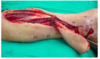
The correct response is Option E.
The reverse-flow sural flap has become one of the more dependable solutions in distal third leg wound and heel reconstruction. Survival of the flap depends on intact flow into the superficial
sural arterial network via perforators from the peroneal system. The largest perforator arises roughly 5 cm cephalad to the lateral malleolus and typically marks the lowest pivot point for the flap.
None of the remaining choices would provide adequate perfusion for this flap.
A 34-year-old man who works as a police officer is brought to the emergency department after sustaining a traumatic avulsion of the right heel. Examination shows a 3 * 3-cm area of exposed calcaneus on the weight-bearing aspect of the heel. Which of the following arteries and nerves supply the flap that will provide the most appropriate sensate coverage?

The correct response is Option B.
Sensate flaps include an intact sensory nerve supplying the overlying skin being transferred or, as in the case of a free tissue transfer, the cutaneous nerve is preserved and reanastomosed to a recipient cutaneous nerve. Sensate flaps are useful for areas where sensation is important for the appropriate function of a reconstructed region. Sensate flaps are beneficial in resurfacing wounds located on weight-bearing areas as in pressure sores or the heel, as in the scenario described.
The most reliable sensate flap for coverage of the plantar calcaneus comes from the medial plantar flap. This flap receives its sensation from the medial plantar nerve (L4-5) and receives its blood supply from the medal plantar artery. This flap comes from the instep of the foot between the head of the first metacarpal and the midpoint of the heel. The size of the flap can be up to 12 * 6 cm. Due to its proximity to the heel and the minimal donor morbidity with loss of sensation to the instep of the foot, this flap is the most appropriate for coverage of this patient €™s defect.
The dorsalis pedis flap receives its blood supply from the dorsalis pedis artery. This flap can be transferred as a sensate flap using the superficial peroneal nerve. However, this flap would not reach the area needed to be covered in this patient. The dorsalis pedis flap is used most frequently for coverage of the anterior ankle and dorsal distal foot.
The medial sural artery supplies the medial gastrocnemius flap. This flap is best used for soft-tissue coverage of the upper third of the leg and the distal knee. Preservation of the saphenous nerve with the medial gastrocnemius musculocutaneous flap will provide a neurosensory flap.
However, due to the distance from the flap, it would require a free tissue transfer and is typically not used for this type of coverage.
The proximal branches of the posterior tibial artery supply the medial soleus muscle flap. The posterior tibial nerve supplies only motor fibers to the soleus muscle and can therefore not be used as a sensate flap. This flap is best used for soft-tissue coverage of the middle third of the leg.
The radial forearm flap receives its blood supply from the radial artery and its septocutaneous perforating vessels. A neurosensory flap can be harvested based on the medial or lateral antebrachial cutaneous nerves to reconstruct the plantar surface of the foot. This would be a second choice to the medial plantar artery flap as it requires free tissue transfer.
A 52-year-old man is brought to the emergency department after sustaining a gunshot wound to the right forearm. History includes well-controlled diabetes mellitus type 2, coronary artery disease, and renal insufficiency. Examination of the forearm shows viable muscle coverage and gross instability. A radiograph is shown. Following debridement and stabilization of the wound, reconstruction of the defect is planned via a vascularized free fibular transfer. In addition to clinical examination, which of the following is the most appropriate preoperative evaluation of this patient €™s lower extremity?
A ) Ankle brachial indices
B ) Color-flow Doppler imaging
C ) CT angiography
D ) Traditional angiography
E ) Transcutaneous partial pressure of oxygen

The correct response is Option B.
While anatomic variants in the vasculature of the leg are rare, failure to recognize these prior to sacrifice of the peroneal artery during harvest of a fibular free flap can result in disastrous ischemic complications. Variations occur more commonly in patients who have vascular insufficiency due to underlying atherosclerotic changes. In these patients, clinical examination alone is insufficient to adequately define blood flow patterns to the leg and foot.
Color-flow Doppler imaging has demonstrated excellent ability to define the presence of flow-limiting lesions in the leg. Monophasic signals in any of the three major runoff vessels strongly suggest that the limb will be at risk for ischemia following sacrifice of the peroneal artery. Comparison of the findings on color-flow Doppler with angiography demonstrates that angiography is an unnecessary addition to the initial study.
Angiography and CT angiography offer excellent road maps of the vasculature of the lower extremity, but each does involve the use of intravenous contrast. The dose of contrast used in CT angiography is significantly lower than that in traditional angiography, but neither would be justified in this patient with pre-existing renal insufficiency.
MR angiography provides a map of the vessels comparable to both angiography and CT angiography without the use of nephrotoxic contrast agents. The additional information gained by seeing the vessels preoperatively (by MRA, CT angiography, or traditional angiography) may be justified when very long segments of the bone need to be harvested or when the perfusion to the overlying skin paddle may be better defined.
Transcutaneous partial pressure of oxygen measures perfusion and does not offer any anatomic detail.
The postoperative image shown demonstrates the free fibula reconstruction of the radius.
A 17-year-old boy sustains a traumatic injury to the right lower extremity. Examination shows weakness of plantar flexion and loss of sensation over the plantar surface of the foot. Which of the following nerves is most likely injured?
A ) Deep peroneal
B ) Femoral
C ) Obturator
D ) Superficial peroneal
E ) Tibial
The correct response is Option E.
The most likely nerve to be injured is the tibial nerve.
The tibial nerve is a branch of the sciatic nerve. It travels through the popliteal fossa and gives off branches to the gastrocnemius, soleus, plantaris, and popliteus muscles. The tibial nerve travels in proximity to the posterior tibial artery. In the leg, it gives off branches to the flexor digitorum longus, tibialis posterior, and flexor hallucis longus. Distally in the foot, it branches to give rise to the medial and lateral plantar nerves, which provide sensation to the plantar surface of the foot. Injury to the tibial nerve results in deficits of plantar flexion, as well as anesthesia to the plantar surface of the foot. Tibial nerve disruption has been considered an indication for amputation as opposed to limb salvage in traumatic injuries of the lower extremity. However, one study suggests that limb salvage can be performed even in the face of absent plantar sensation at the time of presentation. This may indicate that absence of plantar sensation is not a reliable correlate for tibial nerve disruption.
The deep peroneal nerve arises from the common peroneal nerve at the fibular neck. It travels in the anterior compartment of the leg and gives branches to the tibialis anterior, extensor hallucis longus, and extensor digitorum longus and brevis, as well as peroneus tertius. The sensory distribution is in the area of the first web space. Injury to the deep peroneal nerve causes weakness in dorsiflexion of the foot.
The femoral nerve innervates muscles of the anterior thigh, including the quadriceps group, iliacus, and sartorius. Injury to the femoral nerve results in weakness of leg extension.
The superficial peroneal nerve arises from the common peroneal nerve at the fibular neck. It supplies the lateral compartment of the leg, giving motor branches to peroneus longus and brevis, as well as sensory contribution to the lateral aspect of the leg. Injury to the superficial peroneal nerve results in anesthesia of the lateral aspect of the leg and weakness in eversion and plantar flexion of the foot.
The obturator nerve provides innervation to the medial thigh muscles (adductor group), including adductor brevis, longus, and magnus, as well as the gracilis and obturator externus. The cutaneous branch provides sensation of the medial thigh. Injury to the obturator nerve results in weakness in thigh adduction and sensory deficits in the medial thigh.

A 38-year-old man comes to the office for follow-up examination two months after undergoing open reduction and internal fixation of a fracture of the right proximal fibula. A preoperative radiograph is shown. On current physical examination, he can evert the right foot, but he is unable to dorsiflex it. This patient has most likely sustained an injury to which of the following nerves?
A ) Common peroneal B ) Deep peroneal C ) Sciatic D ) Superficial peroneal E ) Sural

The correct response is Option B.
The patient described has sustained an injury to his deep peroneal nerve. An electromyelogram should be ordered to confirm the diagnosis. The common peroneal nerve divides into two branches. The deep branch of the peroneal nerve innervates the extensor hallucis longus and the anterior tibial muscles. The superficial branch of the peroneal nerve provides motor innervation to the peroneus longus and brevis muscles. If the common peroneal nerve had been injured, eversion would not have been likely. A sural nerve injury would have a sensory component, not a motor component. The sciatic nerve injury would have more global consequences, though this pattern is theoretically possible in a Sunderland Grade VI injury. Thus, the most likely injured nerve is the deep peroneal nerve.
A 50-year-old man has noninfected wound dehiscence with exposure of the hardware two weeks after undergoing open reduction and internal fixation of an ankle fracture. A photograph and radiograph are shown above. Following debridement of the wound, which of the following is the most appropriate management?
(A) Skin grafting
(B) Coverage with a free flap
(C) Application of an external fixator and skin grafting
(D) Removal of the hardware and skin grafting
(E) Removal of the hardware and coverage with a free flap

The correct response is Option B.
This patient has early wound dehiscence after undergoing open reduction and internal fixation of a lateral malleolus fracture. Appropriate management involves early coverage of the exposed bone and hardware to promote bony union while preventing bacterial colonization of the hardware, which may lead to the development of osteomyelitis. Because this patient underwent open reduction and internal fixation only two weeks ago, fixation is still required to maintain rigid fracture stabilization. Instead, coverage with a free flap is recommended to provide stability and vascularity and to enhance function once bony union is obtained.
Skin grafting will not provide stable, well vascularized coverage in this patient. External fixation is problematic because of the location of the fracture. Removal of the hardware two weeks after fracture is indicated only for established hardware failure.