Lecture 3 Neuroanatomy Flashcards
Neuroanatomical Directions
Rostral = towards the “beak” or front end
Caudal = towards the tail or back end
Dorsal = top of head
Ventral = part facing the ground
Lateral = towards the side
Medial = towards the midline
Neuroanatomical Planes

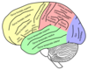
4 Lobes Of The Brain and their main functions
- Frontal lobe - important for cognitive functions and control of voluntary movement or activity.
- Parietal lobe - processes information about temperature, taste, touch and movement.
- Occipital lobe - primarily responsible for vision.
- Temporal lobe - processes memories, integrating them with sensations of taste, sound, sight and touch.
What types of Cognitive Planning does the Frontal lobe help us perform?
- strategic and coordinated planning.
- Verbal activities (such as planning a lecture) involve more left hemisphere regions, whereas spatial activities (such as planning a garden) involve more right hemisphere regions.
- The more challenging the planning of the activity, the greater the number of frontal lobe regions participate in the planning process.
- Example: Child who wants to go skiing.
- Difficulties in planning are common in Alzheimer’s
and other dementias.
- Effects of stress/anxiety, schizophrenia.
What is the role of the Parietal Lobe?
- Processes information about temperature, taste, touch and movement.
- Sensory information goes through the thalamus to the parietal lobe, where it is processed and potentially integrated with the visual system.
- Includes the somatosensory cortex. Basically a map of the sensory representation of body parts.
- Several areas of the parietal lobe are important in language processing eg. Cognitive processing of language in the angular gyrus and sensorimotor control of writing.
Damage to the right parietal lobe can result in:
- Neglecting part of the body or space (contralateral neglect), which can impair many self-care skills such as dressing and washing.
- Can also cause difficulty in making things (constructional apraxia), denial of deficits (anosagnosia) and drawing ability.
What is Gerstmann’s Syndrome?
- Damage to the left parietal lobe:
- Includes right-left confusion, difficulty with writing (agraphia) and difficulty with mathematics (acalculia).
- Can also produce disorders of language (aphasia) and the inability to perceive objects normally (agnosia).
What is Balint’s Syndrome?
- Bi-lateral damage.
- A visual attention and motor syndrome.
- Characterized by the inability to voluntarily control the gaze (ocular apraxia), inability to integrate components of a visual scene (simultanagnosia), and the inability to accurately reach for an object with visual guidance (optic ataxia).
Explain neurological basis for Dyslexia and what it really is.
- Dyslexia is an often-misunderstood, confusing term for reading problems. It is neurobiological in origin.
- One of the most common misunderstandings about this condition is that dyslexia is a problem of letter or word reversals (b/d, was/saw) or of letters, words, or sentences “dancing around” on the page.
- Left parietotemporal system is important for word analysis. This region is critical in the process of mapping letters and written words onto their sound correspondences – letter sounds and spoken words. This area is also important for comprehending written and spoken language.
- People with dyslexia have less gray matter in the left parietotemporal area than nondyslexic individuals. Thought to lead to problems processing the sound structure of language.
- Many people with dyslexia also have less white matter in this same area than average readers; more white matter is correlated with increased reading skill.
Brodmann area for V1 (or primary visual cortex)
Where does it project?
Brodmann area 17
V1 projects to occipital areas of the ventral stream and occipital areas of the dorsal stream

What is the extrastriate cortex and what are these regions for?
•Visually driven regions outside V1 are called extrastriate cortex. There are many extrastriate regions, and these are specialized for different visual tasks, such as visuospatial processing, color differentiation, and motion perception.

main functions of temporal lobe, 3 areas within and their functions
Involved in processing sensory input for the appropriate retention of visual memory, language comprehension, and emotion association
- The temporal lobe holds the primary auditory cortex, which is important for the processing of semantics in both speech and vision in humans.
- Wernicke’s area, which spans the region between temporal and parietal lobes, plays a key role in speech comprehension.
- The medial temporal lobes are thought to be involved in encoding declarative long term memory.
- The medial temporal lobes include the hippocampi, which are essential for memory storage; damage to this area can result in impairment in new memory formation leading to permanent or temporary anterograde amnesia.

Define Gyri and name some
A ridge or fold between two clefts on the cerebral surface in the brain
Define Sulci and name 2 sulci and 3 fissure (4 total with one overlapping)

Define Interneuron
Connectors (relay or association neurons) between the sensory and motor neuron pathways
Found exclusively in the CNS
How many spinal nerves and what do they do?
- Carry motor, sensory, and autonomic signals between the spinal cord and the body.
- In the human body there are 31 pairs of spinal nerves, one on each side of the vertebral column.
Spinal Cord organization
- Spinal cord is organised into dorsal root ganglia (somatosensory) and ventral root ganglia (motor). The dorsal root (posterior) enters the spinal cord and the ventral roots (anterior) exit the spinal cord.
- Each of the 31 dorsal roots innervate different areas of the skin referred to as dermatomes.
- When a dorsal root is cut, the spinal cord can no longer obtain info from that nerve.
- Spinal interneurons – form synapses with motor neurons which can mediate the motor response of sensory stimuli.

What is the brainstem?
What does it do?
- The central trunk of the mammalian brain, consisting of the medulla oblongata, pons, and midbrain, and continuing downwards to form the spinal cord.
- Regulates the central nervous system,
- Provides the main motor and sensory nerve supply to the face and neck via the cranial nerves.
- Plays an important role in the regulation of cardiac and respiratory function.

What are Cranial nerves?
How many originate in the brainstem?
Which two don’t?
- Cranial nerves are the nerves that emerge directly from the brain (including the brainstem), in contrast to spinal nerves (which emerge from segments of the spinal cord).
- 10 of the cranial nerves originate in the brainstem.
From the Cerebrum:
The first and second cranial nerves derive from the telencephalon and diencephalon respectively and are considered extensions of the central nervous system: olfactory nerve (CN I), optic nerve (CN II)

What is the Reticular Formation?
•Clusters of neurons that are interconnected within the brainstem.
•
•Thought to be responsible for mediating quite different behaviour eg. Sleep, respiration, habituation.

What is the Medulla Oblongata?
What happens if it’s damaged?
- Controls autonomic functions such as breathing, digestion, heart and blood vessel function, swallowing, and sneezing.
- Relay of nerve signals between the brain and spinal cord.
- Coordination of body movements.
- Regulation of mood.
Damage:
•May result in a number of sensory-related problems eg. numbness, paralysis, difficulty swallowing, acid reflux, and lack of movement control.
What is the Pons?
- Helps relay messages from the cortex and the cerebellum.
- Basically a messaging station.
What is the Mesencephalon?
- Also known as the midbrain.
- Associated with vision, hearing, motor control, sleep/wake, arousal and temperature regulation.

What is the Diencephalon?
- Includes the thalamus, the hypothalamus, the epithalamus and the subthalamus.
- Information processing and relay centre.
What is the Hypothalamus, Role?
What is it?
- Area of brain that contains a number of small nuclei with a variety of functions.
- Located below the thalamus.
•
What is its role?
- Links the nervous system to the endocrine system via the pituitary gland.
- Releases hormones.
- Regulates body temperature.
- Maintains daily physiological cycles.
- Manages sexual behavior.
- Controls appetite.
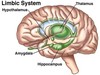
What is the limbic system? (Role and structures)
What is it?
•A collection brain structures including the amygdala, hippocampus, hypothalamus and thalamus.
•
What is its role?
- Supports a wide variety of functions including emotion, behaviour, long-term memory and motivation.
- Integrating the sensory, affective, and cognitive components of pain and processes information regarding the internal bodily state.
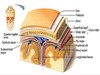
What protects the brain?
- Hair
- Scalp
- Skull
- Meninges – the membranes covering the brain and spinal cord:
- Dura mater – “hard matter”. Tough and covers the inside of the skull. Has indentations which limit the movement of the brain.
- Arachnoid mater – covers over the sulci. Filled with cerebrospinal fluid.
- Pia Mater – immediate covering which covers brain very closely and covers all sulci and fissures.
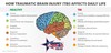
How TBI affects Daily Life
(by area)
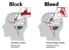
•Stroke:
Acronym of symptoms
•Stroke: a sudden loss of blood supply to the brain, or bleeding into the brain tissue.

Causes of stroke
•Hypertension -an increase in blood pressure due to the constriction of small blood vessels.
•
•Ruptured aneurysm - ballooning/rupture of an artery wall.
•
•Arteriovenous malformation (angioma) - collection of abnormal blood vessels which produce abnormal blood supply.
Causes of Epilepsy:
Mechanism:
- A group of neurons begin firing in an abnormal, excessive and synchronized manner.
- This results in a wave of depolarization.
- This then results in a specific area from which seizures may develop, known as a “seizure focus”.
•
Eliptogenesis:
- Cases which occur as the result of brain injury, stroke, brain tumors, infections of the brain, and birth defects.
- Mechanism thought to result due to up-regulation of excitatory circuits or down-regulation of inhibitory circuits following an injury to the brain.
