AED - Anterior Eye Diseases Slide Exam Revision I: Characteristics - SWOTVAC Flashcards
Name, define, causes (2), features (malignancy, feeder vessels, motility, corneal involvement), biopsy (2).

Conjunctival Papilloma - overgrowth of epithelial cells
Cause: Excess UV or HPV infection (HPV in 39% of cases)
Features: non-malignant, motile, doesn’t involve cornea, modest feeder vessels, pinkish (less red),
Biopsy: BM still intact, non-invasive of stroma
Name, causes (2), features (malignancy, feeder vessels, motility, corneal involvement), biopsy (2).

CIN - conjunctival intraepithelial neoplasia
Cause: Excess UV or HPV (in 39%)
Features: Non-malignant, motile, more marked BV strawberry spots, more lush feeder vessels, invades corneal epithelium (but NOT stroma or substantia propria)
Biopsy: Non-invasive of stroma, plemorphism and metaplasia present
Name, features/biopsy (malignancy, feeder vessels, motility, corneal involvement), requirement for DDx

SCN - squamous cell neoplasia
Features/biopsy: malignant, non-motile, broken through basement membrane and invading stroma/substantia propria, may see ulceration (with white plaques) and small haemorrhages
DDx: requires OCT or biopsy, b/c CIN-like appearance
(note: non-motile as anchored by stromal invasion)
Name each condition and compare: Redness, Feeders, Corneal invasion, Stromal invasion (OCT), Motility (OCT), Malignancy, Pleomorphism, Surgery

Papilloma: redness (+), feeders (+), corneal invasion (-), stromal invasion (-), motile (+++), malignancy (-), pleomorphic (-), surgical removal (optional)
CIN: redness (+++), feeders (+++), corneal invasion (++), stromal invasion (-), motile (++), malignancy (+), pleomorphic (++), surgical removal (YES)
SCN: redness (+++), feeders (++++), corneal invasion (++++), stromal invasion (++++), motile (-), malignancy (++++), pleomorphic (++++), surgical removal (YES)
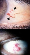
Name, define, features (growth rare, malignancy, onset, elevation, colour, feeder vessels).

Naevus: Benign ocular pigmented lesion
Usually forms in 10-20YO.; Slow growing; 1% become malignant.; Flat or minor elevation.; Colour varies, commonly 1-2 feeder vessels
Name, features (malignancy, motility, layer), Dx requirements (3), Mx (1)

Congenital melanocytosis
.- Pigment in sclera- 2-4% get malignancy.- Not Motile (no conj.)- - -
Dx: F.A.T, annual photos, DFE for choroidal melanoma
Mx: (if malignant) refer for excision + biopsy
Name, characteristics (age group and skin colour, appearance, acquired/congenital, uni/bilateral, elevation, motility, malignancy)

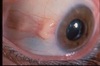
Primary Acquired Melanosis
Older + middle aged + fair skinned at risk.- Diffuse brown colour + acquired.- Unilateral.- Flat + Mobile over sclera.- Concerns for melanoma-
Name, features (growth rate, colour density, edges, boundary, feeder vessels)

Melanoma
- 75% come from PAM.- invasive.- Fast growth.- Dense Colour (may be Diffuse at edge).- Irregular boundary.- Feeders
Name, describe appearance, is it symptomatic?
Telangiectasia:
.- Corkscrew squiggly or masses of BV- Asymptomatic, cosmetic or systemic disease indicator- - - -
Name, define/features (malignancy, disease association, common site)

Kaposi’s sarcoma:
.- malignant tumor (overgrowth) of the blood vessels associated with AIDS (immunocompromised)- - bright red vascular mass - most often inferior fornix- -
Name, characteristics (3). Hallmark sign.

Sturge-Weber Syndrome
Effects seen along trigeminal nerve- Episcleral/conj involvement in 70%.- Congenital conjunctival or Episcleral haemangioma
Portwine stain along CNV hallmark
Name, define, Mx (2).

Cavernous sinus fistula:
.- Break in the cavernous sinus- i.e. “fistula” = break in wall of an artery –> causing higher arterial blood to enter venous space—-
Mx: refer for closure of fistula + mx of IOP
Name, features (fluid, gland, transparency).

Cyst of Moll/sweat glands
- Clear, fluid, translucent balloon; - syringoma (multiple+flat); - milia (multiple hard pimples)
Name, describe (gland, fluid)

Cyst of Zeis Gland - Opaque lesion
- Visible lesion at lash root, often hair follicle, Appears slight yellow/white and opaque/milky (not translucent like Moll)
Name, describe (1), causes (2), a symptom (1).

Stye/Hordeolum
Blockage of duct.- due to bacterial infection (external) or complication of chalazion (internal)- discomfort
Name, describe, symptoms (3), features (gland, cause)

Chalazion
Blockage of MG.- Inflammation- symptoms: cosmesis, generally painless, does not affect V.A (only lower lid)
Name, describe (5), risk group (age/gender, skin type)

Sebaceous gland carcinoma
elderly females (chronic bleph)-yellow and hard - madarosis with thickened, red lid margins (UL) - >2mm 60% mortality—
Name, cause (1), describe/forms (2)

Viral Warts (Verruca)
.- Viral Infection from HPV.- Papule or elongated filliform—-
Name, cause (1), describe (1)

Molluscum contagiosum
Viral Infection from pox virus- Flatter dome-shaped lesion (1-3mm)—-
Name, describe (3), core content (substance), appearance, diagnostic tool, malignancy, management (2)

Keratoacanthoma
- Benign neoplasm, mimics squamous cell carcinoma, resolves spontaneously or may refer for removal/biopsy
Volcano with an ulcer crater
Keratin core - use OCT to find it
Name, describe (1), malignancy.

Xanthelasma
- soft, raised yellow plaques occurring on the skin at the inner corners of the eyes, is benign
Name, describe, malignacy, risk group (age and skin).

Seborrheic Keratosis (SK):
- A superficial benign neoplasm of epidermal cells that presents as a papule or plaque with a characteristic “stuck-on” appearance. These lesions are usually acquired later in life and tend to grow slowly.
Sun-damaged skin/UV damage risk factor
How does Solar or Actinic Keratosis appear? Malignancy, skin appearance, cosmesis, risk factor (skin)
Scaly appearance.- Benign skin tumour.- Just cosmetically unappealing
Sun damage/UV damage risk factor

Epithelial Basement Membrane Dystrophy (EBMD): what is it also known as? How common is it? Describe it’s appearance.
Also known as map-dot fingerprint dystrophy.
Most common corneal dystrophy but often misdiagnosed due to variable appearance

Meesman’s dystrophy, dystrophy of what, characterised by what? Uni/bilateral?
How does it appear?
.- Anterior corneal dystrophy- characterized by extensive, bilateral, clear intraepithelial cysts—-

Describe the appearance of Reis-Buckler’s Dystrophy
- Characteristic appearance where sheet-like connective tissue replaces bowman’s membrane—–

Thiel-Behnke corneal dystrophy (2). What is it similar to and what pattern does it have?
- Similar to Reis-Buckler’s dystrophy but later onset.- Same honeycomb appearance/layers affects so often indistinguishable—-

Lattice Dystrophy: features (layer, distinguishing features (3)), symptom (1)
What appearance does it have?
AD Stromal corneal dystrophy: Amyloid-Refractile branching lines, white dots, and central haze.-stroma develops ground-glass appearance-Decreased vision in 3rd decade–

Granular Dystrophy (appearance, layer, characteristic)
List effect on corneal sensation and VA
What appearance does it have overall?
.- Centrally discrete focal white deposits at all Stromal depths.-“cornflakes”- Area between lesions is clear. Early onset w/ good VAs, RCE rare. Normal or reduced corneal sensation.—

Name, describe, how does it appear, what two other dystrophies is it similar to?
What can it be traced back to?

Avenillino Dystrophy:
.- Combination of Lattice + Granular Dystrophies in one.- Trace back to specific part of Italy—-
Name, desribe (5)
What is the severity of this disease? What appearance does it have?

Macular Dystrophy
- Exceptions to “dystrophy” rule (autosomal recessive, extends to cornea periphery).- Most severe and least common dystrophy- Diffuse “ground-glass” haze lesions, corneal haze between lesions, gray/milky white opacities throughout stroma, and limbus to limbus—
Name, describe (2)
What is it associated with?
What causes it?

Schnyder’s Crystalline Dystrophy:
.- may be associated with systemic hypercholesterolaemia.- Central lipid crystal deposition in the Anterior stroma—-
Fuchs Endothelial Dystrophy: describe (2)
.- Loss of endothelial cells and the resulting oedema and thickening of the stroma- Diffuse thickening and lamination of Descemet’s membrane—-

Name, describe (how common is it? What is happening to endothelium?) (3)

Posterior polymorphous corneal dystrophy
- Rare endothelial dystrophy (also affects descemet’s).- Endothelium begins to show epithelial properties- Grouped (or linear) bubble-like lesions at Descemet’s—
Name, describe (3)

Filamentary Keratopathy
.- Abnormal areas of corneal epithelium + excess mucous in tears . These filaments form and form tails which stick—
Name, describe (2)

Superficial punctate keratopathy (SPK)
- Corneal condition consisting of multiple pinpoint epithelial defects; non-specific sign indicating corneal problem, e.g. dry eye syndrome(refer to lecture for different types!)- —
What’s this? How common is it? What causes it?

Subepithelial Infiltrates
- Relatively common, due to inflammatory response within the anterior corneal stroma—–
Name, causes (8), characteristics (4)

Neurotrophic keratopathy
Causes: *HSV/HZV, DM, LASIK, CL*, recurrent erosions/dystrophies, tumors, strokes, topical drug therapy
Description: Occurs in eyes with decreased corneal sensitivity .- SPK.- Large ulcers (sometimes).- Longest axis horizontal-
Name, describe, how does this affect vision?

Bullous Keratopathy:
- Degenerative process characterized by small blister-like pockets that form in swollen corneal epithelial layers; markedly reduces vision.—–
Name, describe what happens and what it leads to

Interstitial Keratitis
- Opaqueness on cornea that spreads to pupil -> leads to blindness.-Inflammation of the corneal stroma without primary involvement of the corneal epithelium or Endothelium—-
Name, how common? unilateral or bilateral? where/location? any associations? how often?

Marginal Keratitis
- Common, unilateral, peripheral corneal lesion .- CL-associated sometimes—-
Name, appearance, cause (2.5)

Phlyctenulosis
.- small vesicle at lateral limbus caused by Staph or M. tuberculosis.- Type IV reaction
Name, describe, what is unique about it?

Mooren’s ulcer
.- Peripheral corneal ulcer. MUST be independent of any underlying systemic disorder Unique: overhanging edge of corneal defect—-
Thygeson’s Superficial Punctate Keratitis (4)
- *Bilateral gray-white, slightly elevated lesions in white and quiet eye* .-minimal to no staining with NaFl-unknown etiology, but possible viral—

Name, describe, in what condition would you get this? ;)

Vogt’s Striae (it’s me!)
.- Vertical lines in deep stroma.- Keratoconus—
Name, describe

Fleischer’s Ring
- due to iron deposition in basal epithelial cells at the base of keratoconus cone which have degenerated
(note: image on right is a Kayser-Fleisher ring, which is slightly different, arising from copper deficits in descemet’s)
What is Munson’s sign?
.- Used to detect Keratoconus by having the patient look down to see the indentation of the lower lid by the cone of the cornea—–

What is Pellucid Marginal Degeneration? Describe what it looks like. Is it unilateral or bilateral? symmetry? inflamatory or non-inflammatory? What happens to the stroma?
.- Crabclaw on topgraphy (kissing doves)
- Bilateral, asymmetric, noninflammatory peripheral ectatic disorder.
- Stromal thinning in a crescentic band from 4-8’oclock above limbus—

What’s this?

Keratoglobus: .- Globular protrusion of entire cornea—–
What’s this? When is this most common? What other patients may this occur in? (3)

Band keratopathy
- Deposition of calcium in the superficial cornea; most commonly in patient with chronic corneal disease; may occur in patients with hypocalcemia, parathyroidism, and in renal failure—–
What is Phototherapeutic Keratectomy (PTK)?
.- A procedure to treat pathologic conditions of the surface of the cornea using an excimer laser. the laser beam ablation pattern is Flat to create A smoothing of the corneal surface.—–

Name, describe general features (7), cause (1), risk factor (1), outcomes (2)

Salzmann’s Nodular Degeneration
- Elevated, midperipheral, smooth, opaque, blue-white SEI, hyaline nodules .-due to chronic keratitis.-female predilection.-causes Irregular astigmatism and refractive changes–
Name, which gender most commonly affected?, any symptoms? unilateral or bilateral? how fast does it progress? what does this lead to?

Terrien’s Marginal Degeneration
- More common in males (75%), asymptomatic, bilateral- Painless, slowly progressive, thinning of the peripheral corneal stroma –> Corneal gutter.- no NaFl staining.- Can develop astigmatism–
Name, affect on V.A, cause (1), appearance

Spheroidal degeneration
- “Climate droplet degeneration”.- Rarely affects VAs.-
appearance: superficial stroma is golden brown.-
cause: Interpalpebral Actinic (UV) exposure (long term extreme UV)–
Name, where affected? unilateral or bilateral? appearance?

Polymorphic Amyloid Degeneration
- Deep stroma, bilateral.-Appears Similar to Lattice dystrophy.- SL . small Refractile punctate deposits.- Comma-shaped and filamentous Amyloid deposits- -
What is a Corneal arcus?
- Opaque white ring about corneal periphery, seen in many individuals older than 60 years of age. This is due to deposit of lipids in the cornea or to hyaline degeneration. May indicate a lipid disorder, most commonly type II hyperlipidemia if present before the 40 years of age (if seen in younger people, it is called arcus juvenilis).—–

Name, describe. What is this condition characterized by? (4)

Lipid Keratopathy
- Type of degenerative corneal disease characterized by accumulations of lipid within the corneal stroma, corneal epithelium, keratocytes, and/or histiocytes infiltrating the cornea—–
What’s this? Describe it

Vogt’s Limbal Girdle
.- Chalky white deposits along the nasal and temporal limbus in the inter-palpebral fissure (crescent shaped)—–
What’s this? Describe the appearance

Vortex Keratopathy
.- Whorl-shaped golden-brown deposits on the corneal epithelium—–
What is this and why did it occur?

.- Rust ring on cornea due to a metallic foreign body—–
What is a Huson-Stahli Line?
.- epithelial iron deposit Just below mid-point of the Interpalpebral fissure (occurs with age)—–

What is a Stocker’s line?
.- iron line at leading edge of pterygium—–

What is Ferry’s line?
.- iron deposits on the leading edge of A filtering bleb—–

What is Ocular siderosis?
.- Stromal iron deposits from an intraocular FB—–

What are Kayser-Fleischer rings?
.- golden-brown bands in the limbus of the cornea seen on slit lamp exam (due to copper deposition)- Associated with wilson’s disease—-

Name, unilateral or bilateral? describe appearance

Crocodile Shagreen
- Bilateral, gray/white opacification at *Bowman’s layer* (ant) or *deep storma* (posterior) .–mosiac or cracked ice pattern—-
What’s this called? Explain. What might cause this?

Corneal Farinata
.- Farinata = Flour dust deposits in deep corneal stroma.
- May be caused by accumulation of waste (lipofuscin)—
What is this? How does it occur? How would you describe how it looks?

Corneal Guttata.
- endothelial cell abnormalities.- Beaten metal appearance—-
What is this condition? What is it’s defining feature?
Pingueculum
.- Base to limbus-
What is this condition called? What is its defining feature?

Pterygium
.- Base to conj–
What’s this? Describe (3)

Concretions: are deposits on palpebral conj.
- Yellow-white in colour
- Usually <1mm (but up to 4mm).
What is this called? Describe the appearance of this condition (3)

Amyloidosis
- Yellow-ish, avascular, waxy deposits within the bulbar or forniceal conjunctiva—–
Name, describe

Conjunctival cyst/lymphangiectasia
- Typically small clear cyst (bubble) within the bulbar, forniceal or palp. conj.—–
What is this?

Ecchymosis
.- Sub-conjunctival blood anywhere under the bulbar conjunctiva—–
What’s this?

Bitot’s spot
- Pearly looking, foamy mass that forms specifically on a dry patch of bulbar conjunctiva near the cornea—–
What’s this?

Floppy Eyelid Syndrome
.- Upper eyelids are flaccid and Easily everted–
What’s this? Describe it’s characteristics (2)

Conjunctival Papillae
.- characteristic . Central blood vessel.- Usually fine mosaic pattern—-
What’s this? Characteristics (2)

Conjunctival Follicles
.- More common in corniceal conjunctiva.- blood vessel around the Base often surrounding—-
What’s this? Describe it? What happens if you peel it?

Pseudomembrane
- A loose membranous layer of exudate containing organisms, precipitated fibrin, necrotic cells, and inflammatory cells produced during an inflammatory reaction on the surface of a tissue..- Can be peeled without bleeding.- Leaves conjunctival epithelium intact—
What’s this? Describe. How easy is it to peel?

True membrane
.- Coagulated fibrinous exudate anchored to the inflamed conjunctival epithelium (Rare).- Peeling More difficult.- Rips conjunctival epithelium—
What’s this? How common? Acute or chronic? prognosis? unilateral or bilateral?

Bacterial conjunctivitis
- Very common, acute, self limiting, bilateral (start in one and transfer)—–
What’s this? How common? How usually transmitted? What pathogen is to blame?
Bacterial Conjunctivitis (gonococcal)
- Uncommon, often sexually transmitted .- e.g. Neisseria gonorrhoeae—-
What’s this? What pathogen causes it? What age group most common? Describe symptoms (1), transmissability (1) and prognosis (1)

Pharynogoconjunctival fever (PCF)
- Adenovirus serotypes 3/7.- More common in kids.- URTI.- Low grade fever.- Highly contagious- Resolution in 7-14 days
What’s all this? Acute or chronic? What is the main sign?
Trachoma
- Chronic, contagious form of conjunctivitis that typically leads to blindness (chlamydiaaa).
- Main sign = Superior bulbar and palpebral conjunctival follicular response
.- chronic Inflammation over many years—
Name, how transmitted (1), in whom most common

Adult inclusion conjunctivitis (AIC)
.- seen in economically developed countries.- High risk of concurrent chlamydial.- Sexually transmitted.- Most common in young Sexually active adults.- Genital infections (make sure to get tests done)-
Name, describe, where

Anterior blepharitis
.- Very common Inflammation and/or Infection involving the lid margin Anterior to the meibomian glands and including the lashes—–
What’s this? How common? unilateral or bilateral? symmetry? outcomes? (3)

Posterior blepharitis
- Common, bilateral and usually symmetrical dysfunction of the meibomian glands. Results in poor/unstable tear film. - Excess secretion appear as oil droplets, capping MG.- Posterior lid margin thickened with vascular dilation + telangiectasia—
What is this called? What is the classic sign of this?

Ocular rosacea
.- Classic butterfly rash across nose and cheeks—–
What are the main signs of seasonal/perennial allergic conjunctivitis? (7)
Common, seasonal or perennial depending on allergen; Type 1 hypersensitivity (IgE mediated); Usually bilateral, can have conjunctival papillae, hyperaemia, and oedema; lid may be oedematous; Serous + mucoid discharge; Cornea unaffected

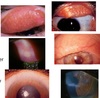
What condition is this? Describe the features seen here (2)

Limbal form of VKC
Limbitis is present with limbal papillae and Horner-Trantas’ dots (mainly eosinophils); There is also psedogerontoxon (cupids bow) which occurs in an area of previously inflamed limbus
(note: horner’s trantas dots can also appear in atopic keratoconjunctivitis)
What condition is this? Who does it occur in? Is it flat or raised? How often does it progress to ____?

CAM = Complexion associated Melanosis. (aka Racial melanosis)
Dark patches usually on dark skinned people
Usually flat and involves the limbus
Rarely progresses to melanoma