Exam 1 (9-12, 18) Flashcards

1
Q

Periodontium

A

Tissues that surround, support and attached to the teeth.

Made of two parts:
1. Gingiva

  1. Attachment apparatus:

periodontal ligament (PDL), cementum, alveolar bone

2
Q

Free gingiva

A

closely adapted around each tooth but not attached
keratinized

3
Q

Parts of free gingiva

A

Gingival margin
gingival sulcus
junctional epithelium
free gingival groove

4
Q

Gingival margin

.

A

the edge of the gingiva nearest to the incised surface of the tooth

5
Q

Gingival sulcus

A

The crevice between the free gingiva and the tooth.

6
Q

Crevicular fluid

A

flows from connective tissue into the sulcus.
cleanses the sulcus transports enzymes and antibodies

7
Q

Junctional epithelium

A

the epithelial attachment provides a seal at the base of the sulcus
non keratinized separates the periodontal ligament from the oral environment

8
Q

Free gingival groove

A

a shallow linear demarcation between free gingiva and attached gingiva

9
Q

Interdental papillae

A

Extensions of unattached gingiva between adjacent teeth
keratinized fills embrasure spaces

10
Q

Col

A

The depression between the lingual and facial papillae that conforms to the proximal contact of posterior teeth.

Non keratinized

11
Q

Attached gingiva

A

Part of the gingiva that is tightly connected to the cementum on the root (cervical third) and to the connective tissue cover of the alveolar bone

12
Q

Mucogingival junction

A

The line that marks the connection between the attached gingiva and the alveolar mucosa on the facial surfaces of all quadrants and the lingual surfaces of the mandibular arch

13
Q

Alveolar mucosa

A

The thin, moveable, loosely attached tissue covering the alveolar bone.
non keratinized

14
Q

Frena

A

Narrow folds of the membrane that pass from a more fixed to moveable mucosa.

ex. from the attached gingiva at the MGJ to lip, cheek, or undersurface of tongue.

15
Q

The periodontal ligament (PDL)

A

Fibrous tissue that surrounds and attaches the alveolar bone to cementum.

16
Q

Interdental fiber

A

Connects adjacent teeth at the CEJ. Cementum to cementum

17
Q

Cementum

A

A thin layer of calcified connective tissue that covers the tooth from CEJ to, and around, the apical foramen

Important function of cementum is to attach periodontal ligament fibers to root surface

18
Q

Cementum functions

A

Seal dentinal tubules provide attachment for the periodontal fiber groups
maintain occlusal relationships

19
Q

Alveolar process

A

The extended areas of bone in each arch that are tooth-bearing

20
Q

Lamina Dura

A

Thin, compact alveolar bone lining the tooth socket

21
Q

Assessment Instruments

A
  1. Mirror
  2. Explorer
  3. Probe
22
Q

The handle of an instrument:

A
  • part that is grasped
  • may be smooth or ribbed with varying diameter
23
Q

Functional Shank

A
  • connects the working end to the handle
  • maybe straight or curved (complex shank)
24
Q

Terminal Shank

A

-the lower shank

25
Q

Working End

A
  • part of the instrument that contacts the tooth or tissue + performs intended function
  • DICTATES THE USE
26
Q

Mouth Mirror Uses (4)

A
  1. Indirect vision
  2. Retraction
  3. Reflection of light
  4. Transillumination
27
Q

Probe Uses

A

-detect periodontal pockets

28
Q

Traditional Probe

A

Williams Probe

thin, round working end

grooves at 1,2,3,5,7,8,9, and 10mm

29
Q

Novatech Probe

A

right angle probe design
-improves adaptation only for posterior teeth

30
Q

Florida Probe

A

-computer-assisted probe with digital readouts and computer data storage

31
Q

Stroke Directions

A
  1. Diagonal/oblique (running across tooth from line angle to line angle)
  2. Vertical (parallel with long axis of the tooth for interproximals)
  3. Horizontal (parallel with occlusal surface for facials or linguals, mesials or distals of teeth with no contact)
  4. Circular (porte polisher)
32
Q

Stroke Selection Determinants

A
  1. Position, Contour, Size of the gingiva
  2. Probing depth
  3. Area of the tooth being worked
  4. Instrument being used
  5. Procedure being used (nature of deposit)
33
Q

Nature of Stroke

A
  1. Grasp: variable
  2. Stability: hand pivots on fulcrum
  3. Motion: wrist activation NOT finger flexion
  4. Length: determined by pocket/sulcus depth, part of tooth being treated, instrument being used, & nature of the procedure (root planing, probing, or exploring)
34
Q

Gingival pocket

A

is deepening of gingival sulcus caused by detachment of coronal portion of junctional epithelium and swelling of tissue

gingivitis

35
Q

Periodontal pocket

A

forms from apical migration of the junctional epithelium and destruction of periodontal fibers and bone

Periodontitis = irreversible

36
Q

Probing Depth

A

The distance in millimeters from the gingival margin to the base of the sulcus or periodontal pocket as measured with a probe

37
Q

Periodontal attachment system

A

is a group of structures that work together to attach the teeth to the maxilla and mandible.

38
Q

Junctional epithelium (JE):

A

attaches the gingiva to the tooth

39
Q

Fibers of the gingiva:

A

a network of fibers that brace the free gingiva against the tooth

40
Q

Loss of attachment (LOA)

A

is damage to the structures that support the tooth.

41
Q

Gingivitis

A

reversible damage to the gingiva

-There is NO damage to the periodontal ligament or alveolar bone

42
Q

Periodontitis

A
  • Results in permanent damage to the periodontal attachment system
  • Damage to gingival connective tissue, periodontal ligament, and alveolar bone
43
Q

Uses for Periodontal Probe:

A
  • Measuring oral deviations
  • Assessing tooth mobility
  • Assessing bleeding on probing
  • Measuring the width of attached Gingiva
44
Q

Levels of the gingival margin

A

Can change over time in response to trauma, medications, or disease.

Three possible levels:

Margin slightly coronal to CEJ (Normal = 0)

Margin significantly covers CEJ (Edema = -negative)

Margin significantly apical to CEJ (Recession = +positive)

45
Q

How to calculate the width of the gingival margin

A

Three Steps:

  1. Measure the total width of the gingiva from the gingival margin to the mucogingival junction.
  2. Measure the probing depth.
  3. Calculate :

total width of the gingiva minus the probing depth

46
Q

Can you measure bone loss with just probing depth?

A

NO!

The probing depth reading alone is NOT ENOUGH to tell us where the bone is located.

47
Q

Probing depth readings accurately indicate the level of the bone only IF______________________________________________

A

the gingival margin is in its normal position, slightly above the CEJ.

48
Q

If the gingival margin is NOT in a normal position (recession or edema)….

A

the bone support must be calculated using two measurements:

1.Probing depth

2.Gingival margin level

49
Q

Clinical Attachment Level (CAL)

A

is the estimated position of the structures that support the tooth as measured with a periodontal probe.

The clinical attachment level provides an estimate of a tooth’s stability and the loss of bone support.

50
Q

Margin at Normal Level

A

If the gingival margin is at the normal level:

The probing depth and the CAL are the same

51
Q

Gingival Margin Receded

A

The gingival margin level reading is added to the probing depth reading.

52
Q

Margin Significantly Above CEJ

(Edema)

A

Subtract gingival margin level reading from the probing depth reading

53
Q

Furcation involvement

A

is a loss of alveolar bone and periodontal ligament fibers in the space between the roots of a multirooted tooth.

54
Q

Root Furcation Morphology

A

Mandibular 1st Molar:

(D) 13mm - 14mm (M) Furcatiion: (M) 3mm - 4mm (D)

Maxillary 1st Premolar:

(L) 12.6mm - 13.4mm (B) Furcation: 7mm

Maxillary 1st Molar:

(DB) 12mm - 14mm (P) Furcation: (MB) 4mm - 7mm (P)

55
Q

A probe’s working end is

A

blunt/rod-shaped

56
Q

Cross-section of a probe may be

A

circular or rectangular

57
Q

Identify the symbol used for periodontal charting

A

Class I

58
Q

Identify the symbol used for periodontal charting

A

Class II

59
Q

Identify the symbol used for periodontal charting

A

Class III

60
Q

Identify the symbol used for periodontal charting

A

Class IV

61
Q

Adaptation

A

is the positioning of the toe-third or tip-third of the working-end against the tooth surface.

62
Q

Psychomotor skills

A

require complex SMALL movements and repeated practice.

-Brain, nervous system, and muscles all work together

63
Q

Muscle memory

A

frequently enacted muscle tasks that are stored in the brain

64
Q

Myelination

A

is the process of forming myelin sheath around a nerve to allow nerve impulses to move more quickly.

With practice movements become smoother, myelination occurs

65
Q

Automaticity

A

is the ability to perform a psychomotor skill smoothly, easily, and without frustration

66
Q

Motion activation

A

is the muscle action used to move the working-end of an instrument across a tooth surface.

67
Q

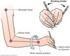
Wrist-Rocking Motion

A

The hand, wrist, arm work as a unit to produce a rotating motion used to move the working-end of an instrument

68
Q

Digital Motion Activation

A

Moving the instrument by flexing the thumb, index, and middle fingers

Used primarily with ultrasonic instruments, also used with periodontal probes, explorers

69
Q

Rolling the instrument handle

A

helps the clinician maintain precise contact of the working-end to the tooth surface.

70
Q

Mandibular roots - anteriors:

A

tilt inwards

(lingually)

71
Q

Mandibular roots - premolars:

A

more vertical

(straight up and down)

72
Q

Mandibular roots - molars:

A

tilt outward

(distally )

73
Q

Assessment Stroke

A

AKA exploratory stroke

Used to evaluate the tooth surface

Used with explorers to locate calculus deposits

74
Q

Root Debridement Stroke

A

Used to remove residual calculus deposits, bacterial plaque, and byproducts from:

(1) root surfaces that are exposed in the mouth because of gingival recession
(2) root surfaces within deep periodontal pockets

75
Q

Classification of Mobility

Class 1

A

Class 1

Slight mobility, up to 1 mm of horizontal displacement in a facial–lingual direction

76
Q

Classification of Mobility

Class 2

A

Class 2

Greater than 1 mm but less than 2 mm of horizontal displacement in a facial–lingual direction

77
Q

Classification of Mobility

Class 3

A

Class 3

Greater than 2 mm of horizontal displacement in a facial–lingual direction or vertical mobility