Neurology: TBL Questions Flashcards
Which lamina of the trilaminar embryo does the nervous system derive from?
A. Ectoderm
B. Endoderm
C. Mesoderm
D. Neural crest
A. Ectoderm
- CNS neurons
- Ependymal cells (inner lining of ventricles, make CSF)
- Oligodendrocytes
- Astrocytes

The neural crest cells go on to form which cell types?
A. Brain
B. Spinal cord
C. Ventral motor neurons
D. Dorsal root ganglia
D. Dorsal root ganglia
In addition to:
- PNS
- Sensory ganglia
- Autonomic ganglia and plexuses
- Neural glia
- Schwann cells
During regionalisation which statement is true?
A. Bone Morphogenic Protein helps to specify the ventral/motor part of the spinal cord.
B. Bone Morphogenic Protein helps to specify the dorsal/sensory part of the spinal cord.
C. Bone Morphogenic Protein is released by the notochord to pattern the sensory nerves.
D. Bone Morphogenic Protein is released by the notochord to specify the identity of motor neurons.
B. Bone Morphogenic Protein helps to specify the dorsal/sensory part of the spinal cord.

The lateral ventricles develop from the cavity of which of the following?
A. Diencephalon
B. Mesencephalon
C. Rhombencephalon
D. Telencephalon
D. Telencephalon
- Cerebral hemispheres
- Basal ganglia
- Lateral ventricles

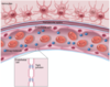
A typical peripheral nerve when observed in cross section in a stained section under the light microscope will be seen to consist of bundles of axons contained within fascicles. Which cell defines or forms the boundary of the fascicle?
A. Endoneurium
B. Epineurium
C. Schwann Cell
D. Perineural cell
D. Perineural cell

The discriminatory function of the blood brain barrier is a consequence of:
A. Capillary fenestrations
B. Tight junctions between endothelial cells
C. Endothelial basement membrane thickness
D. Gap junctions between endothelial cells
B. Tight junctions between endothelial cells
CSF production involves which of the following?

A. Arachnoid granulations
B. Choroid Plexuses
C. Transudate from small cerebral veins in the cerebral hemispheres into ventricles
D. Production by cells within the pia mater on the surface of the brain
B. Choroid Plexuses produced here by specialised ependymal cells
A superior sagittal vein thrombosis is likely to have what effect upon CSF pressure?
A. Reduced absorption of CSF across the arachnoid granulations and a rise in CSF pressure
B. A decrease in the CSF pressure because of a drop in venous pressure inside the skull
C. No change in CSF pressure as there is no connection between the CSF and the cerebral venous system
D. As the production of CSF is pressure dependent any increase in venous back pressure will reduce CSF production. There will be no overall change in CSF pressure

A. Reduced absorption of CSF across the arachnoid granulations and a rise in CSF pressure
CSF production is pressure indepependent, CSF absorption is pressure dependent

How does a Chiari 2 malformation cause hydrocephalus?

A. By causing obstruction to CSF flow at point A
B. By causing obstruction to CSF flow at point B
C. By causing obstruction to CSF flow at point C
D. By causing obstruction to CSF flow at point D
B. By causing obstruction to CSF flow at point B
- Smaller posterior fossa in cranium
- Herniation of cerebellar vermis and tonsils through foramen magnum with aqueductal stenosis: non-communicating hydrocephalus.
- Usually associated with lumbosacral myelomeningocele

Which is correct about hydrocephalus?
A. Hydrocephalus is defined as an increase in CSF volume
B. Hydrocephalus does not cause localising signs
C. In children papilledema is a common sign
D. Hydrocephalus is defined as an increase in CSF pressure
A. Hydrocephalus is defined as an increase in CSF volume
Sympathetic pre-ganglionic neurones are located in which of the following regions?
A. Nucleus of the solitary tract in the medulla
B. Parabrachial nucleus in the pons
C. Paraventricular nucleus in the hypothalamus
D. Preoptic area in the hypothalamus
E. Intermediolateral cell column in the spinal cord
E. Intermediolateral cell column in the spinal cord

Which of the following spinal tracts is most involved in skilled movement?
A. Dorsal column
B. Reticulospinal
C. Vestibulospinal
D. Corticospinal
D. Corticospinal
A 62 year old man complains of trouble walking. On examination, he cannot stand on tip toes and has weakness of dorsiflexion of both ankles. His calves and anterior tibial compartments are wasted. His ankle jerks are absent. These signs are consistent with which type of syndrome?
A. Extrapyramidal
B. Lower motor neurone
C. Neuromuscular junction
D. Upper motor neurone
B. Lower motor neurone
Anterior horn, brainstem or spinal cord out
Which are the three layers of the cerebellar gray matter?
A. Axonal, dendritic and cell body layers
B. Basket cell, interneuron and synaptic layers
C. Dura mater, arachnoid and pia mater
D. Molecular, purkinje and granular layers
D. Molecular, purkinje and granular layers
With reference to the spinal cord image, which of the following would be a likely sign after a lesion to X (shaded area)?
A. Contralateral motor weakness
B. Ipsilateral analgesia
C. Contralateral spasticity
D. Ipsilateral motor weakness
E. Contralateral paraesthesia

D. Ipsilateral motor weakness
A lesion to which of the following ascending tracts in the spinal cord generates the signs or symptoms described: A patient has reduced ability to detect pin-prick on the skin of their right leg.
A. Left spinothalamic tract
B. Right spinothalamic tract
C. Left dorsal column tract
D. Right dorsal column tract
A. Left spinothalamic tract
Which of the following symptoms commonly occurs during the initial stages following a spinal cord injury?
A. Hyperreflexia
B. Hypertension and tachycardia
C. Hypertonia
D. Hypotonia
E. Hypertension and bradycardia
D. Hypotonia
After traumatic injury to the brain or spinal cord, which substance is released from the traumatised tissue that can lead to further damage to surrounding tissue?
A. Glutamate
B. Ketones
C. Lactic acid
D. Prostaglandin E
A. Glutamate
Excitatory neuron: acts on NMDA and AMPA receptors which are over-expressed = excess calcium release
A patient experiences decreased proprioception in the left lower limb. A lesion to which of the following ascending tracts in the spinal cord generates the signs or symptoms described?
A. Left dorsal column tract
B. Right dorsal column tract
C. Left spinothalamic tract
D. Right spinothalamic tract
A. Left dorsal column tract

Hyperalgesia is defined as which of the following?
A. Decreased sensitivity to pain
B. Increased sensitivity to touch
C. Increased sensitivity to pain
D. Pain response to non-noxious stimulus
C. Increased sensitivity to pain
The posterior communicating artery connects which of the following vessels?
A. Vertebral artery to basilar artery
B. Left vertebral artery to right vertebral artery
C. Posterior cerebral artery to anterior cerebral artery
D. Posterior cerebral artery to internal carotid artery
E. Left anterior cerebral artery to right anterior cerebral artery
D. Posterior cerebral artery to internal carotid artery

The major neurotransmitter that characterises the cells of the brainstem raphe nuclei is which of the following?
A. Glutamate
B. Noradrenaline
C. Dopamine
D. Acetylcholine
E. Serotonin
E. Serotonin
- Contributor to feelings of well-being and happiness
- Regulates the sleep cycle along with melatonin
- Regulates intestinal movements.

A 70-year old woman was admitted to the emergency department with a suspected stroke. Radiology showed an infracted region corresponding to the shaded area shown in the photograph. Occlusion of which of the following arteries may have caused infarction in this area?
A. Internal carotid artery
B. Middle cerebral artery
C. Posterior cerebral artery
D. Anterior cerebral artery
E. Lenticulostriate artery

D. Anterior cerebral artery

The non-contrast computed tomography (CT) scan below shows which of the following pathologies?
A. Subdural haemorrhage
B. Intracerebral haemorrhage
C. Ischaemic infarction
D. Extradural haemorrhage
E. Arteriovenous malformation

B. Intracerebral haemorrhage
- Most commonly caused by systemic hypertension
- Amyloid angiopathy (recurrent lobar haemorrhagic stroke in the elderly)
- Vasculitis
- Neoplasm
- May be secondary to reperfusion injury in ischaemic stroke
The age of a cerebral infarct can be estimated pathologically by the cell type that predominates within the infarct. What is the time course of appearance (from earliest to latest) of the following cells (foamy macrophages, neutrophils and reactive astrocytes) in the evolution of a cerebral infarct?
A. Astrocytes then macrophages then neutrophils
B. Astrocytes then neutrophils then macrophages
C. Macrophages then neutrophils then astrocytes
D. Neutrophils then astrocytes then macrophages
E. Neutrophils then macrophages then astrocytes
E. Neutrophils then macrophages then astrocytes

In the midbrain image shown below, what is the name of the structure indicated by shaded region of X?
A. Dorsal motor nucleus of vagus
B. Hypoglossal nucleus
C. Abducent nucleus
D. Oculomotor nucleus
E. Trochlear nucleus

D. Oculomotor nucleus

A patient complains of a hoarse voice, difficulty swallowing and choking when drinking fluids. Examination reveals visible weakness of elevation of the palate, depression of palatal sensation, and loss of gag reflex. Which cranial nerve lesion(s) are responsible for the signs and symptoms described?
A. X lesion
B. V and VII lesion
C. V and X lesion
D. IX and X lesion
E. XII lesion
D. IX and X lesion
A 75-year-old woman is brought into hospital by her husband. He says that she has complained of headache for the last 2 days and has now become confused and is behaving oddly with garbled speech. Examination reveals her to be febrile and drowsy but without motor signs. CSF examination shows an increase in white cells and an EEG reports some focal slowing with epileptiform features in the left temporal lobe. What is the most likely diagnosis?
A. Intracranial tumour
B. Temporal lobe epilepsy
C. Bacterial meningitis
D. Left middle cerebral artery infarct
E. Herpes simplex encephalitis
E. Herpes simplex encephalitis
- Likely reactivation of HSV-1
- Infection primarily involves temporal lobes
- Increase in WCC (majority monocytes)
- EEG: paroxysmal lateralised epileptiform (PLED) discharges in temporal lobe (characteristic of herpes simplex encephalitis)

A 32-year-old woman had separate episodes of unilateral visual loss, ataxia, and altered left-sided bodily sensation. She made a sub-total recovery from each of these episodes. She died six months after the last episode in a motor vehicle accident. Many lesions were found in the white matter of her brain at post-mortem. Which of the following is correct concerning this type of lesion (arrow)?

A. Axons are likely to be mostly intact in these lesions
B. The lesions are found in men more often than women
C. Myelin loss in these lesions occurs secondary to axonal loss
D. These lesions are often due to infection with JC virus
A. Axons are likely to be mostly intact in these lesions