Lecture 6- Embryology part I Flashcards
primitive atrium
forms the atria
Achievements of early embryonic development
- First two weeks created tissues of the future embryo and future placenta
- The third week created the three germ layers
- Ectoderm, mesoderm and endoderm – primordia of all tissue
- Fourth week created recognisable body form and the mesoderm begins to organise
when does the CVS begin development
10th day after fertilisation- first functional organ to develop
when can the heart be heard
- Can be heard beating on a sonography by week 6
outline the key 5 stages of embryonic development of the CVS
(1) Formation of the primitive heart tube
(2) Cardiac looping
(3) Development of the atria
(4) Formation of the Great vessels
(5) Septation
At the start of this development the CVS system exists as two regions
near the cranial (head end) end of the embryo- cardiogenic fields (derived from the mesoderm)
Cardiogenic fields consist
consists of blood islands which are primitive tissue and mark the beginning of blood, vessel and heart development
Blood islands develop further and
fuse to form two tubes which are called endocardial tubes – one on each side of the embryo

how are the two endocardail tubes brough together
Folding
In the 4th week the embryo begins to folding which puts the heart tissue in the correct position to form the primitive heart tube surrounded by the pericardial sac
how many ways does folding occur
2
- Cephalo-caudal folding
- Lateral folding
Cephalo-caudal folding
Brings cardiogenic filed from cranial entre towards the centre of the embryo to sit in the thoracic region where the heart will be

Lateral folding
- Fuses the two lateral sides of the embryo
- Brings two cardiogenic fields into the midline so they can fuse and form the primitive heart tube

what day is the ptimitive ehart tube form
day 25
characteristics of the primitive heart tube
- 6 parts
- no valves
- no barriers between structure
name the 6 parts of the primitive heart tube
- Aortic roots- forms arteries of the aortic arch
-
Truncus arteriosus- outflow of blood
- Involved in the formation of the pulmonary trunk and aorta
- Bulbus cordis- Involved in the formation of the pulmonary trunk and aorta
- Primitive ventricle- forms ventricles
- Primitive atrium- forms the atria
- Sinus venosus- forms part of the right atrium and vena cave

aortic roots
formed at the top of the primitive heart tube
- forms arteries of the aortic arch
Truncus arteriosus
outflow of blood
involved in the formation of the pulmonary trunk and aorta
bulbus cordis
- Involved in the formation of the pulmonary trunk and aorta
primitive ventricles
forms ventricles
sinus venosis
acronymn to learn parts of the primitive heart tube
All The Best Vaccums Are Silver
what happens after the primitive heart tube is formed
(2) Cardiac looping
why must the heart loop
The newly formed heart tube is surrounded by the pericardial sac- as the heart tube grows and elongates, it gets too long for the sac. This means that to fit, it must loop
outline cardiac looping
- The primitive ventricle moves ventrally (coming forward) and to the right
- The primitive node moves dorsally (behind) and to the left
- This puts the inflow portion of the heart (veins and atria) behind the outflow portion (ventricles and arteries) the same shape and orientation as mature hearts

the looping creates a space behind the arteries (aorta and pulmonary trunk) and in front of the superior vena cava called the
transverse pericardial sinus

at the end of the looping what has happened to inflow and outflow
in the correct orientation with respect to eachother
what occurs after (2) cardiac looping
(3) development of the atria)
after looping the atria are a
single space
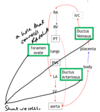
(3) Development of the atria: develoment of the sinus venosus
- Right and left sinus horns are equal size
- Then venous return shifts to the right sinus horn
- Left sinus horn recedes
- Right sinus horn is absorbed by the enlarging right atrium becoming the vena cava

pulmonary veins begins as a
a single vein entering the left atrium
- This vein is formed from four branches which converge to form one vein draining into the developing left atrium
- As the left atrium grows, its absorbs the single pulmonary veins, absorbing all the way to the four branches
- This means that when the left atrium has finished growing it is receiving blood from four pulmonary veins as seen in the mature heart

absorption of the pulmonary vein
xThe proximal parts of the pulmonary veins become absorbed into the wall of the left atrium. The contribution of the pulmonary veins is shown in blue, and the primitive left atrium is shown in pink

Oblique sinus
Oblique pericardial sinus formed as left atrium expands absorbing the pulmonary veins

Name the shunts in foetal circulation
ductus venosus
foramen ovale
ductruc arteriosus

Outline foetal circulation (warning v long winded)
- Oxygenated bloods is carried from the placenta into the foetal circulation via the umbilical vein
- Oxygenated blood enter the inferior vena cava and mixes with deoxygenated blood- bypasses the developing liver via the ductus venosus
- The blood enter the right atrium and passes into the left atrium via the foramen ovale (when its closes become fossa ovalis) thereby bypassing the pulmonary circulation (not full developed yet so wouldn’t tolerate the pressure)
- The blood can shunt from right to left side of the heart because the pressure in the right side of the heart is higher than the left in the foetus
- The baby is not breathing and instead the foetus’ blood is being oxygenated by the mothers blood
- Therefore blood doesn’t have to go to the alveoli for oxygenation- so pulmonary circulation can be btpassed
- The blood can shunt from right to left side of the heart because the pressure in the right side of the heart is higher than the left in the foetus
- The blood is pumped from the left ventricle into the aorta
- Blood that doesn’t pass through the foramen ovale, and instead is pumped into the pulmonary trunk from the right ventricle, enter systemic circulation at the arch of the aorta via the ductus arteriosus

Simple diagram of shunts in the foetal heart
shunts after birth
- Baby takes first breath and pO2 increases- Ductus arteriousus contracts
- More blood now flows through pulmonary circulation as blood in the pulmonary trunk cannot leave via the ductus arteriosus anymore
- This causes increased venous return to the left atrium, leading to an increase in left atrial pressure. When the pressure in the LA exceeds the RA the foramen ovale closes becomes fossa ovalis
- When the umbilical cord is cut, there is no longer blood flowing through the umbicilical vein, causing the ductus venosus to collapse

outline the formation of the great vessels
- Truncus arteriosus: divided by the aorticopulmonary septum to form the pulmonary trunk and the aorta.
-
Sixth arch: goes on to form the right and left pulmonary (R VI and L VI) arteries arising from the pulmonary trunk. The left artery maintains its connection to the rest of the vessels via the ductus arteriosus, however the right artery loses this connection. When the baby is born and the ductus arteriosus closes, this separates the pulmonary circulation from the systemic circulation.
- The sixth arch is also known as the pulmonary arch.
- 4th arch: on the left side becomes the arch of the aorta, and on the right becomes the right subclavian.
- 3rd arch: becomes the common carotids and the first part of the internal carotids.
- Second and first arches: disappear.

Types of septation:
- Interatrial septum
- Interventricular septum
- Septation of ventricular outflow tract (pulmonary trunk and aorta)

WHat are key to the separation of the left and right side of the heart?
Endocardial cushions
- The lining of dorsal and ventral aspects of the developing heart grow endocardial cushions.
- These grow to meet in the middle of the heart and are key in the separation of the left and right sides of the heart.
- They act as a target for the septa that develop as each septum will grow towards the cushions

(5) Septation I- The Inter-atrial septum
- Septum of tissue grows from the top of developing atria towards the endocardial cushions. This is the septum primum, as it grows down the communicating hole between the atria is called the ostium primum
- Just before the septum primum meets the endocardial cushions and the ostium primum is closed, a hole forms in the middle of the septum- ostium secundum
- The septum primum meets the endocardial cushions, and the ostium secundum allows blood to continue to move from the right to left atrium
- Another septum grows down from the top of the atria called the septum secundum
- As the septum secundum grows down, it leaves another hole just below the ostium secundum
- These two septa, and two holes together form the foramen ovale - a right to left shuntallowing blood to flow from the right atrium into the left. This shunt will reverse and close after birth

Ventricular septation
The formation of the ventricular septum take place in two steps:
- A muscular portion of the heart tissue grows upwards from the floor of the primitive ventricle towards the endocardial cushions. It doesn’t quite reach the cushions, forming the primary interventricular foramen
- A membranous portion then grows down from the endocardial cushions to meet the muscular portion and close the foramen (window)

Septation of the Outflow tract
The bulbis cordis and truncus arteriosus form one tube allowing outflow from the heart. This tube needs to be split in order to form the aorta and pulmonary trunk.
- In the beginning, there are two lines of proliferations of neural crest cells (these cells appear in the neural tube during neurulation and migrate to contribute to the development of a wide range of structures) on the walls of the outflow tract.
- This two lines of the cells spiral around and grow towards each other to meet in the middle
- This form a single spiral septum called the aorticopulmonary septum – consequently forming the aorta and pulmonary trunk.
