9-Histology brainscape Flashcards

(75 cards)

1
Q
A
2
Q
A
3
Q
A

1= Stratum Basale 2= Stratum Spinosum 3= Stratum Granulosum 4= Stratum Lucidum 5= Stratum Corneum

4
Q
A

Stratum Basale (note Desmosomes & Hemidesmosomes)

5
Q
A
6
Q
A
7
Q
A
8
Q
A

Left= Thin Skin Right= Thick Skin

9
Q
A

Melanin granules of stratum spinosum with light coming from top down

10
Q
A
11
Q
A
12
Q
A
13
Q
A
14
Q
A
15
Q
A
16
Q
A

*Note underneath the nail plate is the hyponychium Matrix: part of epidermis responsible for nail formation Bed: Stratum basale & spinosum- physical support for nail plate Eponychium: cuticle, stratum corneum to protect underlying nail matrix from infection

17
Q
A
18
Q

paste-36597416329758.jpg

A

paste-36610301231663.jpg

19
Q

paste-36644660969990.jpg

A

paste-36657545871878.jpg

20
Q

paste-36713380446750.jpg

A

paste-36726265348696.jpg

21
Q

paste-37344740639262.jpg

A

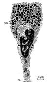
Sebaceous gland - simple branched acinar gland *Stem cells at bottom

22
Q

paste-37379100377630.jpg

A

paste-37391985279518.jpg

23
Q

paste-37417755083294.jpg

A

paste-37430639985182.jpg

24
Q

paste-37464999723364.jpg

A

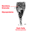
Screen Shot 2014-03-30 at 3.09.15 PM.png Eccrine Glands

25
paste-37688338022774.jpg
Clear cells of Eccrine glands - Fluid Transport
26
paste-38074885079582.jpg
Eccrine Sweat Gland \*Duct lined by stratified cuboidal epithelium
27
paste-38100654883358.jpg
paste-38113539785246.jpg
28
Afferent Nerve Fibers & Sensory Structures of the Skin
1) Free nerve endings (from Merkel's Cells) - extend into epidermis 2) Meissners Corpuscles - light touch mechanoreceptors, prominent on thick skin 3) Pacinian Corpuscle- deep pressure & vibrations 4) Krause (bulboid corpuscles) - cutaneous corpuscles
29
Albinism
lack of tyrosinase synthesis or activity
30
Apocrine Sweat Glands (Distribution, Structure, Function)
Distribution: found in axilla & genito-anal region, open directly into hair follicle Structure: simple coiled tubular gland, only dark cells, secrete via eccrine mode Function: unclear
31
Arrector Pili Muscle
oblique smooth muscle innervated by sympathetics ---\> goose bumps
32
Balding
Conversion of scalp terminal hairs to vellus type- requires genetic background and levels of androgenic hormones
33
Cells of the Epidermis
\*Keratinocytes (most popular) \*Langerhan's \*Merkel cells \*Melanocytes
34
Derma-Epidermal Junction
Epidermal ridges interdigitate with dermal papillae creates a Basal Lamina \*Basement Membrane formed with laminins, Type IV collagen, fibronectin, and type VII collagen
35
Dermatoglyphics
Finger prints- surface pattern of papillary ridges found only on thick skin
36
Eccrine (Merocrine) Sweat Glands (Distribution, Structure, Function)
Distribution: Most numerous, found in most skin Structure: Simple coiled tube with secretory & duct portions Function: hypotonic watery secretion (sweat)
37
Eccrine Sweat Gland Cells & Ducts
Cells: Simple epithelium: 1) Dark cells = mucoid antibacterial secretions 2) Clear cells = secreting sweat for evaporative cooling Ducts: Stratified cuboidal epithelium 1) Excretory Duct: secretion, mainly protein free filtrate 2) Secretory Duct: absorption, mainly Na+ & Cl-
38
Function of sweat
1) Thermoregulation 2) Moistens skin surface 3) Excretion of electrolytes & protein metabolites
39
Functions of Epidermis:
1) Physical/chemical barrier 2) Water proofing 3) Abrasion resistance 4) Genesis of Vitamin D 5) Cytokine production (especially interleukins)
40
Functions of the Dermis
1) Physical support 2) Vascular support 3) Neural receptors 4) Controls epidermis movement
41
Germinal Matrix
sits on top of papilla and is responsible for producing keratin that creates the hair
42
Glands of the Skin
Sebaceous Eccrine (Merocrine) Apocrine
43
Hair Follicle
Epidermal invagination that extends into the dermal layer of the skin and terminates at the hair bulb (deepest portion)
44
Hair Follicle Growth Cycle
1) Anagen Phase = hair growth 2) Catagen Phase = regression 3) Telogen Phase = Inactivity
45
Hair Papilla
dermal CT which provides blood supply & nerves to hair follicle essential \*\*Very sensitive to chemo
46
Hypodermis
superficial fascia= fibro fatty tissue layer below skin, not part of integument. Fat cells, insulation
47
Keratinocytes
Produce keratin (form intermediate filaments which anchor to desmosomes) \*Attached to one another via desmosomes, and attached to basement membrane via hemidesmosomes
48
Keratohyaline Granules
No membrane, made predominantly of filaggrin, Very basophilic
49
Lamellar Granules
\*Membrane Coated \*Lipid sheets secreted into intracellular space to form hydrophobic barrier between cells (contains tonofibrils)
50
Langerhan's Cells
\*Dendritic cell found in stratum spinosum \*Derived from monocytes and act as antigen presenting cell (APC) in the epidermis
51
Layers of epidermis (thick skin)
1) Stratum Basale (aka stratum Germinativum) 2) Stratum Spinosum 3) Stratum Granulosum 4) Stratum Lucidum 5) Stratum Corneum
52
Layers of Integument
Epidermis Dermis Skin Appendages
53
Layers of the dermis
1) Papillary (attaches to epidermis) 2) Reticular layer (attaches to hypodermis)
54
Layers of the Hair Follicle (from external to internal)
Glassy Membrane (separates follicle from membrane) External Root Sheath (continuous with stratus corneum) Internal Root Sheath (does not extend beyond sebaceous glands) Hair Cuticle/Shaft
55
Main types of cancer
1) Basal cell carcinoma (\>75%) 2) Squamous cellc arcinoma (20%) 3) Melanoma (\<5%)
56
Melanin granules
dense collection of melanin with no tyrosinase activity
57
Melanin Synthesis
1) Tyrosine --\> Melanin using enzyme Tyrosinase 2) Melanin is injected into keratinocytes to create barrier around nucleus and protect it from sunlight: more exposure to sunlight, more melanin that is produced
58
Melanocytes
\*Located in stratum Basale \*Derived from NC cells and produce Melanin
59
Merkel's Cells
\*Clear cells along stratum basale \*Sensor cells, have free nerve endings
60
Most serious type of skin cancer?
Melanoma
61
Origin of hair & nails
skin appendages of the epidermis
62
Papillary Layer
\*Loose CT (type III collagen) elastin \*Meissner's Corpuscles - nerve endings, light touches \*Capillaries
63
Reticular Layer
\*Leather \*Dense irregular CT (Type I Collagen) \*AV shunts to regulate heat loss & BP \*Pacinian Corpuscles \*Smooth muscle bundles that insert into hair follicles
64
Sebaceous Gland (Distribution, structure & Function)
Distribution: most but not all thin skin Structure: Simple branched acinar gland Function: holocrine secretion of lipid containing cells --\> results in sebum, enhanced by androgens and inhibited by estrogens
65
Stratum Basale
single layer of cuboidal stem cells on basement membrane with intense mitotic activity- desmosomes between cells, and hemidesmosomes to the BM
66
Stratum Corneum
\*Flat dead cells filled with mature keratin \*Continously sloughed off as desmosomes are lost \*\*Thickness of this is the defining difference between thick andthin cells
67
Stratum Granulosm
\*Last lyaer of living cells \*Layer of flattened cells with no mitotic activity \*Numerous Granules: Keraothyaline & Lamellar
68
Stratum Lucidum
\*Only in thick skin \*Cellular organelles not visible, translucent layer
69
Stratum Spinosum
cuboidal cells with central nuclei and low mitotic activity \*Spiny appearance \*Tonofibrils here- keratin fibers that extend out of desmosomes to anchor desmosome to the cytoskeleton
70
Tanning process
1) Physiochemical reaction that darkens existing melanin 2) Increase in tyrosinase
71
Thick vs Thin Skin
Thin= covers most of body, contains hair Thick= restricted to volar surfaces of hands & feet, lacks hair, restists frictional forcesThin skin has attenuated layers of Stratums: \*Spinosum, granulosum, and corneum) and Absent stratum lucidum
72
Types of Hairs
Lanugo- fine hairs on fetus Vellus- short, ifne, soft non-pigmented, over general body Terminal- long, coarse, pigmented - scalp, eyebrows, pubic
73
Types of Melanin
Eumelanin= black Pheomelanin= red
74
What accounts for the difference in hair length from region to region?
Difference in growth rates and cycles (shorter anagen phases)
75
What factors control race?
Size, number, and distribution of melanin granules not number of melanocytes