Muscles of the Hip and Thigh Flashcards
Quadratus femoris m.

Deep to Biceps femoris m.
Attachments:
(O): Ventral surface of caudal ischium
(I): Intertrochanteric crest
Action:
- Extend the hip joint
- Rotate the pelvic limb laterally
Innervation:
- Sciatic n.
Blood Supply:
- Cranial gluteal a.

Cranial Muscles of the Thigh
Quadriceps femoris m.
- 4 heads of origin
- Fused distally
- Rectus femoris m.
- Vastus lateralis m.
- Vastus intermedius m.
- Vastus medialis m.
Iliopsoas m.
Gluteobiceps m.
(Ruminant Only)**
Combination of Superficial gluteal m. + Biceps femoris m.
Attachments:
(O): Ischiatic tuberosity
(O): Gluteal fascia
(O): Intermuscular septum separating Semitendinosus m.
(I): Patella and cranial surface of Tibia
(I): Common calcanean tendon
Actions:
- Extend the hip joint
- Extend OR flex the stifle
- Weight bearing or not
- Extend the tarsal joints (Hock)
Innervation:
- Sciatic n.

Fasciae of The Hindlimb
Superficial Fascia of the Trunk:
- Gluteal Fascia
- Superficial Caudal Fascia
Deep Fascia of the Trunk:
- Thoracolumbar Fascia
- Medial and Lateral Femoral Fascia
- Fascia Latae

Semitendinosus m.
Sandwiched btwn Semimembranosus m. and Biceps femoris m.

Attachments:
(O): Ischiatic tuberosity
(I): Distocranial border of Tibia
(I): Body of Tibia (medial surface)
(I): Tuber calcanei (Common calcanean tendon)
Action:
- Extend the hip
- Flex the stifle
- Extend the tarsal joints
Innervation:
- Sciatic n.
Blood Supply:
- Distal caudal femoral a.

Iliopsoas m.

Represents a fusion of the Psoas major m. and Iliacus m.
- Can’t see these two mm.
Attachments:
(O): Psoas major: lumbar vertebrae
(O): Iliacus: cranioventral ilium
(I): lesser trochanter
Action:
- Flex the hip joint
Innervation:
- Femoral n.
Blood Supply:
- Iliolumbar a.
Sartorius m. of Equine
Caudal Muscles of the Thigh
3 Primary Muscles:
- Laterally
- Biceps femoris m.
- Caudally
- Semitendinosus m.
- Medially
- Semimembranousus m.
Femoral triangle

Shallow triangular space
Femoral vessels run to and from pelvic limb
Boundaries:
- Cranial:
- Caudal belly of Sartorius m.
- Caudal:
- Pectineus m.
- Adductor m.
- Base:
- Abdominal wall

Muscles of the Caudal Hip
4 Muscles that lie caudal to the hip
Extend from the inner and outer surfaces of ischium and femur
All rotate the limb laterally
- Internal obturator m.
- Gemelli m.
- Quadratis femoris m.
- External obturator m.
Quadriceps femoris m.
Head #3: Vastus medialis

Attachments:
(O): Proximal femur
(I): Tibial tuberosity
Action:
- Extend the stifle
Innervation:
- Femoral n.
Blood Supply:
- Lateral circumflex femoral a.
Quadriceps femoris m.
Most powerful extensor of the stifle joint
Necessary for animal to support weight
Gluteobiceps m.
(Ruminant Only)**
Combination of Superficial gluteal m. + Biceps femoris m.

Common Calcanean Tendon
Biceps femoris m. & Semitendinosus m.
- Minor contributors
Achilles tendon in humans
Action:
- Extend the tarsal joints

Biceps femoris m.
Attachments:
(O): Sacrotuberous ligament
(O): Ischiatic tuberosity
(I): Patella
(I): Patellar ligament
(I): Cranial border of Tibia
(I): Tuber calcanei (common calcanean tendon)
Action:
- Extend the hip, stifle and tarsal joints
- Flex the stifle joint
- Caudal part only
Innervation:
- Sciatic n.
Blood Supply:
LOTS

Adductor m.

Attachments:
(O): Pelvic symphysis
(O): Adjacent Ischiatic arch
(O): Ventral surface of Pubis and Ischium
(I): Lateral lip of caudal rough surface of femur
Action:
- Adduct the limb
- Extend the hip joint
Innervation:
- Obturator n.
Blood Supply:
- LOTS

Pectineus m.

Attachments:
(O): Iliopubic eminence
(O): Pubic tubercle
(I): Distal end of medial lip of rough face of femus
Action:
- Adduct the limb
Innervation:
- Obturator n.
Blood Supply:
- Medial circumflex femoral a.

Quadriceps femoris m.
Head #2: Vastus lateralis
Attachments:
(O): Proximal femur
(I): Tibial tuberosity
Action:
- Extend the stifle joint
Innervation:
- Femoral n.
Blood Supply:
- Lateral circumflex femoral a.

Middle gluteal m.
Attachments:
(O): Crest and gluteal surface of Ilium
(I): The greater trochanter
Actions:
- Extend and abduct the hip joint
- Rotate the pelvic limb medially
Innervation:
- Cranial gluteal n.
Blood Supply:
- Cranial gluteal a.
- Lateral circumflex femoral a.

Trochanteric Bursitis
(Large Animals Only)**
Inflammation of the Trochanteric Bursa
Causes lameness
Most common in standardbreds

Sacrotuberous Ligament
Collagenous band
- Runs from Sacrum to lateral angle of Ischiatic Tuberosity
Superficial gluteal m. arises from proximal half

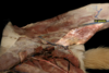
Cow Lateral Pelvis Image

Quadriceps femoris m.
Head 1: Rectus femoris

Only head that crosses the hip and stifle joint
Attachments:
(O): Ilium
(I): Tibal tuberosity
Action:
- Extend the stifle joint
- Flex the hip joint
Innervation:
- Femoral n.
Blood Supply:
- Lateral circumflex femoral a.
- Superficial circumflex iliac a.

Semimembranosus m.

Attachments:
(O): Ischiatic tuberosity
(I): Distal medial lip of femur
(I): Medial condyle of Tibia
Actions:
- Extend the hip
- Flexes OR Extends the stifle
- Depends on limb position
Innervation:
- Sciatic n.
Blood Supply:
- Distal caudal femoral a.

Gracilis m.
Attachments:
(O): Pelvic symphysis by symphysial tendon
(I): Cranial border of Tibia
(I): Tuber calcanei (common calcanean tendon)
Actions:
- Adduct the limb
- Flex the stifle
- Extend the hip and tarsal joints
Innervation:
- Obturator n.
Blood Supply:
- Proximal caudal femoral a.

Lateral Muscles of the Pelvis
Gluteal Muscles
- Superficial Gluteal m.
- Middle Gluteal m.
- Piriformis m. * (Dogs only)
- Deep Gluteal m.
Accessory Gluteal m.
- Large animals only **
Gluteal Biceps (Glutobiceps) m.
- Ruminany only**
Tensor Fasciae Latae m.

Sacrosciatic Ligament
(Large Animals Only)**
Broad sheet of connective tissue
- Forms dorsal, softer part of the lateral wall of the pelvis
Formation of Greater and Lesser Ischiatic Foramina
Need to reflect the accessory gluteal m. and middle gluteal m. to see it

Quadriceps femoris m.
Head #4: Vastus intermedius

Attachments:
(O): Proximal femur
(I): Tibial tuberosity
Action:
- Extend the stifle
Innervation:
- Femoral n.
Blood Supply:
- Lateral circumflex femoral a.

Accessory Gluteal m.
(Large Animals Only!!) **
Possibly analogous to the Piriformis m.
Deep to the middle gluteal m.
- Need to reflect the middle gluteal m. to see it
Actions:
- Extend and abduct the hip joint
- Rotate the pelvic limb medially
Innervation:
- Cranial gluteal n.
Blood Supply:
- Cranial gluteal a.
- Lateral circumflex femoral a.

Superficial Gluteal m.
Attachments:
(O): Lateral border Sacrum
- By Sacrotuberous Ligament
(O): Cranial Dorsal Iliac Spine
(I): Third Trochanter
Action:
- Extend the hip joint
- Abduct the limb
Innervation:
- Caudal gluteal n.
Blood Supply:
- Caudal gluteal a.
- Lateral circumflex femoral a.
Deep gluteal m.
Attachments:
(O): The body of the Ilium
(O): Ischiatic spine
(I): Cranial aspect of the greater trochanter
Action:
- Extend and abduct the hip
- Rotate the pelvic limb medially
Innervation:
- Cranial gluteal n.
Blood Supply:
- Cranial gluteal a.

Sartorius m.

Cranial and Caudal parts
Attachments:
- Cranial:
- (O): Crest of Ilium
- (O): Thoracolumbar fascia
- (I): Patella
- Caudal:
- (O): Cranial ventral iliac spine
- (O): Ventral border of Ilium
- (I): Cranial border of Tibia
Actions:
- Flex the hip joint
- Cranial: extend stifle
- Caudal: flex stifle
Innervation:
- Femoral n.
Blood Supply:
- Superficial circumflex iliac a.

Tensor Fasciae Latae m.
Attachments:
(O): The tuber coxae
(O): Ilium
(O): Aponeurosis of middle gluteal
(I): The lateral femoral fascia
Actions:
- Tense the lateral femoral fasciae
- Flex the hip joint
- Extend the stifle
Innervation:
- Cranial gluteal n.
Blood Supply:
- Iliolumbar a.
- Superficial circumflex iliac a.

Popliteal Lymph Node
Palpable in dog
Caudal border of Biceps femoris m.
Directly caudal to the stifle

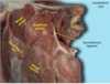
Equine Lateral Pelvis Image

Internal obturator m.

(NOT IN RUMINANT)
Attachments:
(O): Symphysis pelvis
(O): Dorsal surface of ischium and pubis
(I): Trochanteric fossa of femur
Action:
- Rotate the pelvic limb laterally at the hip joint
Innervation:
- Sciatic n.

External obturator m.
Attachments:
(O): Ventral surface of pubis and ischium
(I): Trochanteric fossa of femur
Action:
- Rotate pelvic limb laterally at hip joint
Innervation:
- Obturator n.
Blood Supply:
- Obturator a.

Gemelli m.
2 muscles fused together
Lie under the tendon of Internal obturator m.
Attachments:
(O): Lateral surface of Ischium
(O): Caudal to Acetabulum
(O): Ventral to lesser ischiatic notch
(I): Trochanteric fossa of femur
Action:
- Rotate the pelvic limb laterally at the hip joint
Innervation:
- Sciatic n.
Blood Supply:
- Iliolumbar a.
