Muscles of the Crus Flashcards
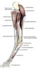
Craniolateral Muscles of the Leg
- Cranial tibial m.
- Long digital extensor m.
- Peroneus (fibularis) longus m.
- Lateral digital extensor m.
- Peroneus brevis m.
Lots of species differences on the size of muscles
Species difference on whether they are present or absent
Cranial tibial m.
(DOG)

Most cranial, most medial muscle in the dog
Tendon goes down to the tarsal region
Attachments:
(O): Extensor groove of tibia
(O): Lateral edge of cranial tibial border
(I): The plantar surface of the base of Metatarsals 1 and 2
Action:
- Flex the tarsocrucal joint
- Rotate the paw laterally
Innervation:
- Peroneal (Fibular) n.
Blood Supply:
- Cranial tibial a.

Long digital extensor m.
(Dog)
Attachments:
(O): Extensor fossa of the femur
(I): Extensor process of distal phalanges 2
(I): Extensor process of distal phalanges 3
(I): Extensor process of distal phalanges 4
(I): Extensor process of distal phalanges 5
Action:
- Extend the digital joints
- Flex the tarsal joints
- Has to happen together
Innervation:
- Peroneal (Fibular) n.
Blood Supply:
- Cranial tibial a.
** Need to remove the cranial tibial m.**

Crural extensor retinaculum
Long digital extensor runs under in the distal crus

Peroneus longus m.
DOG
(Fibularis longus m.)

Attachments:
(O): Lateral condyle of the tibia
(O): Proximal end of the fibula
(O): Lateral epicondyle of the femur
(I): 4th Tarsal bone
(I): Plantar aspect of the base of the metarsals
Action:
- Flex the tarsal joints
- AKA the “hock”
- Rotate the paw medially
Innervation:
- Peroneal (Fibural) n.
Blood Supply:
- Cranial tibial a.

Peroneus brevis m.
DOGS ONLY!!!

- Don’t need to disect
Arises in distal 2/3 of fibula
Actions:
- Flex the hock
Innervation:
- Peroneal (Fibular) n.

Popliteus m.

Fasciae of the leg
- Similiar to the superficial fascia
- Corresponds to the region
- Superficial crural
- Tarsal
- Metatarsal
- Digital fasciae

Tarsal extensor retinaculum
Long digital extensor runs under in the tarsus

Lateral digital extensor m.
(DOG)
(Very small in the dog)
Attachments:
(O): Proximal lateral tibia and fibula
(I): Digit 5
Action:
- Flex the tarsal joints and extend digit 5
Innervation:
- Peroneal (Fibular) n.
Blood Supply:
- Cranial tibia a.

Long digital extensor m.
(Horse)

Most cranial muscle in the horse
- Large muscle located cranially and superficially
- Peroneus tertius m. (underneath)
Attachments:
(O): Extensor fossa (distal femur)
(I): Dorsal surface of phalanges, primairly on P3 (extensor process)
Action:
- Extend the digital joints
- Flex the tarsal joints
- Has to happen together
Innervation:
- Fibular n. (Peroneal n.)
Blood Supply:
- Cranial tibial a.

Species difference for Horses
Absent in horses:
- Peroneus brevis m.
- Peroneus longus m.
Deep to Long digital extensor m. is Peroneus tertius m.
Peroneus tertius m.
(Horses)

- Deep to the long digital extensor m.
- Looks like a tendon
- Entirely tendinous in adult horse
- Partially wrapped in the long digital extensor m.
Attachments:
(O): Extensor fossa of the femur
(I): Dorsal tendon:
- Proximal extremity of metatarsal bone & 3rd tarsal bone
(I): Lateral tendon:
- Calcaneus & 4th tarsal bone
Action:
- Link stifle and hock
- Flex the hock
Innervation:
- Peroneal n.
Clinical Correlation:
Horse with a ruptured Peroneus tertius m.

The horse is able to extend the hock when the stifle is flexed
Result = Overextension of the hock
Shouldn’t happen because the hock (ankle) should be flexed
Actions are linked!
Lame on trot

Cranial tibial m.
(Horses)
Deep to Peroneus tertius m.
Largely covered by the long digital extensor m.
Attachments:
(O): Tibial tuberosity and lateral condyle of tibia
Inserts with 2 tendons:
(I): Dorsal Tendon
- Dorsal aspect of the proximal end of the 3rd metatarsal bone
(I): Medial Tendon/Cunean Tendon
- Fused 1st and 2nd tarsal bones
Innervation:
- Peroneal (Fibular) n.
Blood Supply:
- Cranial tibial a.

Bone Spavin

Bony growth within the lower hock joint of horse or cattle
Most common cause of clinical lameness of the tarsus (“hock”) in horses
Caused by osteoarthritis
Enlargement of medial aspect of the hock, under the cunean tendon
Repair:
Cunean tenectomy
- Cut tendon
- Really severe osteoarthritis cases –> joint fusion
Lateral digital extensor m.
(Horses)

Caudal to the long digital extensor m.
Actions:
- Flex the hock
- Extend the tarsus
Innervation:
- Peroneal n.
Blood Supply:
- Cranial tibia a.

Stringhalt
(Horses)
- Exaggerated flexion of the tarsal joints (“hock”)
- Horse may strike ventral abdomen with foot
- Injury to the digital flexor muscles
Repair: Remove distal portion of the lateral digital extensor

Extensor Retinacula
(Horses)

Proximal extensor retinaculum
Middle extensor retinaculum
Distal extensor retinaculum
Wraps around the long digital extensor m.
Peroneus tertius m.
(Ruminant)
Large muscle located most cranial and superficial

Long digital extensor m.
(Ruminant)

2 digits = 2 tendons = 2 heads
Medial & lateral head
Medial head:
- (I): Middle and distal phalanges of digit 3 (only!)
Lateral head:
- (I): Extensor process of distal phalanges of 3 & 4
- Common head
Look for the tendon split in lab
Action:
- Extend the digital joints
- Flex the tarsal joints
- Has to happen together
Innervation:
- Fibular n. (Peroneal n.)
Blood Supply:
- Cranial tibial a.

Cranial tibial m.
(Ruminant)

Deep to long digital extensor m.
Caudal to peroneus tertius m.
** No image**
Lateral digital extensor m.
(Ruminant)

Caudal to peroneus longus m.
Attachments:
(O): Proximal lateral tibia and fibula
(I): Middle and distal phalanges of digit 4
Actions:
- Extend the digits
Innervation:
- Peroneal n.
Blood Supply:
- Cranial tibia a.
Short digital extensor m.
(Ruminant)

Muscles of the crus
(Species differences)

Gastrocnemius m.
(DOG)

2 heads
- Medial and lateral heads
Attachments:
(O): Medial supracondylar tuberosity of the femur
(O): Lateral supracondylar tuberosity of the femur
(I): Proximal dorsal surface of tuber calcanei
Action:
- Extend the tarsal joints and flex the stifle joints
Innervation:
- Tibial n.
Blood Supply:
- Distal caudal femoral a.

Gastrocnemius m.
(Horse and Cow)

3 heads
Medial, lateral, and soleus heads
Attachments:
(O): Medial supracondylar tuberosity of the femur
(O): Lateral supracondylar tuberosity of the femur
(I): Proximal dorsal surface of tuber calcanei
Action:
- Extend the tarsal joints and flex the stifle joints
- Soleus- helps extending the hock
Innervation:
- Tibial n.
Blood Supply:
- Distal caudal femoral a.

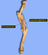
Superficial digital flexor m.
(Dog, Horse, Cow)

Attachments:
(O): Lateral supracondylar tuberosity of the femur
(I): Tuber calcanei & base of middle phalanges of digits 2-5
Action:
- Flex the proximal and middle digital joints of digits 2-5
- Flex the stifle joint
- Extend the tarsal joint
Innervation:
- Tibial n.
Blood Supply:
- Distal caudal femoral a.
** Both heads make up large portion of common calcanean tendon**

Comon Calcanean Tendon

Achilles tendon in humans
Combination of multiple tendons for insertion on tuber calcanei
Continious muscles to tendon:
- Major:
- Gastrocnemius m.
- Superficial digital flexor m.
- Minor:
- Biceps femoris m.
- Gracillis m.
- Semitendinosus m.

Flexor Manica

Similiar to forelimb
Superficial digital flexor m. (attaches here)
Deep digital flexor m. passes through distal phalange
Deep Digital Flexor m.
(Dogs)

2 Heads:
- Lateral digital flexor m.
- Covered by the flexor retinaculum
- Larger than medial head
- Joins medial head at the level of the distal tarsal bones
- Forms common tendon
- Medial digital flexor m.
Attachments:
(O): Caudal aspect of proximal 2/3 of tibia, proximal 1/2 of fibula and interosseous membrane
(I): Flexor tubercle of plantar surface of distal phalanges
Actions:
- Flex the digits
- Extend the tarsal joints
Innervation:
- Tibial n.
Blood Supply:
- Distal caudal femoral a.

Deep digital flexor m.
(Horses and Cows)

3 Heads:
- Medial digital flexor m.
- Lateral digital flexor m.
- Caudal tibial flexor m.
Lateral head fuses with caudal tibial
- Medial joins further distally
Attachments:
(O): Caudal aspect of proximal 2/3 of tibia, proximal 1/2 of fibula and interosseous membrane
(I): Flexor tubercle of plantar surface of distal phalanges
Actions:
Flex the digits
Extend the tarsal joints
Innervation:
Tibial n.
Blood Supply:
Distal caudal femoral a.

Tarsal Tunnel

Allows for passage of:
- Lateral digital flexor m.
- Medial and lateral plantar n.
- Caudal branch of saphenous v.
Borders:
- Sustenaculum Tali (dorsally)
- Calcanean tuber (laterally)
- Flexor retinaculum (plantar side)
Popliteus m.

Covered by Gastrocnemius m. & Superficial digital flexor m.
Medial muscle!
Attachments:
(O): Lateral epicondyle of the femur
(I): Proximal 1/3 of caudal surface of tibia
Action:
- Rotate the leg medially
Innervation:
- Tibial n.
Blood Supply:
- Popliteal a.

Interosseous m.

Similiar to forelimb
Action:
Flexor of the metatarsophalangeal joint

Digital annular ligaments
(Dog)

Brace the digital flexor tendons
- Plantar annular ligament
- Proximal digital annular ligament
- Distal digital annular ligament
Digital Annular Ligaments
(Horse and Cow)
Horse:
Plantar Annular Ligament
Proximal digital annular ligament
Cow:
Proximal digital annular ligament
Distal digital annular ligament
Distal interdigital ligament
