Forearm and Wrist SDL Flashcards
The forearm bones are the radius and ulna. What do these bones articulate with?
Proximally with the humerus at the elbow joint
Proximally with each other at the proximal radioulnar joint
Distally with with each other at the distal radioulnar joint
Radius - distally with the carpal bones to form the radiocarpal/wrist joint
In life, a tough, fibrous membrane lies between the radius and ulna. What is this membrane called?
Interosseous membrane
Bony landmarks of radius and ulna

What muscles is supination of the arm achieved by?
- Supinator (a deep posterior forearm muscle)
- Biceps brachii (an anterior arm muscle) acts as powerful supinator especially when the elbow is flexed

What muscle is pronation of the forearm achieved by?
2 anterior forearm muscles:
- Pronator teres (superficial)
- Pronator quadratus (deep)
What type of joints are the joints within the hand?
Synovial
The human thumb is opposable. What benefit is this?
Allows us to manipulate all manner of objects with great dexterity, and form various grips, including a precision grip.
How are the 8 carpal bones arranged?
In 2 rows; proximal and distal
What bones makes up the proximal carpal row?
- Scaphoid, lunate, triquetral, pisiform (from radial to ulnar side)
What bones make up the distal carpal row?
- Hamate, capitate, trapezoid, trapezium (from ulnar to radial side)
Which carpal bones articulate with the radius to form the wrist (radiocarpal) joint?
Scaphoid and lunate
The scaphoid bone is the most commonly fractured carpal bone. What can happen as a result of a missing a scaphoid fracture?
Arthritis, wrist fusion, death of bone tissue, non union
What bones does the first carpometacarpal joint involve?
The first metacarpal and the trapezium
What movements is the thumb capable of?
- Abduction
- Adduction
- Flexion
- Extension
- Opposition
- Reposition
What movements is the wrist capable of?
- Flexion
- Extension
- Abduction
- Adduction
All 4: circumduction
What movements are the fingers capable of?
- Abduction
- Adduction
- Flexion
- Extension
Why is the (ante)cubital fossa important in clinical practice?
- Contains several important neurovascular structures
- Superficial veins that overlie the cubital fossa are routinely accessed for venepuncture and intravenous cannulation.
What is the superior boundary of the cubital fossa?
An imaginary line between the medial and lateral epicondyle of the humerus
What makes up the floor of the cubital fossa?
Brachialis
What makes up the medial and lateral borders of the cubital fossa?
2 forearm muscles: pronator teres and brachioradialis

If the skin that overlies the cubital fossa is removed, what do we see?
Fascia and superficial veins overlying the cubital fossa
When accessing the superficial veins in the cubital fossa, why must care be taken to keep the needle superficial?
The brachial artery and median nerve lie deeper in the cubital fossa

How many layers of muscles are there in the anterior forearm?
3; superficial, middle, deep
What muscles make up the superficial layer of the anterior forearm?
- Flexor carpi radialis
- Flexor carpi ulnaris
- Palmaris longus
- Pronator teres
What muscles make up the middle layer of the anterior forearm?
Flexor digitorum superficialis
What muscles make up the deep layer of the anterior forearm?
- Flexor digitorum profundus
- Flexor pollicis longus
- Prontor quadratus
Where do the superficial flexors of the anterior forearm arise from?
The medial epicondyle - ‘common flexor origin’
Location of superficial flexors

Which superficial anterior forearm muscle is found most laterally? What is its action?
Pronator teres - pronator (and weak flexor) of the forearm
Where does palmaris longus lie?
Between the flexor carpi ulnaris and radialis
Where does palmaris longus arise from? Where does it insert?
Arises from ‘common flexor origin’ and inserts into palmar aponeuorsis
What is palmar aponeurosis?
A thick band of connective tissue in the palm
Action of palmaris longus?
Flexes the wrist
Distal insertion of pronator teres?
Lateral surface of radial midshaft
Distal insertion of flexor carpi radialis?
Metacarpals of 2nd and 3rd digits
Distal insertion of flexor carpi ulnaris?
Base of metacarpal 5
Distal insertion of palmaris longus?
Palmar aponeurosis
Insertions of superficial anterior forearm muscles

Other than flexion, what actions do FCR and FCU have at the wrist?
- Flexor carpi ulnaris adducts
- Flexor carpi radialis abducts
Which nerves innervate the superficial flexors of the wrist?
Median nerve - innervates flexor carpi radialis, palmaris longus and pronator teres
Ulnar nerve - innervates flexor carpi ulnaris
How many tendons does flexor digitorum superficialis give rise to? Where do these travel?
4 - these travel under the flexor retinaculum to digits 2-5
Onto which phalanges of digits 2-5 do the FDS tendons insert?
Each tendon of FDS then splits into 2, inserting either side of the middle phalanx of digitis 2-5
What happens to the FDS tendons just before they insert upon these bones?
They split into 2
What is the action of FDS and what is its innervation?
Actions:
- Flexor of digits at PIPJ, MCPJ and wrist
Innervation:
- Median nerve
Diagram of origin and insertion of FDS

Where do the ulnar and radial neurovascular bundles lie in relation to FDS?
Ulnar neurovascular bundle - lies medial
Radial neurovascular bundle - lies lateral
Where does the median nerve lie in relation to FDS?
Deep to FDS
Where do the deep flexors of the anterior forearm arise from?
The shaft of the radius and ulna and the interosseous membrane
Where do the tendons of flexor digitorum profundus insert? How are they related to the tendons of FDS?
- Insert on the distal phalanges of digits 2-5
- Run deep to FDS
- The 4 tendons of FDS then split into 2 and FDP tendons travel between this split

Diagram of carpal tunnel

Diagram of origin and insertion of superficial flexors

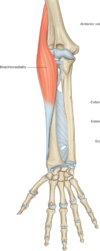
Where does brachioradialis originate from? Where does it insert?
The supracondylar ridge of the humerus (not the common extensor origin) and extends to the distal radius
Distal insertion of:
- EI
- EPL
- EPB
- APL
- EI - inserts on extensor expansion of index finger
- EPL - inserts on distal phalanx of thumb
- EPB - inserts on proximal phalanx of thumb
- APL - inserts on lateral aspect of 1st metacarpal

What tendons are involved in forming the ASB?
APL, EPL, EPB

What is E?

Styloid process of the ulna
What is C?

Flexor carpi radialis
What is B?

Brachioradialis
Name B-F

B - extensor carpi ulnaris
C - extensor digitorum
D - Abductor pollicis longus
E - Extensor pollicis brevis
F - Extensor digiti minimi
what is C?

Extensor pollicis longus
Which carpal bone articulates with the distal radius at region C

Lunate
Muscle B is innervated by which nerve?

Radial
What is E?

Flexor digitorum superficialis
Which bony landmark lies under region A?

Medial epicondyle - We can see the posterior forearm muscles and the dorsum of the hand, hence this is the posterior aspect of a right forearm. A is the medial epicondyle. The posterior forearm muscles can be seen origination form the region directly opposite region A - the lateral epicondyle.
Identify A and B.

A - Flexor pollicis longus
B - Flexor digitorum profundus
Muscle A inserts on the:

distal phalanx of the thumb
Nerve E innervates which muscles in the anterior forearm?

Pronator teres
Flexor carpi radialis
Flexor digitorum superficialis
Lateral half of flexor digitorum profundus
Muscle B is innervated by which nerve?

Posterior interosseous nerve
The red arrow indicates the:

Lateral epicondyle
This image shows branches of which nerve innervating the skin of the region circled?

Superficial radial
What is artery A?

Radial artery
The tendons of which muscles travel through the carpal tunnel in the region circled B?

Flexor digitorum profundus
Flexor digitorum superficialis
Flexor pollicis longus
What is A-E?

A - ulnar nerve
B - ulnar artery
C - median nerve
D - tendon of flexor pollicis longus
E - radial arery
Muscle A is innervated by which nerve?

Anterior interosseous
Identify the tendon indicated by the letter C.

Extensor pollicis longus
Letter C indicates the common flexor origin. Is this true or false?

True
Identify the vein indicated by letter C.

Basilic