Vessels of the Lower Limb Flashcards
What is the main arter of the lower limb?
The femoral artery
What is the femoral artery a continuation of?
The external iliac artery (terminal branch of the abdominal aorta).
When does the external iliac artery become the femoral artery?
when it crosses under the inguinal ligament and enters the femoral triangle
What branch arises from the femoral artery in the femoral triangle?
Profunda femoris (and perforating branches)
Where do the perforating branches of profunda femoris pass?
perforate the adductor magnus, contributing to the supply of the muscles in the medial and posterior thigh
The femoral artery also gives rise to the medial and lateral circumflex arteries. What do these suppy?
These anastamose around the femur and supply the neck and supply its head and neck.

How can a fracture of the femoral neck affect the blood supply?
In a fracture of the femoral neck the medial circumflex artery can easily be damaged, and avascular necrosis of the femur head can occur.
Where does the femoral triangle pass after exiting the femoral triangle?
Continues down the anterior aspect of the thigh. (During its descent, the artery supplies the anterior thigh muscles.) Then passes through the adductor hiatus and enters the posterior compartment of the thigh.
What is the adductor hiatus?
An opening in adductor magnus
After passing through the popliteal fossa, what does the femoral artery become?
Popliteal artery
Where does the obturator artery arise from?
The internal iliac artery in the pelvic region
How does the obturator artery enter the thigh?
It descends via the obturator canal to enter the medial thigh
Where do the superior and inferior gluteal arteries arise from?
The internal iliac artery
How do the superior and inferior gluteal arteries enter the gluteal region?
Via the greater sciatic foramen
Where does the superior gluteal artery leave the foramen in relation to piriformis? How about the inferior gluteal artery?
Superior - leaves the foramen above the piriformis muscle
Inferior - leaves the foramen below piriformis
What does the popliteal artery divide into?

- Anterior tibial artery
- Posterior tibial artery (continuation of popliteal artery)

Where does the anterior tibial artery then travel? What does it become when it reaches the foot?
Passes anteriorly through apertures in IOM (between tibia and fibula) and then moves inferiorly down the leg. It runs down the entire length of the leg, and into the foot, where it becomes the dorsalis pedis artery.

What branch does the posterior tibial artery give rise to?
Fibular artery

What does the fibular artery supply?
Muscles in the lateral compartment of the leg
Where does the posterior tibial artery travel? How does it enter the foot?
Continues inferiorly, along the surface of the deep posterior leg muscles (such as tibialis posterior). It enters the sole of the foot via the via the tarsal tunnel, accompanying the tibial nerve.
Where is dorsalis pedis palpable?
On the dorsum of the foot
How is arterial supply to the foot delivered?
Via 2 branches:
- Dorsalis pedis (a continuation of the anterior tibial artery)
- Posterior tibial
What branch does does dorsalis pedis give in the foot?
Deep plantar - travels deep into the foot.
The dorsalis pedis then moves inferiorly towards the sole of the foot

How does the posterior tibial artery enter the sole of the foot?
Via the tarsal tunnel
What does the posterior tibial artery then split into in the foot?
into the lateral and medial plantar arteries
What does the lateral plantar atery continue on to form?
The plantar arch

What does the plantar arch anastamose with?
The deep plantar artery (travelled down from dorsum of foot)
Why is the femoral artery easy to access?
The femoral artery is located superficially within the femoral triangle, and is thus easy to access. This makes it suitable for a range of clinical procedures.
When would the femoral artery need to be accessed?
- To obtain an arterial blood gas in emergencies if poor peripheral perfusion / pulses
- To undertake minimally invasive processes
- E.g. coronary angioplasty
What is the general procedure for accessing the femoral artery in minimally invasive procedures?
A catheter can be placed into the femoral artery which can then be advanced up the arterial tree to target organ
What is coronary angiography?
Here, the femoral artery is catheterised with a long, thin tube. This tube is navigated up through the external iliac artery, common iliac artery, aorta, and into the coronary vessels. A radio-opaque dye is then injected into the coronary vessels, and any wall thickening or blockages can be visualised via x-ray.
What is ischaemia?
When perfusion fails to meet the demands of tissues; tissue hypoxia and anaerobic metabolism result, leading to tissue damage (and death, if adequate perfusion is not restored)
What is lower limb ischaemia most commonly caused by?
Atherosclerotic disease
What is chronic ischaemia caused by?
A gradual process caused by atherosclerosis
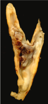
What is atherosclerosis?
- Atherosclerosis = disease process - lipids deposited in arterial walls
- Plaques may remain stable but partially occlude the artery and cause symptoms
- Plaque may become unstable and rupture

Image of an atherosclerotic plaque at bifurcation of the common carotid:
What is the Fontaine classification of chronic limb ischaemia?
- Asymptomatic
- Intermittent claudication
- Ischaemic rest pain
- Ulceration / gangrene ( = critical ischaemia)

What is ulceration and gagrene signs of?
Critical ischaemia

What is ‘dry’ gangrene?
Tissue necrosis without infection. Toes are usually first – black, dry, shrunken

What is ‘wet’ gangrene?
Tissue necrosis + infection with the affected part being black, soft and putrid. Can rapidly lead to sepsis and death.

What are the signs and symptoms of acute limb ischaemia?
The 6 P’s:
- Painful
- Pulseless
- Perishingly cold
- Pallor (pale)
- Paraesthesia (reduced or abnormal sensation)
- Paralysis (difficulty moving the limb)

Where is the small saphenous vein seen?
It moves up the posterior side of the leg, passing posteriorly to the lateral malleolus, empties into the popliteal fossa

Where is the great saphenous vein seen? What does it drain into?
It ascends up the medial side of the leg, passing anteriorly to the medial malleolus at the ankle, and posteriorly to the medial condyle at the knee.
Drains into the femoral vein.

What vein is preferred for central lines?
Internal jugular vein

Where can the great saphenous vein be accessed?
Anterior to the medial malleolus –> site can be used in emergencies to obtain IV access

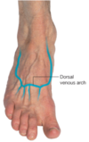
Can you cannulate the dorsal veins of the foot?
Yes (not ideal though)
What are varicose veins?
- If valves in the veins become incompetent, blood can flow back into the superficial veins.
- This results in an increased intra-luminal pressure, which the veins cannot withstand, causing them to become dilated and tortuous.

Signs and symptoms of DVT?
- limb is swollen, red, warm and painful
- extent of swelling depends on location of DVT
- tender on examination
- always ask patient about any symptoms of chest pain and SOB, take O2 sats
- 50% of those with DVT have long term pain and swelling in the affected leg

General areas innervated by major nerves of the lower limb

Motor and sensory function of femoral nerve?
Motor: anterior thigh muscles
Sensory: anteromedial thigh and the medial side of the leg and foot

What 2 maps are commonly used to show dermatomes of lower limb
- More closely related to embryology
- More closely related to clinical findings
