Indirect Ophthalmoscopy Flashcards
What are some advantages of direct ophthalmoscopy?
- High Magnification
- Relatively good image with small pupil (elderly people have smaller pupils)
- Portable
- What you see is where it is
What are some disadvantages of direct ophthalmoscopy?
The magification is affected by the refractive error
Small FOV (10 degrees in emetropes)
Short working distance- be mindful of PPE and corona
Px might feel uncomfortable
Image degrades with lens opacities e.g. with a nucelar cataract
No stereopsis (depth perception) thus you can’t check for any raised section of the fundus or for things such as raised disc margins
What are the two methods of binocular ophthalmoscopy?
Slitlamp Binocular Ophthalmoscopy (using either a volk lens or a high positive lens)
Headset Binocular indirect Ophthalmoscopy (also using a positive lens but one thats slightly lowered powered than the one used in slit lamp)
What does indirect ophthalmoscopy using both a slit lamp and headset produce?
An ariel image
A real image
An inverted image
Stereo (3D) image
With a magnification of x2 to x5 (headset)
[Magnification with slit lamp can vary]
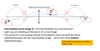
Why is indirect ophthalmoscopy called indirect ophthalmoscopy?
Basically you keep in mind you are looking at an image of the the patient’s retina (O’ on the diagram) thus it’s called indirect ophthalmoscopy.
As the image produced by the volk lens is inverted how would you record what you see?
You could just turn ur page upside and start drawing that way everything is in the right position.
What affects the field of illumination in indirect ophthalmoscopy?
Patient pupil size affects the field of illumination as a smaller pupil means they let less light through.
What affects the field of view in indirect ophthalmoscopy?
The size of the lens and the examiner’s pupil size affects the field of view.
What’s a key bit of optics to do with indirect ophthalmoscopy?
That the Px pupil plane [circled in red] and the examiner’s pupil plane [circled in red] are conjugate points.
Conjugate points are are “corresponding pairs of object and image points and planes”.

Why are volk lenses aspheric?
Volk lenses are aspheric to try and make the lens as large as possible while trying to minimise the optical aberrations ☺
How is FOV affected by lens diameter and what considerations are associated with this?
Larger diameter lens = Larger Field of View (FoV)
- HOWEVER Larger lens= Greater optical aberrations
- Volk lenses are aspheric to try and make the lens as large as possible while trying to minimise the optical aberrations ☺
[Keeping the lens diameter the same, but moving lens closer to the eye would in theory lead to a larger FoV, but pupils would no longer be conjugate thus image would be blurry.
The only way to move the lens closer and still see an image is to change the lens power]
As the lens power increases in indirect ophthalmoscopy what happens to FOV , magnification and working distance?
FOV - Increases with higher positive power.
Magnification decreases with higher positive power.
Working distance decreases wioth larger positive power.
How would you carry out binocular indirect ophthalmoscopy using a slit lamp?
- Ensure it is safe to dilate (van Herick)
- Document the dilating drug details
- Room lights off
- Set-up slit lamp
- Keep lamp intensity to a minimum
- Begin with low magnification
- Slit beam height usually same height as pupil and width approx. 2mm
- Illumination system in line with viewing system
- Px fixates distant at a target straight ahead (so eyes are aligned and steady)
- Focus on the surface of the cornea (on tearfilm) from the slitlamp
- Present the Volk lens in front of pupil ~5mm away
- Now move the SL joystick straight back towards you (keep lens where it is and ensure light is still travelling through centre of the pupil)
- At first, the surface of the Volk lens surface will come into focus – thus you might see scratches or even fingerprints!!
- Then blurred red reflex of the retina shall be seen
- Then the image of the fundus will come into focus
If you are carrying out binocular indirect ophthalmoscopy using a slit lamp, without dilating the pupil of the patient which lenses may you want to use?
LOW MAG LENSES
What are common mistakes with indirect ophthalmoscopy via a slit lamp?
- Students often mistake the iris for a blurred fundus image. The fundus image will be orange, not blue, brown, or green!
- Another common error is not aligning the observation and illumination systems-make sure they are straight and in line with the pupil

How do you use a headset for indirect ophthalmoscopy?
