15 - T&O Shoulder and Arm Flashcards
How are rotator cuff tears classified?
Acute (<3 months) or Chronic (>3 months)
Partial or Full Thickness
Full thickness small (<1cm), medium (1-3cm), large (3-5cm) or massive (>5cm)

What is the pathophysiology and some risk factors for rotator cuff tears?
Pathophysiology
Acute: in tendons with pre-existing degeneration with minimal froce
Chronic: degeneratice microtears from overuse
Risk Factors
- Age
- Trauma
- Overuse
- Repetitive overhead shoulder motions
- BMI>25
- Smoking
- Diabetes

How do rotator cuff tears present?
- Pain over the lateral aspect of the shoulder
- Inability to abduct the arm over 90 degrees
- Tenderness over greater tuberosity
Special Test: empty can, lift off test, posterior cuff test

What are some differential diagnosis for a rotator cuff tear?
- Fracture
- Glenohumeral subluxation
- Radiculopathy
- Brachial plexus injury
How do you investigate a rotator cuff tear?
- Urgent plain film radiograph to exclude fracture (unremarkable unless chronic tear then may be reduced acromiohumeral distance, sclerosis or cyst formation)
- Once fracture excluded can do US or MRI

How are rotator cuff tears managed?
Depends on tear and functional status of patient
Conservative
- For those presenting within 2 weeks of injury, unsuitable for surgery or not limited by loss of function
- Analgesia, physiotherapy, corticosteroid injections into subacromial space
Surgical
- Presenting over 2 weeks since injury or remaining symptomatic despite conservative treatment or large/massive tears
- Surgical repair

What is the prognosis with rotator cuff tears?
- Main complication is adhesive capsulitis
- If age related tears will often become larger within 5 years and become symptomatic
What is the pathophysiology and risk factors of a shoulder fracture?
Usually proxmal humerus from low energy injuries (FOOSH) with osteoporosis
Risk factors (any OA risk factors): female gender, early menopause, prolonged steroid use, recurrent falls, frailty
What are the clinical features of a shoulder fracture and what should you check on examination?
- Pain around upper arm and shoulder
- Inability to abduct arm
- Swelling and bruising of the shoulder
Check for loss of sensation in lateral shoulder and loss of power in deltoid muscle due to close relationship with axillary nerve and circumflex vessels

What investigations are done if you suspect a shoulder fracture?
- Urgent bloods with coagulation and G+S
- If suspect pathological look at serum Ca and myeloma screen
- AP, lateral scapular and axillary view plain radiographs
- CT for intraoperative planning

How are proximal humeral fractures classified?
Neer Classification
Articular segment is anatomical neck and humeral shaft is surgical neck

How are shoulder fractures managed?
Conservative (if minimally displaced and no neurovascular compromise)
- Immobilisation with polysling as gravity will help reduction
- Early mobilisation with pendular exercise at 2-4 weeks
Surgical (if displaced, open or neurovascular compromise)
- If multiple segments do ORIF or intermedullary nailing
- Hemiarthroplasty if complex

What are some complications of a shoulder fracture?
- Reduce range of motion: need to do extensive physio and rehabilitation can take about a year
- Risk of AVN of humeral head: from anterior and posterior humeral circumflex arteries. Needs hemiarthroplasty or reverse shoulder arthroplasty
What are scapular fractures and how are they managed?
- High energy trauma as lot of protection from muscles so difficult to break
- Most treated non-operatively

What is the aetiology of shoulder dislocations?
Most common is anteroinferior (anterior)
Anterior: force applied to extended, abducted, externally rotated humerus
Posterior: seizures or electrocution

What are the clinical features of a shoulder dislocation?

- Painful, reduced motility and feeling of instability
- Often asymmetry on examination with loss of shoulder contours with anterior bulge
ASSESS NEUROVASCULAR STATUS ESPECIALLY AXILLARY AND SUPRASCAPULAR NERVES

What are some associated injuries with a shoulder dislocation?
Bone:
- Bankart Lesion (fracture of anterior inferior glenoid)
- Hill Sachs defect (of humeral head)
- Fractures of greater tuberosity and surgical neck of humerus
Labral, Ligaments, Rotator cuff
- Bankart lesion (avulsion of anterior labrum)
- Glenohumeral ligament avulsion
- Rotator cuff injuries (usually in anterior and younger patients

What investigations do you do if you suspect a shoulder dislocation and what are the findings?
- Trauma shoulder series of radiographs: AP, axial, Y-scapular
- Y view helps differentiate between anterior and posterior dislocations
- Can do MRI if labral or rotator cuff injuries

How are shoulder dislocations managed?
NON-OPERATIVELY

- A to E
- Analgesia
- Closed reduction with pre and post neurovascular assessment
- Broad arm sling for around 2 weeks
- Physiotherapy to strengthen rotator cuff
- May need future surgical treatment if Hill Sachs or Bankart
What is the prognosis with a shoulder dislocation?

- Chronic pain, limited mobility, stiffness
- High rate of recurrence
- Adhesive capsulitis
- Nerve damage
- Rotator cuff injury
- OA (especially if labral tear)

What is the aetiology and risk factors of a humeral shaft fracture?
- Bimodal distribution younger and older
- Often damage radial nerve due to spiral groove disruption
Risk factors: osteoporosis, increasing age, previous fractures, pathological
How do humeral shaft fractures present?
- Pain and deformity following FOOSH or falling laterally onto adduct limb
- If radial nerve involved will have reduced sensation over 1st dorsal webspace and a weakness in wrist extension
- Always check neurovascular status, any open wounds and any concurrent injuries
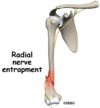
What is a Holstein-Lewis Fracture?
Fracture of the distal third of the humerus resulting in entrapment of the radial nerve
Will lead to loss of sensation in radial nerve distribution and a wrist drop deformity
Needs surgical intervention

What investigations are done for a suspected humeral shaft fracture?
AP and Lateral radiographs

How is a humeral shaft fracture managed?
Conservative (more common)
- Realignment of limb
- If broken at diaphysis give humeral brace
- Follow up with repeated imaging and will heal after about 8-12 weeks
Surgical
- ORIF with a plate
- Intramedullary nailing if pathological, polytrauma or severely osteoporotic

What are some complications of a humeral shaft fracture?
- Nonunion/malunion
- Varus angulation especially with transverse fractures
- Radial nerve injuries (often improve after 3 months)

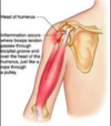
What is the pathophysiology of biceps tendinopathy?
Can occur proximal or distal.
Common in young sporty individuals acutely and older individuals as degenerative disease
Leads to painful, swollen, structurally weaker tendon at risk of rupture
What are the clinical features of biceps tendinopathy and what are some special tests you can use to aid diagnosis?
- Pain made worse when stressing the tendon and alleviated by rest and ice
- Weakness of flexion and supination
- Tenderness over affected tendon
- Disuse atrophy due to pain
Speed and Yergason’s test
(pain in anterior shoulder is positive test)

How is biceps tendinopathy investigated and managed?
Ix

- Often clinical
- FBC (CRP and ESR) and radiographs can be taken to exclude differentials
- US and MRI sometimes
Mx
- Analgesia and ice therapy
- Physiotherapy
- US guided steroid injections if above fails
- Final line is arthroscopic tenodesis or tenotomy
What are some risk factors for biceps tendon rupture?
Usually following forced extension of flexed elbow
Risks: previous episodes of biceps tendinopathy, steroid use, smoking, CKD, fluroquinolones

What are the presenting features of a biceps tendon rupture and what special test can you do on examination?
- Sudden onset pain and weakness at affected are (can still flex at elbow due to action of supinator muscles and brachialis)
- Marked swelling and bruising in antecubital fosse
- Reverse pop-eye sign
- Hook test (Distal rupture): actively flex elbow to 90 degress supinated and examiner hook index finger under lateral edge of biceps tendon, cannot be done if ruptured

How are bicep tendon ruptures managed?
Conservative
- If lower demand patient as leaving can cause weakness and fatigueability
- Analgesia
- Physiotherapy
Surgical
- Anterior single-incision or dual incision technique
- Done within few weeks of initial injury otherwise tendon will retract and scar

How is a biceps tendon rupture investigated?
- Often diagnosed clinically
- Can do US imaging
- If US inconclusive can do MRI

What is the pathophysiology of adhesive capsulitis (frozen shoulder)?
- Glenohumeral joint capsule becomes contracted and adherent to humeral head
- Often affects woman between 40-70 years old
- Associated with inflammatory diseases so may have autoimmune element

What are the three stages of adhesive capsulities?
- Initial painful
- Freezing
- Thawing

What are the clinical features of adhesive capsulitis?
- Generalise deep constant pain of the shoulder that disturbs sleep
- Joint stiffness
- Loss of arm swing
- Atrophy of deltoid muscle
- Generalised tenderness on palpation
- Limited ROM, especially external rotation and flexion

What are some differential diagnoses for adhesive capsulitis?

What investigations are done if you suspect adhesive capsulitis?
- Usually clinical diagnosis
- X-ray normally unremarkable but can rule out differentials
- MRI will show thickening of glenohumeral joint capsule and can rule out differentials
- Measure HbA1c and blood glucose as more common in diabetics

How is adhesive capsulitis managed?
- Self limiting and often has recurrence so educate and reassure
- Physiotherapy
- Analgesia and possible corticosteroid injections
- If conservative not working and affecting QoL then joint manipulation under GA, arthrogaphic distension or surgical release of glenohumeral joint capsule

What is subacromial impingement syndrome?
Inflammation and irritation of the rotator cuff tendons as they pass through the subacromial space resulting in pain, weakness and reduced ROM
Usually in active people or in manual occupations

What is the pathophysiology of subacromial impingement syndrome?
Intrinsic (pathology of rotator cuff tendons due to tension)
- Muscular weakness
- Shoulder overuse
- Degenerative tendinopathy
Extrinsic (pathology of rotator cuff tendons due to external compression)
- Anatomical factors
- Scapular musculature
- Glenohumeral instability

What are the clinical features of subacromial impingement syndrome and what tests can be used on examinationt to aid diagnosis?
- Progressive pain in anterior superior shoulder
- Pain worse on abduction
Neers Impingement Test and Hawkin’s Test

What are some differential diagnoses for subacromial impingement syndrome?
- Muscle tear
- Neurological pain
- Frozen shoulder
- Acromioclavicular pathology

How is subacromial impingement syndrome investigated and diagnosed?
- Often clinical
- MRI: subacromial osteophytes and sclerosis, subacromial bursitis, humeral cystic changes, narrowing of subacromial space

How is subacromial impingement syndrome managed?
Conservative

- NSAIDs
- Physiotherapy
- Corticosteroid injections
Surgical (6 months without response to conservative)
- Surgical repair of muscle tendons to improve ROM
- Surgical removal of subacromial bursa to increase subacromial space
- Acromioplasty to increase subacromial space
How do you perform a shoulder exam for an OSCE?

If a patient presents with shoulder pathology from the following age groups, what is the most likely diagnosis?
- Under 30
- 40-60
- Over 65

What are the special tests for a shoulder exam?
- Hawkins: tests external impingement, forced internal rotation at different levels of abduction/adduction
- Scarf: flex patients shoulder 90 degrees then maximally adduct to other shoulder, if pain then AC joint pathothology
- Empty can: testing supraspinatus
- Apprehension: tests the integrity of the glenohumeral joint capsule and anterior stability. passively externally rotate shoulder and push anteriorly on posterior humeral head

What are the different stages of adhesive capsulitis?

What is the pain distribution in adhesive capsulitis?
- Progressive loss of ROM of shoulder joint
- Lot of pain at night so lose sleep

What is muscle patterning instability and what are some differentials you should consider?
Usually younger patients who can voluntarily slip the shoulder out of joint as a trick movement, but may then go on to dislocate repeatedly involuntarily
Needs physio
Consider Marfan’s and EDS

How do you manage cubital tunnel syndrome?
- Do NCS and EMG to diagnose
- Rest, splint at night, protect in sling for 2-4 weeks
- If above doesn’t work can try steroid injections or decompression surgery

What is a special test used to diagnose a distal biceps rupture?
- Hook Test
- Also another test is weaker supination on resistance on side of rupture (gym lad in clinic)
