Abdominal Flashcards
(33 cards)
What are the main types and locations of primary colorectal cancers?
The majority of colorectal cancers are adenocarcinomas derived from epithelial cells. Less common types of primary malignant colorectal tumours are carcinoid tumours, GI stromal cell tumours, and lymphomas.
The rectum is the most common site in the large bowel, accounting for 1/3 of all large bowel cancers, although the sigmoid colon in the most common site in the colon.

What are the risk factors for Colorectal Cancer?
- Increasing age is the greatest risk factor for sporadic colorectal adenocarcinoma, with 99% occurring in people aged 40 or over.
- Family history (APC mutation)
- Lynch syndrome (Hereditary Non-Polyposis Colorectal Cancer) accounts for 5% of colorectal cancers.
- Familial adenomatous polyposis (FAP)
- Diet (high fat, low fibre)
What are the clinical features of Colorectal Cancer?
Red flag symptoms are common. They include:
- Rectal bleeding
- Change in bowel habit - increased frequency or looser stools is the most common symptom
Although weight loss and anorexia are uncommon.
Right sided carcinomas present with iron deficiency anaemia, usually with dark blood mixed in with stool. There may also be a RIF mass.
Left sided (commonest) carcinomas present with increasing frequency/looser stools, rectal bleeding (bright red blood coating stool) with or without mucus. Can also present as intestinal obstruction due to constricting neoplasm.
Rectal: Tenesmus, worm-like stool with other signs? There is a palpable rectal mass in up to 80% of patients.
Anal: Pain, pruritus ani, and mass felt.
Describe the NHS colorectal cancer screening programme
The NHS now has a national screening programme offering screening every 2 years to all men and women aged 60 to 74 years in England.
- Eligible patients are sent Faecal Immunochemical Test (FIT) tests through the post
a type of faecal occult blood (FOB) test which uses antibodies that specifically recognise human haemoglobin (Hb)
Patients with abnormal results are offered a colonoscopy:
- 5 out of 10 patients will have a normal exam
- 4 out of 10 patients will be found to have polyps which may be removed due to their premalignant potential
- 1 out of 10 patients will be found to have cancer
What are the investigations for Colorectal Cancer?
FBC may show microscopic anaemia (iron deficiency). LTFs and U&Es are often normal.
- CEA (Carcinoembryonic Antigen) is a tumour marker elevated in only 40%-80% of patients.
Occult blood is often present and detected by faecal immunochemical test (FIT).
Sigmoidoscopy will reveal tumours in the rectosigmoid region and allow biopsy. Even if tumour is not detected, the presence of blood or slime coming down from above is strongly suspicious of a malignant disease.
Colonoscopy and biopsy is necessary.
CT thorax, abdomen and pelvis may be indicated in some patients.
Describe the management of Colorectal Cancer
Management centres around surgery:
- Pre-operative: The bowel is cleared by enemas and oral stimulant laxatives (e.g. Picolax). Metronidazole and gentamicin are given at time of surgery.
-
Operative: The principle of operative treatment is wide resection of the growth together with its regional lymphatics.
- In the unobstructed case, the bowel can be prepared beforehand and primary resection with restoration of continuity can be achieved.
- In cases of obstruction, the primary goal is to relieve obstruction and so a Hartmann’s procedure can be performed. Primary restoration can occur at a different time.
- Post-operative: Adjuvant chemotherapy with 5-fluorouracil (5-FU) in combination with folinic acid may reduce the risk of recurrent disease, and may prologue survival for metastatic disease.
Describe the surgical options for of colorectal cancer
- Typically for a lesion in the right colon a right hemicolectomy is performed, with an ileocolic anastomosis.
- For a lesion in the left colon, a left hemicolectomy or sigmoid colectomy is performed, with anastomosis of the colon to the rectum.
- In an emergency situation, with unprepared bowel, a Hartmann’s operation can be performed with the formation of an end colostomy and the rectum oversewn or brought to surface.
What is the definition and classification of Mechanical Intestinal Obstruction?
Intestinal obstruction is a restriction to the normal passage of intestinal contents. It may be divided into two main groups: paralytic and mechanical. This page will discuss mechanical intestinal obstruction.
Classification
Mechanical bowel obstruction can be classified in various ways:
-
Speed of onset:
- Can be acute which has a rapid onset and severe symptoms
- Can be chronic which has insidious onset and less severe symptoms, for example due to carcinoma.
- Can be acute-on-chronic which is the rapid development of obstruction in the background of chronic bowel obstruction.
-
Site:
- Can be low/large bowel
- Can be high/small bowel
- Simple (no damage to blood supply) or strangulated (blood supply compromised)
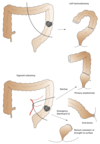
What is the aetiology of Mechanical Intestinal Obstruction?
The aetiology can also be structured in different ways. One way is thinking about it by causes in the lumen, in the wall, or outside the wall:
-
Intraluminal causes include:
- Impaction of faeces or worms
- Intussusception
- Gallstone ‘ileus’ (remember not really ileus)
-
Intramural causes include:
- Tumours
- Strictures such as from IBD, surgery, diverticulitis.
- Congenital atresia
-
Extramural causes include:
- Hernias
- Volvulus
- Adhesions
- Extrinsic compression due to abscesses, haematomas, non-colonic tumours etc.
What are the common causes of small and large bowel obstruction?
Small bowel obstruction is commonly due to:
- Adhesions (60%) - abdominal surgery is becoming increasingly common, which can commonly lead to adhesions.
- Hernias
Large bowel obstruction is commonly due to:
- Tumours (60%)
- Diverticular stricture (20%)
- Volvulus (5%)
What are the causes of intestinal obstruction in neonates and infants?
- Neonatal: congenital atresia and stenosis (e.g. duodenal atresia), imperforate anus, volvulus neonatorum, Hirschsprung’s disease and meconium ileus.
- Infants: intussusception, Hirschsprung’s disease, strangulated hernia and obstruction due to Meckel’s diverticulum.
What are the causes of intestinal obstruction in young adults/middle aged and the elderly?
- Young adults and middle age: strangulated hernia, adhesions and bands, Crohn’s disease.
- The elderly: strangulated hernia, carcinoma of the colon, colonic diverticulitis, impacted faeces.
What are the cardinal symptoms of Mechanical Intestinal Obstruction?
There are four cardinal features of bowel obstruction:
- Colicky abdominal pain: usually the first symptom. Small bowel pain tends to be periumbilical, whereas large bowel tends to be suprapubic.
- Abdominal distention: usually more pronounced in large bowel obstruction. With a small bowel obstruction that has little proximal bowel there may not be much to distant.
- Absolute constipation is an early feature in large bowel obstruction, but a late feature in small bowel obstruction - in fact the patient may pass a single normal stool after onset of small bowel obstruction as the distal bowel empties.
- Vomiting is more a early feature of small bowel obstruction and may be absent large or chronic bowel obstruction. In the late stages of intestinal obstruction, the vomiting becomes faeculent but not faecal. The faeculent vomiting is due to bacterial decomposition of the stagnant contents of the obstructed small intestine.
What are the examination features of intestinal obstruction?
- The patient may be dehydrated if vomiting has been copious. They are in pain and may be rolling about with colic.
- The pulse is usually elevated, but the temperature is frequently normal.
- A raised temperature and a tachycardia suggest strangulation.
- The abdomen is distended and visible peristalsis may be present.
- The abdomen is tender and you may feel a mass.
- Bowel sounds are usually accentuated and tinkling.
What must you look out for/perform if suspecting intestinal obstruction?
During inspection it is important to look carefully for three features:
- The presence of a strangulated external hernia, which may require a careful search in the case of a small strangulated femoral hernia in a very obese and distended patient, and
- The presence of an abdominal scar. Intestinal obstruction in the presence of this evidence of a previous operation immediately suggests adhesions or a band as the cause.
- Perform a rectal examination. It may reveal an obstructing mass in the pouch of Douglas, the apex of an intussusception or faecal impaction.
What are the investigations for Mechanical Intestinal Obstruction?
Describe the management of Mechanical Intestinal Obstruction
Conservative
- Gastric aspiration by means of nasogastric suction. This helps to decompress the bowel and prevent asipration.
- Intravenous fluid replacement - a lot of fluid may be required. Hartmann’s solution or normal saline are given, with potassium if this is low and renal function satisfactory.
- Antibiotic therapy is commenced if intestinal strangulation is likely (or is found at operation).
Gastrografin can be therapeutic and help resolve bowel obstruction. If the contrast does not pass the through the bowel in 8 hours, it is unlikely conservative management would suffice, and surgery should be considered.
Operative
The affected bowel is carefully inspected to determine its viability. Doubtful bowel may recover after relief of the obstruction. It should be reassessed after it has been left for a few minutes wrapped up in a warm wet pack.
- Small bowel: Conservative management is successful in 65- 80% of cases and surgical intervention is only considered for those patients who do not improve with conservative management. Generally small bowel can be resected and primary anastomosis performed with safety because of its excellent blood supply.
- Large bowel obstruction is treated by resection of the obstructing lesion, with a primary ileocolic anastomosis in the case of obstructing lesions proximal to the splenic flexure. Left-sided lesions are managed by excision of the affected segment and exteriorizing the two ends of colon as a temporary colostomy and mucous fistula(Hartmann’s procedure). This is due to poorer blood supply of the large bowel and the fact that a colonic primary anastomosis is very liable to leak in the presence of obstruction.
What are the complications for mechanical bowel obstruction?
Bowel ischaemia
Bowel perforation leading to faecal peritonitis (high mortality)
Dehydration and renal impairment
What is the aetiology of peritonitis?
What are the clinical features of peritonitis?
When the visceral peritoneum becomes inflammed, it presents as a generalised and severe constant abdominal pain due to the poor localisation of pain from the visceral peritoneum. The pain may become localised after (with the involvement of the somatically innervated parietal peritoneum).
Irritation of the diaphragm may be accompanied by shoulder tip pain.
Vomiting is frequent.
The pain is exacerbated by movement and coughing.
On Examination
Peritonitis is associated with guarding or rigidity of the abdominal muscles. If peritonitis involves the whole abdomen, the patient would typically present with a board-like, rigid, tender abdomen with absent bowel sounds. Rebound tenderness is a sign of peritonitis, as sudden removal of the hand causes pain due to movement of the peritoneum.
Patient also tends to be generally unwell and may have signs of dehydration.
What are the potential complications of peritonitis?
Early: Septic shock, respiratory or multiorgan failure, paralytic ileus, would infection, abscesses.
Late: Incisional hernia, adhesions.
What are the investigations for peritonitis?
Blood tests and ABG should be ordered, looking for acidosis or respiratory failure). Amylase may be raised in acute pancreatitis and prevent unnecessary surgery.
CXR may show pneumoperitoneum indicating a Gastrointestinal Perforation.
AXR may reveal cause such as Intestinal Obstruction.
USS or CT abdomen and laparotomy can be done to investigate underlying cause.
If the patient has ascites, perform an Ascitic tap and cell count (diagnostic of SBP if >250 neutrophils/mm3). The bacteria should be gram stained and cultured.
Describe the management of peritonitis
Localised
Treatment will depend on underlying cause, may be surgery or I.V antibiotics.
General
- A-E resuscitation to restore fluid and electrolyte status and oxygenation.
- I.V antibiotics is necessary - specificity to treat broad spectrum of bowel organisms such as a cephalosporin and metronidazole.
- NG tube to aspirate the gastric contents, preventing inhalation of vomit.
Any localised collection of pus requires drainage, and later surgery may be required for the evacuation of residual abscesses, e.g. subphrenic or pelvic collections.
Treat the cause and perform a peritoneal washing with copious irrigation to remove all seropurulent exudate.
Spontaneous Bacterial Peritonitis
Medical treatment with quinolone antibiotic or cefuroxime and metronidazole combination.
What is the aetiology of gastrointestinal perforation?
- Large bowel mainly caused by diverticulitis and colorectal carcinoma. Perforated appendix is the most common complication of appendicitis. Other causes include volvulus, ulcerative colitis (toxic megacolon), trauma, radiation enteritis and complications of operations.
- Gastroduodenal is most commonly caused by a perforated gastric or duodenal ulcer and rarely gastric carcinoma.
- Small bowel (rare) caused by trauma, infection, Crohn’s disease, lymphoma, vasculitis, radiation enteritis.
- Oesophagus - Boerhaave’s syndrome is rupture following forcible vomiting against a closed glottis. OGD can also rarely cause perforation.
