Hepatobiliary Flashcards
(36 cards)
Describe the serum markers of liver damage and the causes of their release
-
Transaminases such as alkaline transaminase (ALT) and aspartate transaminase (AST) - contained within the hepatocyte, increases in levels when hepatocytes die.
- In alcoholic liver damage AST is raised more than ALT (AST:ALT >1).
- In viral hepatitis, ALT (VirAL) is raised higher than AST (AST:ALT <1).
- Alkaline phosphatase (ALP) - found in sinusoidal and canaliculi membranes.
- ALP is raised in obstructive jaundice and bile duct damage. Less elevated in viral and alcoholic liver disease.
- Also raised in bone and metastatic disease.
-
Gamma glutamyl transferase (GGT) - also found within hepatocytes and small bile duct epithelium.
- GGT is raised in chronic alcohol use, liver metastases (not viral) and bile duct disease.
What are the serum markers of liver function?
-
Albumin
- Low production in chronic liver disease and malnutrition
- Increase loss from Gut and kidney (nephrotic syndrome)
- Low albumin also in sepsis due to movement into third space.
-
Pro-thrombin time
- Acute marker of liver function
- Bilirubin:
What is a serum tumour marker that rises in hepatocellular carcinoma?
alpha-fetoprotien may rise in hepatocellular carcinoma.
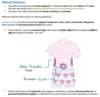
Describe the common approach to evaluating the type of jaundice

What is a cholecystectomy and what are its indications?
A cholecystectomy is the surgical removal of the gallbladder. Can be done openly or laparoscopically (gold standard). It is one of the most commonly performed abdominal surgical procedures.
Indications:
Cholelithiasis (gall stones), causing biliary colic or pancreatitis
Cholecystitis
Gall bladder cancer
What are the specific complications of a cholecystectomy?
- Gallbladder perforation which results in the spillage of bile and/or stones. Stones left in the abdomen can later become a site of infection.
- Vascular injury - bleeding from cystic or hepatic artery can be much more difficult to deal with laparoscopically
- Bowel injury
- Bile duct injuries can lead to the leaking of bile and cause a painful and potentially dangerous infection.
What is an ERCP and its indications?
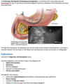
What are the complications of an ERCP?
The major risk of an ERCP is the development of pancreatitis, which can occur in 5% of all patients.
Smaller risks includes of intestinal perforation (especially if a sphincterotomy is performed). There is also risk of an allergic reaction to the contrast dye which contains iodine.
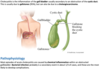
What is biliary colic?

What clinical features can help distinguish biliary colic caused by temporary gallstone obstruction from other causes of biliary colic?
Biliary colic is its own disease entity, as well as a presentation (I think). Distinguish biliary colic from acute cholecystitis, acute pancreatitis, obstructive jaundice, ascending cholangitis by:
-
Nature and characteristics of pain.
- Biliary colic is more colicky, whereas cholecystitis can be more constant and radiate to the shoulder.
- Presence/absence of jaundice; obstructive jaundice is caused by obstruction to the common bile duct. Therefore caused by cholangitis, common bile duct stone and acute pancreatitis.
- Presence/absence of fever (and leukocytosis). A fever points to an infective process such as cholecystitis (though this is more inflammatory) or cholangitis.
Describe the investigations for biliary colic
These are the same investigations as cholecystitis as it may be difficult to differentiate clinically.
Bloods:
- FBC is important as leukocytosis points to cholecytitis rather than biliary colic
- U&Es
-
LFTs
- Elevated ALP suggests obstruction of the cystic or common bile duct.
- Serum lipase and amylase (to investigate pancreatitis)
- Group and save in case surgery needs to be performed.
- Acute phase reactants (CRP)
Imaging:
- AXR - only 10% of stones are radio-opaque
- Erect CXR to look for perforation
-
Ultrasound may show:
- Dilated ducts
- Stones seen as acoustic shadow
- Inflammed gallbladder seen as wall oedema
If dilated ducts are still not seen on ultrasound, an MRCP (Magnetic resonance cholangiopancreatography) can be performed. However an ERCP (Endoscopic retrograde cholangiopancreatography) is the preferred way to visualise gallstones as therapy can be provided at the same time.
Describe the management of biliary colic
Conservative management includes rehydration and keeping the patient NBM. Opioid analgesia is effective at reducing pain. However due to high recurrence rate surgical management is preferred. Cholecystectomy is commonly performed laparoscopically.
What is the epidemiology and risk factors for cholelithiasis?
Gall stones are very common in the west - 20% of adults have them while 85% of patients remain asymptomatic. It is more common in women (2:1). It is not possible to predict which subgroup of patients will become symptomatic and, therefore, cholecystectomy cannot be recommended routinely in asymptomatic patients
Risk factors include:
- Age
- Gender (females > males)
- Hereditary - disorders of bile metabolism
- Drugs - such as oral contraceptives
- Rapid weight loss
- Haemolytic disease (pigment gallstones)
What are the features of the two main types of gallstones?
There are basically two types of gallstones:
- Cholesterol (75%)- contain more than 50% of cholesterol. They are usually single and radiolucent (so will not be seen in an X-ray)
- Pigment (25%)- contain calcium salts of unconjugated bilirubin (calcium bilirubinate). There may be multiple and are mostly radio-opaque.
What are the complications of gallstones?

What is cholecystitis?
What are the clinical features of cholecystitis?
Most patients have a history of biliary colic, (epigastric to RUQ pain, nausea and vomiting) but with the pain becoming more severe and localised to the right upper quadrant. Importantly the presence of a fever indicates infection or inflammation. Furthermore, unlike biliary colic, patients will prefer to lie still and take shallow breaths (now a form of local peritonitis, not colic).
On examination, the patient presents with tenderness and muscle guarding or rigidity.
A positive Murphy’s sign can also be elicited. This is when fingers are placed on the right costal margin by the midline, and asking patients to inspire. A positive Murphy’s sign is the cessation of inspiration when the gallbladder descends and hits the fingers. Murphy’s sign is highly sensitive, but not very specific. You must also perform this test on the left costal margin to exclude nonspecific reactions due to other pathology.
Describe the investigations for cholecystitis
These are the same investigations as biliary colic as it may be difficult to differentiate clinically.
Bloods:
- FBC is important as leukocytosis points to cholecystitis rather than biliary colic
- U&Es
- LFTs - marginally raised bilirubin, ALT and AST may also be seen.
- Serum lipase and amylase (to investigate pancreatitis)
- Group and save in case surgery needs to be performed.
- Acute phase reactants (CRP) are raised
Imaging:
- AXR - only 10% of stones are radio-opaque
- Erect CXR to look for perforation
- An abdominal ultrasound is the single most useful investigation when taken with a history of fever and a positive Murphy’s sign. The US can show a biliary stone, but also gallbladder wall thickening and oedema.
If dilated ducts or gallstones are still not seen on ultrasound, an MRCP (Magnetic resonance cholangiopancreatography) can be performed. However, an ERCP (Endoscopic retrograde cholangiopancreatography) is the preferred way to visualise gallstones as therapy can be provided at the same time.
Only very occasionally a HIDA (radioisotope) scan is used to help confirm or exclude cholecystitis.
Describe the management of cholecystitis
It is first critical to stabilise the patient with I.V fluids (keeping them nil by mouth). I.V antibiotics is also administered to prevent sepsis although cholecystitis (in comparison to cholangitis) is primarily an inflammatory and only secondarily an infective process. Opiates can be given to help with pain.
Once the patient is stabilised, a cholecystectomy is the treatment of choice for virtually all patients to prevent recurrence. This can be done laparoscopically.
What are the complications of cholecystitis?
- Gallbladder perforation
- Empyema
- Gallbladder carcinoma
- Gallstone ileus
What do you know about gallbladder perforation?
Cholecystitis may progress to gangrene followed by perforation.
- Patients present with sepsis and peritonitis.
- Treat by resuscitation followed by urgent open cholecystectomy.
What do you know about gallbladder empyema?
Clinical features similar to cholecystitis, except patients are more unwell with high fever and tender, palpable GB. Some sources say a swinging fever.
- Treat as for cholecystitis plus consider US-guided aspiration of pus, followed by emergency cholecystectomy.
Differential diagnosis is a mucocoele: this is when patients have a tender palpable GB (due to mucus causing distention), but no signs of sepsis.
What do you know about gallstone ileus?
This is a misnomer; it is small bowel obstruction and not ileus (cessation of peristalsis). A rare complication of cholecystitis in which a large gallstone (usually >2.5cm) erodes directly through the gallbladder wall into the duodenum. This process takes a very long time, after which the gallstone travels down the intestinal tract getting stuck in the narrowest part after the gastro-oesophageal junction. Usually occurs in the elderly.
Gas in the biliary tree in the presence of features of small bowel obstruction on a plain abdominal X-ray is diagnostic.
Treat by urgent laparotomy. The gallstone is retrieved via a longitudinal enterotomy (closed transversely to prevent stricture formation) in the distal ileum. Presence of gangrene +/− perforation may require small bowel resection +/− primary anastomosis. Cholecystectomy is contra-indicated in patients with a cholecystoduodenal fistula.
What are the hallmark features of obstructive jaundice?
Jaundice
Dark urine and pale stools
Pruritus (bile salts)