Abdominal Radiology Flashcards
(30 cards)
Is this x-ray noral or abnormal?

normal
Is this normal or abnormal?
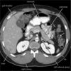
Identify the labelled structures:

NORMAL
1 11th rib.
2 Vertebral body (TH 12).
3 Gas in stomach.
4 Gas in colon (splenic flexure).
5 Gas in transverse colon.
6 Gas in sigmoid.
7 Sacrum.
8 Sacroiliac joint.
9 Femoral head.
10 Gas in cecum
11 Iliac crest.
12 Gas in colon (hepatic flexure).
13 Psoas margin.
What abnormality does this show?

adynamic ileus (localized)
What abnormality does this show?

adynamic ileum (generalized)
What abnormality does this show?

mechanical small bowel obstruction (supine view)
What abnormality does this show?

mechanical small bowel obstruction (supine view)
What abnormality does this show?

small bowel obstruction (pinpointing location)
What abnormality does this show?

gallstone ileus
(in last part of small bowel)
What abnormality does this show?

mesenteric ischemia
What abnormality does this show?

Superior Mesenteric Artery Occlusion (clot)
mesenteric ischemia
What abnormality does this show?

intestinal pneumatosis
mesenteric ischemia
What abnormality does this show?

portal venous air
mesenteric ischemia
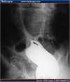
What abnormality does this show?

sigmoid volvulus
What abnormality does this “bird beak sign” show?
sigmoid volvulus
What abnormality is indicated by the arrows?

cecal volvulus
What abnormality does this show?
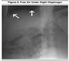
extraluminal air, free air
(beneath diaphragm)
What abnormality does this “RIGLER’S SIGN” show?
extraluminal (pneumoperitoneum) air
What abnormality does this “falciform ligament sign” or “football sign” show?

extraluminal (pneumoperitoneum) air
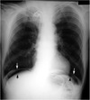
What abnormality does this show?

Pneumoperitoneum free air
Arrowheads point to free air.
Arrows points to collection of fluid around bowel loops.
Black arrows point to pericolonic fascial infiltration consistent with abscess.
What abnormality does this show?

free air in retroperitoneum
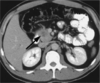
What abnormality does this show?

calcifications
on rim of Abnormal Aortic Anurism (AAA)
What abnormality does this show?

renal stone
What abnormality does this show?

appendicolith
(calcification in appendix)
What type of imaging was used?
CT computerized tomography
(view from feet-up)