Advanced Abdominal Exam COPY Flashcards

1
Q

Peritonitis

A

Inflammation of the peritoneum that results in intense abdominal pain and a board-like abdomen to palpation.

2
Q

Rebound tenderness

A

Peritoneal inflammation causes increased pain when the examiner takes away his/her hand abruptly from the abdomen compared to the initial pressing down on the abdomen

3
Q

Percussion tenderness of the liver, spleen or kidney

A

Gentle fist percussion using the heel of the hand on the ribs over the liver, spleen or kidney may elicit pain generally caused by stretching or inflammation of the capsule surrounding the organ

4
Q

Traube’s Space

A

Traube’s space is defined superiorly by the left 6th rib, inferiorly by the left costal margin, and laterally by the left anterior axillary line.

If dullness is noted upon percussion of Traube’s space, this finding is suggestive of splenomegaly and should be confirmed with palpation for an enlarged spleen.

5
Q

Hypoactive bowel sound

A

May be normal or suggest an ileus (obstruction of the intestine due to paralysis of the intestinal muscles.)

6
Q

Hyperactive bowel sounds

A

May be normal or suggest an inflammatory or infectious process in the intestines.

7
Q

High pitched, tinkling bowel sounds

A

Suggestive of small bowel obstruction

8
Q

Tympany of the abdomen

A

Indicates that there is gas in loops of bowel or in the peritoneal space itself.

9
Q

Shifting dullness on percussion of the abdomen (especially supine vs lateral decubitus)

A

Suggests fluid in the abdomen (ascites).

10
Q

Rigidity on palpation of the abdomen

A

Points toward peritoneal inflammation due to a perforation or rupture of an intraabdominal organ such as stomach, gallbladder, small bowel or colon.

11
Q

If contraction of the rectus abdominus on palpation is voluntary, it is ____. If it is involuntary, it is ____.

A

If contraction of the rectus abdominus on palpation is voluntary, it is guarding. If it is involuntary, it is rigidity.

12
Q

Midline abdominal bruit

A

Suggests abdominal aortic narrowing or stenosis

13
Q

Suprapubic abdominal bruit

A

Suggests iliac artery narrowing or stenosis

14
Q

Lateral umbilical bruit

A

Suggests renal artery narrowing or stenosis

15
Q

Costovertebral angle tenderness

A

If tenderness is elicited with palpation in the costovertebral angle, it can indicate inflammation in the kidney (pyelonephritis)
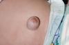
16
Q

Umbilical hernia

A

A protrusion through a defective umbilical ring is most common in infants but also occurs in adults. In infants, it usually closes spontaneously within 1–2 yrs

May be seen more clearly while the patient is standing.

17
Q

Inguinal hernia

A

Presence is noted best in the male patient standing up. The examiner’s finger invaginates the most dependent part of the scrotum to insert the finger in the canal laterally and cephalad.

Ask the patient to cough or strain. A small hernia causes an impulse felt on the fingertip. A larger hernia feels as if there is a mass in the canal.
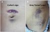
18
Q

Caput Medusa

A

Sign of portal hypertension. Indicates chronic portal vein obstruction. It is a dilatation of the paraumbilical vein that gives the appearance of a rosette around the umbilicus.

19
Q

When inferior epigastric veins are visible over the abdomen, ___ should be suspected.

A

When inferior epigastric veins are visible over the abdomen, IVC obstruction should be suspected.

20
Q

McBurney’s Point

A

Lies one-third of the distance between the between the anterior superior iliac spine and the umbilicus and is located over where the appendix comes off the base of the cecum.

The pain of acute appendicitis is commonly located at McBurney’s Point.

21
Q

Rovsing’s Sign

A

When pressing in on the left lower quadrant elicits pain in the right lower quadrant.

This is an indication of a possible appendicitis.

22
Q

Rectal tenderness

A

May indicate a thrombosed hemorrhoid, anal fissure, inflammatory bowel disease, infectious colitis, appendicitis, ischemic colitis, ulcerated rectal cancer or inflamed prostate.
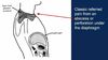
23
Q

Psoas sign

A

It is performed by having the patient lie on his/her left side and extending the right leg at the hip to stretch the psoas muscle. If significant pain results, this is a positive psoas sign.

May support the diagnosis of appendicitis, or irritation of any inflamed organ proximal to the psoas.

24
Q

Obturator sign

A

It is performed by flexing the patient’s right thigh at the hip with the knee bent and then rotating the leg internally which stretches the obturator muscle. If significant pain results, this is a positive obturator sign.

May support the diagnosis of appendicitis, or inflammation of any pelvic sturcture.

25
Q

Normal frequency of bowel sounds

A

Bowels sounds should occur a few times over the course of a minute. Listen for 15 seconds or so in each of the four quadrants. Appreciate whether they seem normal, increased or decreased.

If you think they are decreased, really spend a few minutes listening, and check how the patient is feeling. If bowel sounds are absent and the patient doesn’t feel poorly, it is probably fine. However, if there is severe pain, then suspicion of paralytic ileus is warranted.

26
Q

When a patient presents for abdominal examination with abdominal pain, you should start the exam . . .

A

. . . by looking everywhere ELSE on the abdomen.

If you start with the painful site, the patient may guard, which will interfere with the exam. So, counterintuitively, you really want to examine the affected area last.

27
Q

The integrity of the sacral nerves that innervate the bladder can be tested by assessing . . .

A

The integrity of the sacral nerves that innervate the bladder can be tested by assessing perirectal and perineal sensation in the S2, S3, and S4 dermatomes

28
Q

Presentation of peptic ulcer and dyspepsia

A

Epigastric pain, may radiate straight to the back. Variable in quality. Intermittent timing, may happen for several weeks, disappear, and reappear months later. More likely to occur at night than other GI pain.

Food and antacids may bring relief (more likely for duodenal ulcers, less likely for gatric ulcers). Often accompanied by vomiting, belching, nausea, bloating, heartburn.

May be gnawing or burning (dyspepsia); may also be boring, aching, or hungerlike. No symptoms in up to 20%.

29
Q

Dyspepsia

A

aka indigestion

Term that describes post-prandial discomfort or pain in the upper abdomen.

30
Q

Presentation of gastric cancer

A

>90% due to adenocarcinoma. Pain often in cardia and GE junction.

Pain is persistent, slowly progressive; duration of pain is typically shorter than in peptic ulcer. Not relieved by food or antacids. Associated with anorexia, nausea, early satiety, weight loss, and bleeding (could be hematemesis or melena)

31
Q

Presentation of acute appendicitis

A

Presents with poorly localized periumbilical pain, usually migrates to the right lower quadrant. Mild but increasing, possibly cramping. Steady and more severe. Can last 4-6 hours depending on intervention. Movement or cough provoke pain.

Associated with anorexia, nausea, emesis (usually following pain onset), and low fever.

If it subsides temporarily, suspect perforation of the appendix.

32
Q

Presentation of Acute cholecystitis

A

Right upper quadrant or epigastrium; may radiate to right shoulder or interscapular area. Steady, aching pain with gradual onset, course longer than in biliary colic. Aggravated by deep breathing (Murphy’s sign takes advantage of this).

Associated with anorexia, nausea, vomiting, fever; but not jaundice (it is acute!)

33
Q

Presentation of biliary colic

A

Caused by sudden obstruction of the cystic duct or common bile duct by a gallstone. Epigastric or right upper quadrant; may radiate to the right scapula and shoulder. Steady, aching; not colicky. Rapid onset over a few min, lasts one to several hrs and subsides gradually; often recurrent.

Supposedly aggrivated by fatty meals but also fasting. Often precedes cholecystitis, cholangitis, pancreatitis. Associated with anorexia, nausea, vomiting, restlessness.

34
Q

Presentation of acute pancreatitis

A

Epigastric, may radiate straight to the back or other areas of the abdomen; 20% with severe sequelae of organ failure. Pain us usually steady and aggravated by lying supine. Leaning forward with flexed trunk often lessens pain.

Associated with nausea, vomiting, hypovolemia, abdominal distention, fever. Often recurrent. History often includes alcohol use, and other cases are often gall stone-mediated.

35
Q

Presentation of chronic pancreatitis

A

Epigastric pain, radiating to the back. Severe, persistent, deep in quality with a chronic or recurrent course. Aggravated by alcohol or fatty meals.

Leaning forward with trunk flexed may alleviate some pain, but often pain is intractable. Associated with pancreatic enzyme insufficiency, steatorrhea, and type I diabetes mellitus.

36
Q

Steatorrhea

A

Diarrhea with fatty stools. Occurs when pancreatic lipases are absent or non-functional.

37
Q

Presentation of pancreatic cancer

A

Predominantly adenocarcinoma (>95%), poor prognosis. If cancer in body or tail, epigastric, in either upper quadrant, often radiates to the back.

Steady, persistent, deep pain. Smoking aggravates. Leaning forward with trunk flexed may alleviate some pain, but often intractable. Associated with painless jaundice, anorexia, weight loss, fever, glucose intolerance, depression.

38
Q

Presentation of Acute diverticulitis

A

Left lower quadrant pain. May be cramping at first, then steady. Often a gradual onset.

Pain responds to analgesia and antibiotics, and is lessened with bowel rest. Associated with fever, constipation, nausea, vomiting, and abdominal mass with rebound tenderness.

39
Q

Presentation of Acute Bowel Obstruction

A

May be caused by adhesion, hernia, diverticulitis, or cancer. If in the small bowel, periumbilical or upper abdominal. If in the colon, lower abdominal or generalized. Cramping pain. Onset is paroxysmal.

Aggravated by ingestion of food or liquid. Assocaited with vomiting of bile and mucous if obstruction is high, and with fecal vomiting if obstruction is low. Obstipation may develop.

40
Q

Obstipation

A

Severe form of constipation, where a person cannot pass stool or gas. Prolonged obstipation puts the patient at risk for bowel perforation.

41
Q

Presentation of mesenteric ischemia

A

Abrupt, crampy, periumbilical pain that progresses to diffuse, persistent, steady pain disproportionate to exam findings. May be postprandial, classically inducing “food fear.”

Associated with vomitting, bloody stool, and peritonitis signs. May lead to shock. Often associated with coronary artery disease.

42
Q

Types of visceral pain

A
43
Q

Colicky pain

A

Experiencing or denoting severe pain in the abdomen (colic) due to gas or intestinal obstruction.

44
Q

Presentation of an umbilical hernia

A

A protrusion through a defective umbilical ring is most common in infants but also occurs in adults. In infants, it usually closes spontaneously within 1–2 yrs.

45
Q

Presentation of an incisional hernia

A

This is a protrusion through an operative scar. Palpate to detect the length and width of the defect in the abdominal wall.

46
Q

When it comes to hernia, how should you think about size?

A

A small defect, through which a large hernia has passed, has a greater risk for complications than a large defect.

It’s all about the risk of the defect cutting off the circulation of the herniated tissue.

47
Q

Presentation of an epigastric hernia

A

A small defect, through which a large hernia has passed, has a greater risk for complications than a large defect.

48
Q

Presentation of Diastasis Recti

A

Separation of the two rectus abdominis muscles, through which abdominal contents form a midline ridge typically extending from the xiphoid to the umbilicus and seen only when the patient raises the head and shoulders. Often present in patients with repeated pregnancies, obesity, and chronic lung disease. It is clinically benign.

49
Q

Summary of all hernia sites

A
50
Q

Presentation of a Lipoma

A

Common, benign, fatty tumors usually in the subcutaneous tissues almost anywhere in the body, including the abdominal wall. Small or large, they are usually soft and often lobulated. Press your finger down on the edge of a lipoma. The tumor typically slips out from under your finger and is well demarcated, nonreducible, and usually nontender.

Lipomas may occur anywhere on the body, and are often localized to the sub-dermal fat. In the abdomen, another prominent source is the greater omentum.

51
Q

How to assess the anatomic level (superficial vs deep) of an abdominal mass

A

Ask the patient to tighten their rectus abdominus.

If the mass is within the rectus abdominus or above it, it will become more prominent and more easily felt. If the mass is below the rectus abdominus, it will become difficult to appreciate.

52
Q

Friction rub on RUQ auscultation

A

Possible liver capsule inflammation, especially if associated with RUQ pain. Beware that a low pleural rub may mimic this sound, and so you should also listen to the right lung carefully to rule this out.

53
Q

Assessing for focal tenderness of the liver capsule

A

Fist-on-hand technique. This projects the force deeper into the abdomen to reach the liver, and can reach the deeper portions of the liver capsule.

54
Q

Assessing for rebound tenderness

A

Press on a different site of the abdomen from the location of the pain. Release quickly. If this generates referred pain in the other location, rebound tenderness is present, indicating peritonitis.

Be careful how you prompt the patient. Ask if when you release your hand, the pain feels better. This prompts in the other direction and increases your specificity.

55
Q

Three levels of abdominal pain

A
  • Abdominal wall pain
  • Intraabdominal pain
  • Retroperitoneal pain
56
Q

Carnet’s sign

A

Examine first with abdominal muscles relaxed and then tensed – ask patient which is worse

Contracting abdominal muscles guards against pressure transmission to
intra-abdominal organs
, but itdoes not reduce tenderness located in the abdominal wall

57
Q

Assessing the three levels of the abdomen for pain

A
  1. Abdominal wall: Have patient contract chest muscles. If still tender on palpation, lesion is within or above the abdominal wall.
  2. Intraabdominal: If pain is induced only while abdominal muscles are relaxed, pain is intra-abdominal
  3. Retroperitoneal: If both of the above fail to induce pain, but percussion of the costovertebral angle produces pain, then the pain is retroperitoneal. The Psoas stretch test and Obturator stretch test may also be used to assess retroperitoneal pain.
58
Q

Succussion splash

A

Detects fluid in stomach. This may be abnormal if it’s more than 2 hours after eating, indicating an outflow obstruction or gastric motility disorder.

Shake the patient and listen for splashing of fluid in the stomach with your headphone over the costovertebral area/epigastric region.

59
Q

What is this umbilical finding?

A

Sister Mary Joseph nodule

Metastatic cancer to the umbilicus

60
Q

Things to check all hernias for

A
  1. Contents (bowel? fat?)
  2. Redubilility (Can you push it back in?)
  3. Opening size (smaller opening is more dangerous)
61
Q

Courvoisier’s sign

A

An enlarged, palpable gallbladder in patients with obstructive jaundice caused by tumors of the biliary tree or by pancreatic head tumors

62
Q

Signs of hepatocellular disease

A
  • Hepatomegaly
  • Coagulopathy
  • Encephalopathy (mental status change, asterixis)
  • Portal hypertension
63
Q

Fluid wave test (for ascites)

A

Have another participant place their hand on the patient’s mid-abdomen as the patient is lying supine. Take one of your hands and tap on one side of the abdomen, with your other hand pressed against the opposite side. If you can feel a pressure wave produced by your tap, the fluid wave test is positive, suggesting free fluid in the abdomen.

64
Q

Shifting dullness in ascites

A

Tympanic boundary shifts when moving from supine to lateral decubitus position.

65
Q

Signs of hemorrhagic pancreatitis

A

Retroperitoneal blood seeping through abdominal wall

66
Q

Dermoid cyst

A

A dermoid cyst is a saclike growth that is present at birth. It contains structures such as hair, fluid, teeth, or skin glands that can be found on or in the skin

67
Q

What quadrant are ileostomy bags usually placed in?

A

Right lower quadrant

68
Q

Classic referred pain locations

A

Memorize these! They will be on boards!

69
Q

Phrenic and just infra-phrenic pain

A
70
Q

Why is it so difficult to localize visceral pain?

A
  1. Unmyelinated C fibers that carry these signals are slow/poor conductors
  2. There is a relative paucity of nerve endings in comparison to the somatic and parietal systems
  3. Bilateral Symmetrical Innervation: Embryology of Foregut, Midgut and Hindgut
  4. No tidy ordering of nerves as in somatic afferents. One nerve carries fibers from several different organs
  5. Cranial and caudal extension of nerves once in spinal cord
71
Q

Kehr’s sign

A
72
Q

A 55 year-old man sustains an abdominal laceration after a motor vehicle accident, requiring multiple stitches. One week later, he is seen in clinic. His incision site under the umbilicus is erythematous, and palpation is notable for the following crepitus (aubile without a stethoscope). What is the likely diagnosis?

A

Infection with a gas-forming organism

73
Q

Colicky pain definition

A

Starts and stops abruptly

74
Q

Gnawing pain definition

A

Steady, hunger-like pain

75
Q

Fat stranding on CT

A

Stranding seen in fat on CT. Indicates inflammation

76
Q

Bezoar

A

Collection of vegetable matter (phytobezoar) or hair (philobezoar) that gets stuck in the antrum and causes GI tract obstruction.