Anatomy Flashcards
(1225 cards)
What is the origin and insertion of the extrinsic shoulder muscles
originate from the torso, and attach to the bones of the shoulder (clavicle, scapula or humerus).
What is the origin and insertion of the intrinsic shoulder muscles?
They originate from the scapula and/or clavicle, and attach to the humerus.
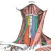
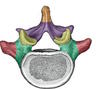
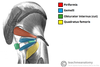
What is the origin and insertion of the trapezius muscle?
Originates from the skull, spinous processes of C7-T12 and the ligamentum nuchae. The fibres attach to the clavicle, acromion and the scapula spine.
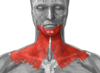
What is the innervation of the trapezius muscle?
Motor innervation is from the accessory nerve. It also receives proprioceptor fibres from C3 and C4 spinal nerves.
What does the trapzius muscle do?
The upper fibres of the trapezius elevates the scapula and rotates it during abduction of the arm. The middle fibres retract the scapula and the lower fibres pull the scapula inferiorly.
How do you test the accessory nerve?
To test the accessory nerve, trapezius function can be assessed. This can be done by asking the patient to shrug his/her shoulders.
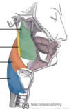
What is the origin and insertion of the latissimus dorsi?
Has a broad origin – arising from the spinous processes of T6-T12, iliac crest, thoracolumbar fascia and the inferior three ribs. The fibres converge into a tendon that attaches to the intertubecular sulcus of the humerus.
What is the innervation of the latissimus dorsi?
Thoracodorsal nerve
What is the function of latissimus dorsi?
Extends, adducts and medially rotates the upper limb.
What is the origin and insertion of the levator scapullae?
Originates from the transverse processes of the C1-C4 vertebrae and attaches to the medial border of the scapula.
What innervates the levator scapulae?
Dorsal scapular nerve
What is the function of the levator scapulae
Elevates the scapula
What is the origin and insertion of the rhomboid major?
Originates from the spinous processes of T2-T5 vertebrae. Attaches to the medial border of the scapula, between the scapula spine and inferior angle.
What innervates the rhomboid major?
Dorsal scapula nerve
What does the rhomboid major do?
Retracts and rotates the scapula.
What is the origin and insertion of the rhomboid minor?
Originates from the spinous processes of C7-T1 vertebrae. Attaches to the medial border of the scapula, at the level of the spine of scapula
What is the innervation of the rhomboid minor?
Dorsal scapula nerve
What is the function of the rhomboid minor?
Retracts and rotates the scapula
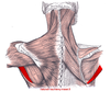
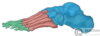
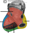
What is this muscle?

What is this muscle?

Latissimus Dorsi
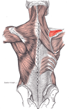
What is this muscle

The levator scapulae
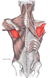
What is this muscle?

Rhomboid major
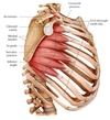
What is this muscle

The Rhomboid minor
What is the origin and insertion of the deltoid?
Originates from the scapula and clavicle, and attaches to the deltoid tuberosity on the lateral surface of the humerus