anatomy of GI tract Flashcards

(58 cards)

1
Q

structures of GI tract

A

oesophagus, stomach, small intestine (duodenum, jejunum, ileum), large intestine (cecum, ascending, transverse, descending, sigmoid), rectum, anal canal

2
Q

primary functions of GI system

A

motility, secretion, digestion and absorption

3
Q

accessory organs

A

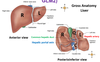
salivary glands, pancreas, liver, gallbladder, appendix

4
Q

what is peritoneum

A

Serous membrane made of simple squamous epithelium with underlying thin layer of connective tissue

5
Q

types of peritoneum

A
  1. Parietal peritoneum - lines abdominal and pelvic cavities
  2. Visceral peritoneum - covers external surfaces of most abdominal organs, including intestinal tract
  3. Mesentery - double layer of peritoneal membrane from the body wall to the organ - gives passage to blood vessels, nerves and lymphatics
  4. Omentum - double layer of peritoneal membrane from organ to organ
6
Q

function of peritoneum

A
  • Form covering (partial or complete) for abdominal organs
  • Smooth lining - frictional surface
  • Hold organs in position
  • Omentum and mesentery serve as fat store
  • Fats of peritoneum prevents infections of abdominal organs
7
Q

3 main subdivisions of abdominal GI blood supply

A
  • Celiac trunk supplies foregut with blood
  • Superior mesenteric supplies midgut with blood
  • Interior mesenteric supplies hindgut with blood
8
Q

divisions of celiac trunk

A

common hepatic, left gastric and splenic arteries

9
Q

divisions of superior mesenteric artery

A

intestinal arteries, ileocolic artery, colic artery

10
Q

divisions of inferior mesenteric artery

A

left colic artery, sigmoid arteries, superior rectal artery

11
Q

why is there large blood supply to small intestine

A

maintain concentration gradient for absorption of nutrients from GI tract

12
Q

4 layers of gut tube

A
  1. mucosa
    • has gland ducts
  2. submucosa
    • connective tissue
    • glands
    • nerves called meissners plexus
  3. muscularis
    • smooth muscle
    • myenteric plexus
    • inside = circular
    • outside = longitudinal
  4. adventitia
    • FCT
13
Q

3 layers of mucosa

A
  1. epithelium - mucus secreting
  2. lamina propria (loose FCT)
    • lymph nodes
    • nerve fibres
    • blood vessels
  3. muscularis mucosa (thin layer of smooth muscle)
14
Q

control of digestion

A
  • enteric NS (primary control - independent, short, local reflexes)
  • CNS (modulates activity of ENS - long neural reflexes)
  • hormones
  • receptors
15
Q

types of epithelium in GI tract

A
  • Simple squamous in peritoneum
  • Simple cuboidal lining ducts
  • Simple columnar lining stomach to rectum - this is modified to carry out specific functions
  • Stratified squamous lining oesophagus and anal canal (hardwearing protects against abrasion)
  • Glandular epithelium secrete mucus in small intestine
16
Q

how are epithelial cells in GI tract joined

A

joined by tight junctions, zonula adherens and spot desmosomes to form a continuous and relatively impermeable membrane

17
Q

function of mouth in GI tract

A

mechanical digestion via mastication (chewing)

18
Q

3 pairs of salivary glands, where they are and what they secrete

A
  1. sublingual - under tongue, mainly mucous, 3-5% of total saliva
  2. parotid - anterior and inferior to ear, serous fluid, 25-30% of total saliva
  3. submandibular - floor of mouth, both mucous and serous fluid, 60-70% of totoal saliva
19
Q

how are salivary glands controlled

A

parasympathetic NS

20
Q

function of saliva

A
  • moisten ingested material
  • moistens, cleanses and lubricates structures of oral cavity
  • begins chemical digestion of carbohydrates with amylase
  • antibacterial action with lysozyme
  • dissolves food to stimulate taste receptors
21
Q

structure of oesophagus

A
  • appears collapsed when there is no food going down
  • mucosal epithelium - protective stratified squamous
  • muscularis externa - move food bolus, transitions between skeletal and smooth
22
Q

structure of oropharynx

A
  • posterior to oral cavity
  • contains palatine and lingual tonsils
  • stratified, squamous epithelium for protection
23
Q

regions of stomach

A
  • fundus is round bit at top
  • cardia is where oesophagus joins
  • body is main region
  • pylorus is where it leads into duodenum
  • greater omentum attached to greater curvature
  • lesser omentum attached to lesser curvature
24
Q

greater omentum structure and function

A
  • large apron-like fold of visceral peritoneum that hangs down from the stomach over the small intestines and doubles back up to the transverse colon
  • functions are fat deposition, immune contribution, infection isolation
25
structural features of stomach
* pyloric sphincter separates stomach and duodenum - controlled release * lower oesophageal sphincter separates stomach from oesophagus - prevents gastric juices flowing up * 3 layers of smooth muscle: oblique (inside), circular and longitudinal - mechanical digestion * rugae are folds in inside of stomach - allow expansion
26
mucosal layer of stomach
* infolding of epithelium forms pits and glands * glands contain mucous neck cells, chief cells, parietal cells and endocrine cells
27
functions of stomach
1. storage of food - rugae 2. mechanical digestion - 3 layers of muscularis externa 3. chemical digestion - breakdown of proteins 4. some absorption (fat soluble substances e.g. alcohol)
28
function of liver
accessory organ that produces bile salts, which emulsify lipids, aiding their digestion and absorption
29
chief cell function and structure
* secretes enzymes – pepsinogen converted to pepsin * Has many rough ER * Tight junctions between them to prevent acid moving between them * granules near apical surface
30
parietal cell function and structure
* Pumps H+ * Lots of mitochondria * Large SA * Folded with microvilli
31
endocrine cells function
* Secrete hormones into blood stream * Gastrin - acts on parietal cells to increase HCl release * Ghrelin - stimulates appetite * nervous control - receptors
32
mucous neck cell function
secrete mucous for protection from acid
33
features of enteric nervous system in stomach
Myenteric plexus (Auerbach) - neuronal cell bodies and nerve fibres between Oblique and Circular muscle layers
34
35
anatomy of small intestine
1. pyloric sphincter 2. duodenum 3. duodenojejunal flexure 4. jejunum 5. ileum 6. ileocecal junction
36
how is small intestine epithelium protected from stomach acid
* mucous secreted by epithelium and alkaline mucous secreting glands in submucosa * bicarbonate from pancreas neutralises pH
37
modification of SI required for digestion and absorption
* Secretion of enzymes for digestion * Large surface area for absorption * Protection from acidic chyme * Mixing and movement along tube
38
how is surface area increased in small intestine
* Plicae circulares - folds in inner surface of SI (submucosa and underlying mucosa) * Villi cover surface of each plicae circulares * Microvilli cover villi to increase SA by around 600x
39
structure of villi
* core of lamina propria (FCT) * lacteals which collect fat * capillary network with rich blood supply to absorb products of digestion * covered with enterocytes (columnar) and goblet cells to secrete mucous for protection
40
what is bottom of space between villi called
intestinal crypts (crypts of lieberkuhn)
41
what does glycocalyx on microvilli do
hold brush border enzymes for contact digestion
42
what effect does plasma membrane of enterocytes have on absorption of nutrients
* acts as a barrier with channel and transporter proteins which are selectively permeable to nutrients. * tight junctions can allow small molecules to diffuse through
43
process of segmentation
* occurs mainly in the small intestine and consists of localised contractions of circular muscle of muscularis layer * These contractions isolate small sections of the intestine, moving their contents back and forth while continuously subdividing, and mixing the contents
44
function of segmentation
* segmentation mixes food with digestive juices and facilitates absorption * slows progression of chyme through the system allowing time for digestion and absorption
45
function of gallbladder
accessory organ that stores, concentrates, and releases bile in response to hormonal signals
46
function of pancreas
accessory organs that produces digestive enzymes and bicarbonate to help neutralize acidic chyme. Sphincter of Oddi controls release of secretions. * endocrine pancreas is 5% of cells and produces hormones that regulate blood sugar and pancreatic secretions * exocrine pancreas is 95% of cells and produces and releases digestive enzymes and bicarbonate
47
function of large intestine
* Absorbs water and ions and compacts undigestible wastes and solidifies them into faeces * Stores faeces until defecation
48
structure of large intestine
* surrounds 3 sides of small intestine. * order is cecum, ascending colon, hepatic flexure, transverse colon, splenic flexure, descending colon, sigmoid colon, rectum and anal canal * wider in diameter but shorter than small intestine * covered by greater omentum
49
how is colon attached to back wall of abdomen
* Transverse mesocolon anchors transverse colon to the back wall * Ascending and descending colon are retroperitoneal - against the back abdominal wall
50
which abdominal viscera are retroperitoneal
S - suprarenal glands A - aorta/IVC D - duodenum except proximal 2cm P - pancreas except tail U - ureters C - colon (ascending and descending) K - kidneys E - Esophagus R - rectum
51
what does large intestine have that small intestine doesnt have
* taeniae coli - 3 longitudinal bands of outer smooth muscle * haustra - contraction of taeniae coli to bunch large intestine up into many sacs * epiploicae appendices - fat storage * semilina folds between haustra * appendix
52
wall of large intestine
* has 4 basic layers (mucosa, submucosa, muscularis and adventitia) * Simple columnar epithelium with abundant goblet cells * lacks plicae circulares and villi of small intestine * inner muscularis is circular and outer muscularis forms 3 longitudinal taeniae coli bands
53
anal sphincters
* external anal sphincter is skeletal muscle which allows for voluntary control - relax when empty, contract when full but you dont want to defecate * internal anal sphincter is smooth muscle - involuntary control, contract when rectum is empty, relax when full
54
structure of liver
split into right lobe, left lobe, caudate lobe (inferior) and quadrate lobe (superior)
55
liver blood supply
Portal system carries venous blood (rich in nutrients that have been extracted from food) to the liver for processing
56
processing of blood in liver
The hepatic artery and portal vein divide into interlobular arteries and veins to supply each of the lobes of the liver. The arterial and venous blood from these smaller vessels mix in hepatic sinusoids and are oxygen and nutrient rich. When the blood reaches the hepatic vein, it is nutrient poor and deoxygenated
57
blood supply to liver and from liver
to: * hepatic portal vein from intestines * hepatic artery which supplies O2 from: * hepatic vein to IVC
58
cellular structure of liver
Rows of hepatocytes Sinusoids between rows Bile canaliculi between cells Central vein