Anatomy- wrist Flashcards
(27 cards)
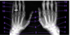
ID these bones


ID these bones


ID these bones


ID these bones


ID these bones


ID these bones


ID this structure


ID this radiograph


ID this radiograph


what is the significance of human HAR2 (HACNS1)
enhancer that may play a role human bipedalism and tool making. expressed in human hands and feet but not primates
what are the 3 classic injuries to the radius and the ulna
- Monteggia fracture is a fracture of the proximal ulna and an anterior dislocation of the head of the radius at the elbow
- Galeazzifracture is a fracture of distal radius associated with subluxation (partial dislocation) of the head of the ulna (distal ulna) at the wrist joint
- Colles’ fracture is a fracture, and posterior displacement, of the distal end of the radius
ID this fracture

galeazzi fracture
fracture of distal radius associated with subluxation (partial dislocation) of the head of the ulna (distal ulna) at the wrist joint
ID this fracture

Monteggia fracture
is a fracture of the proximal ulna and an anterior dislocation of the head of the radius at the elbow
ID this fracture

colles fracture
posterior displacement, of the distal end of the radius
protocol for imaging a wrist injury
image in multiple positions, AP, PA and orthogenal (oblique and lateral, which is 90 degrees)
CTs may also be used to visualize complex fractures, especially around joints
ID this structure

What are the bones of the hand
how many?
there are 27 bones in the hand
8 carpel
5 metacarpel
14 phalanges (proximal, distal and medial for the 4 fingers and proximal and distal for the thumb)
What are the carpal bones
proximal
(PTLS, place top lover straight)
Pisiform
Triquetrum
Lunate
Scaphoid
distal
(HCTT, handle caringly tom tingle)
Hamate
Capitate
Trapezoid
Trapezium
ID these carpal bones

proximal
Pisiform
Triquetrum
Lunate
Scaphoid
distal
Hamate
Capitate
Trapezoid
Trapezium

which bone is the most commonly fractured and often involes avascular necrosis
how long does it show up on imaging
what is the Tx
scaphoid
10-14 days to show up on imaging
•Often need to re-image
thumb spica cast
which bone is commonly fractured when striking a hard surface with a stick (or golf club) or catching baseballs
hamate
What are the joints of the hand
distal and proximal interphalangeal
metacarpophalangeal (MCP) (knuckles)
Carpometacarpaljoints (CMC)
carpal (intercarpal and midcarpal and radiaocarpal)
- intercarpal are joints between adjacent carpal bones in a single row (radial and ullnar flexion abduction and adduction i think)
- midcarpal are joints between the proximal and distal rows (flexion of the hand)

distinguish metacarpophalangeal (MCP) and carpometacarpal joints (CMC)
Carpometacarpal joints(CMC)
plane type synovial joints, except for the carpometacarpal joint of thumb which is a saddle type
allow: flexion, extension, abduction, adduction, circumduction, and opposition
•Not all CMC joints permit all of these movements. Some are restricted due to the shape of the articulating carpal bone.
A metacarpophalangeal joints (MCP or MP) or “knuckle” is found where the head of a metacarpal articulates with the base of a proximal phalanx.
•Collateral ligaments tighten during flexion. Abduction, and adduction are only possible in the extended position.(Try it!)
•The metacarpophalangeal joints are biaxial condylar joints (ellipsoid joints) that allow abduction, adduction, flexion, extension, and circumduction
what phenomenon underlies the ability to adduct further than the ability to abduct the wrist
radial styloid process extends further distally than does the ulnar styloid process,

