Anterior Neck and Thorax Flashcards
(74 cards)
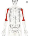
1
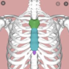
Q
Humerus
A
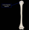
2
Q
Crest of greater tubercle
A
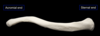
3
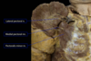
Q
Clavicle
A
4
Q
Scapula
A

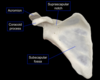
5
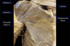
Q
scapula, incl. coracoid process
A
6
Q
Hyoid
A

7
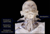
Q
hyoid, detailed
A

8
Q
sternum
A
9
Q
sternum details
A

10
Q
cranium, incl. details
A

11
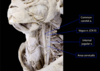
Q
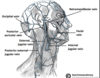
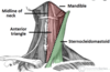
temporal bone
A

12
Q
mastoid process
A

13
Q
pectoralis major
A

14
Q
deltopectoral triangle
A
15
Q
pectoralis minor
A

16
Q
pectoralis minor and serratus anterior
A

17
Q
Sternocleidomastoid
A

18
Q
infrahyoid strap
A
missing sternothyroid

19
Q
sternocleidomastoid 2
A

20
Q
sternocleidomastoid 3
A

21
Q
omohyoid and sternohyoid
A
22
Q
infrahyoid strap 2
A

23
Q
thyrohyoid and sternothyroid
A

24
Q
vagus nerve
A
25
medial pectoral nerve and lateral pectoral nerve
26
ansa cervicalis nerve
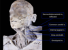
27
lateral pectoral nerve 2

28
accessory nerve

29
brachial plexus

30
cervical plexus

31
thoraco-acromial artery

32
common carotid artery

33
common carotid artery 2

34
carotid artery details

35
inferior thyroid artery
???
36
cephalic vein
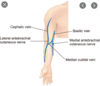
37
internal jugular vein (superior thyroid vein and middle thyroid vein)

38
inferior thyroid vein
39
external jugular vein
40
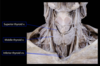
thyroid gland
41
jugular veins
42
thyroid

43
parathyroid gland
???
44
anterior cervical triangle
45
proximal attachments of the pectoralis major
* Clavicular (clavicle)
* Sternocostal (sternum and costal cartilages)
46
distal attachments of pectoralis major
* Shared tendon on crest of greater tubercle
* Crosses the glenohumeral (shoulder) joint
47
actions of pectoralis major
* Both heads contracting simultaneously:
* Adduction & medial rotation of glenohumeral joint
* Clavicular head independently:
* Flexion of glenohumeral joint
* Sternocostal head contracting independently
* Extension of glenohumeral joint (from flexed position)
48
innervation of pectoralis major
Two nerves derived from the brachial plexus
* Lateral pectoral n.
* Medial pectoral n.
49
dominant arterial supply of pectoralis major
Thoraco-acromial a. (branch of axillary a.) branches
50
anatomical relationships of pectoralis major
* Forms anterior wall of axilla
* Forms inferior border of deltopectoral triangle/groove
* Invested in pectoral fascia
* Breast is located superficial to pectoral fascia
51
clinical considerations of pectoralis major
* Clavicular fractures
* Can pull clavicular fractures with contraction, which may slow healing & damage deep structures
* Important to immobilize area
* Breast pathology & surgery
* Due to the close anatomical relationship, pectoral fascia and muscle can be affected
52
proximal attachment of pectoralis minor
anterior ribs 3-5
53
distal attachment of pectoralis minor
coracoid process of scapula
54
actions of pectoralis minor
* Stabilization of scapula to allow for efficient movements at other joints, specifically the glenohumeral joint
* Protraction (moving the scapula anteriorly)
55
innervation of pectoralis minor
medial pectoral n.
56
dominant arterial supply of pectoralis minor
thoraco-acromial a. (branch of axillary a.) branches
57
anatomical relationships of pectoralis minor
* Medial pectoral n. pierces the pectoralis minor m.
* Important anatomical landmark locating branches of the axillary a. both in the lab, in imaging, and in surgeries
* Subdivides the artery into 3 parts based on relationship to the muscle (medial, deep, or lateral to)
58
clinical considerations of pectoralis minor
Breast pathology & surgery
## Footnote
Due to the close anatomical relationship, pectoral fascia and muscle can be affected or resected
59
proximal attachment of serratus anterior muscle
Ribs 1-8
60
distal attachment of serratus anterior muscle
anterior surface of scapula
61
actions of serratus anterior muscle
* Protraction of scapula
* Upward rotation of glenoid fossa
* Important for allowing full range of motion for the glenohumeral (shoulder) joint, particularly in abduction and flexion
* Stabilization of scapula
62
innervation of serratus anterior muscle
long thoracic n.
## Footnote
The placement of this nerve is unique in that it is superficial to the muscle
63
anatomical relationships of serratus anterior muscle
Forms the medial wall of the axilla
64
clinical considerations of serratus anterior muscle
* With injury to the long thoracic n., the scapula may develop a wing-like appearance (‘winged scapula’)
* The medial border of the scapula is displaced posteriorly and laterally in comparison to the uninjured side. This is particularly evident when the shoulder joint is flexed (as in pushing against a wall with the upper limb).
* This affects scapular motion, but (more importantly) prevents full range of motion at the glenohumeral joint
65
superior attachments of sternocleidomastoid muscle
* Mastoid process of temporal bone
* Superior nuchal line of occipital bone
66
inferior attachments of sternocleidomastoid muscle
Manubrium of sternum
Medial clavicle
67
actions of the sternocleidomastoid muscle
* Bilateral contractions: flexes cervical vertebrae
* Unilateral contraction: lateral flexes the neck & rotates the face in the opposite direction
68
innervation of the sternocleidomastoid muscle
* Efferent: Accessory n. (CN XI)
* Afferent: C2 & C3 fibers
* It is uncommon that separate nerves provide afferent and efferent innervation to a muscle.
69
anatomical relationships of sternocleidomastoid muscle
* Forms the lateral boundary of the anterior cervical triangle and anterior boundary of posterior cervical triangle
* Prominently visible and palpable landmark in the neck
* External jugular v. typically sits anterior to this muscle
70
infrahyoid muscle actions
Stabilization or depression of hyoid
Dependent on muscles, will pull larynx either superiorly, or inferiorly
71
innervation of infrahyoid muscles
* Ansa cervicalis (C1,2,3) for 3 of the 4 muscles
* Thyrohyoid m. is innervated by fibers of C1 traveling with hypoglossal n. (CN XII)
72
anatomical relationships of infrahyoid muscles
Arranged in two layers
Superficial: sternohyoid & omohyoid
Deep: sternothyroid & thyrohyoid
To access the thyroid gland, these muscles have to be reflected or moved
73
arterial supply of thyroid gland
* Superior thyroid a.
* Typically the first branch of the external carotid a.
* Inferior thyroid a.
* Branch of thyrocervical trunk of the 1st part of the subclavian a.
* Typically crosses the recurrent laryngeal n. deep to the thyroid gland in the vicinity of the larynx
* Clinical consideration: when ligating this artery during a thyroidectomy, care must be taken to not damage the recurrent laryngeal n., which innervates most intrinsic laryngeal muscles and inferior larynx.
74
venous draining of the thyroid gland
* Superior thyroid v.: typically drain into the internal jugular v.
* Middle thyroid v.: typically drain into the internal jugular v.
* Inferior thyroid v.: typically drain into the L. brachiocephalic v.
