Arm and Elbow Flashcards
Segments of the spinal cord innervating specific muscular groups:
C5
C6
C7
C8 - T1
L2
L 3,4
S1
L 5
S 1,2
C 5 – Upper extremity abduction
C 6 – Flexion of arm and forearm
C 7 – Extension of forearm
C 8,T1 – Intrinsic hand muscles
L 2 – Flex thigh
L 3,4 – Extend leg
S 1 – Flex leg
L 5 – Dorsiflexes foot
S 1,2 – Plantar flexes foot

The intertubercular sulcus lies between the greater and lesser tubercle. The long head of the biceps muscle lies in the groove.
The capitulum articulates with the head of the radius, while the trochlea articulates with the trochlear notch of the ulna.
With flexion of the forearm, the coronoid process of the ulna moves into the coronoid fossa, while the head of the radius occupies the radial fossa.
The medial epicondyle serves as a common site for the origin of the flexor muscles of the forearm and hand, while the extensor muscles of the hand originate on the lateral side.


The radial groove is located on the posterior surface of the humerus, and the radial nerve and the deep (profunda) brachial vessels accompany the radial nerve.
The olecranon fossa is occupied by the olecranon process of the ulna on extension of the forearm.


The ulnar nerve as it is derived from the medial cord and continues through the arm region, passing posterior to the medial epicondyle.


** Posterior cord supplies primarily extensors of the arm


Cutting the lateral head diagonally following the path of the radial groove reveals the deep brachial artery (first large branch of the brachial artery) accompanying the radial nerve.
- The medial head of the triceps is located medial to the radial groove.
- The long head of the triceps takes origin from the infraglenoid tubercle.
- The lateral head takes origin from the posterior surface of the humerus lateral to the radial groove.


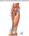
The biceps long head takes origin from the supraglenoid tubercle, while the short head takes origin from the coracoid process of the scapula.
The two muscle bellies join and form a tendon that inserts on the radial tuberosity. Also, the biceps muscle forms a dense band of connective tissue called the bicipital aponeurosis.
The muscle is innervated by the musculocutaneous nerve.
The coracobrachialis muscle attaches superiorly to the coracoid process and inferiorly on the humerus at its midpoint on the medial side.
The musculocutaneous nerve innervates the muscle and usually passes through the muscle before coming to lie between the biceps and brachialis muscles that it also innervates.


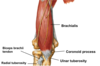
The brachialis muscle takes origin from the anterior lower one-half of the humerus and attaches distally to the coronoid process and tuberosity of the ulna.
Like the other muscles in the anterior or flexor compartment, it is innervated by the musculocutaneous nerve.
Note that the musculocutaneous nerve passes distally lying on the brachialis muscle and covered by the biceps brachii.
The musculocutaneous nerve exits from the lateral border of the biceps and continues into the forearm as the lateral cutaneous nerve of the forearm (lateral antebrachial cutaneous nerve).

What are the cubital fossa borders?
The cubital fossa is a triangular space defined by an imaginary line that extends between the medial and lateral epicondyles.
The lateral border of the fossa is the brachioradialis muscle and the medial border is the pronator teres muscle. T
he bicipital aponeurosis together with other deep fascia forms the roof of the fossa, while the floor is formed by the brachialis and supinator muscles.


The brachial artery begins at the lateral border of the teres major muscle and is a continuation of the axillary artery.
There are many small muscular branches supplying the arm, but three major branches are usually recognized.
- The first branch is the deep brachial artery. It passes posteriorly and accompanies the radial nerve in the radial groove.
- The second branch is the superior ulnar collateral artery. It accompanies the ulnar nerve and passes behind the medial epicondyle.
- The third and smallest branch is the inferior ulnar collateral artery. It supplies the area of the elbow joint and anastomoses with recurrent branches of the ulnar and radial arteries.
The brachial artery accompanies the median nerve in the arm.


The brachial artery brachnes:
- The first branch is the deep brachial artery. It passes posteriorly and accompanies the radial nerve in the radial groove.
- The second branch is the superior ulnar collateral artery. It accompanies the ulnar nerve and passes behind the medial epicondyle.
- The third and smallest branch is the inferior ulnar collateral artery. It supplies the area of the elbow joint and anastomoses with recurrent branches of the ulnar and radial arteries.


The deep brachial artery accompanies the radial nerve in the radial groove.


The bicipital aponeurosis is an expansion and thickening of the brachial fascia extending into the medial side of the proximal forearm. Contraction of the biceps muscle extends its force of contraction into the antebrachial (forearm) fascia.


In the area of the cubital fossa the brachial artery divides into the radial and ulnar arteries. They can branch before the forearm begins.

After the radial nerve has pierced the lateral intermuscular septum, it lies beneath the brachioradialis muscle. It then divides into a superficial branch that is entirely sensory and lies beneath the brachioradialis muscle to pass distally to reach the dorsum of the hand, and a deep branch that pierces the supinator muscle to reach the posterior forearm compartment containing the extensor muscles.


The radial tuberosity serves as the attachment for the biceps brachii tendon, while the coronoid process of the ulna and the ulnar tuberosity provide an attachment site for the tendon of the brachialis muscle.

The glenohumeral joint has lax glenohumeral (capsular) ligaments that permit extensive movement of the joint.
The joint is reinforced anteriorly by the tendon of the subscapularis, superiorly by the supraspinatus tendon, and posteriorly by the tendons of the infraspinatus and teres minor muscles.
No muscle tendons are found inferiorly, and the head of the humerus usually dislocates inferiorly, and then is usually pulled anteriorly.


The subacromial or subdeltoid bursa is located below the deltoid muscle, and the acromion and coracoacromial ligament.


The subacromial or subdeltoid bursa is located below the deltoid muscle, and the acromion and coracoacromial ligament.

