Axillary Region Flashcards
(21 cards)
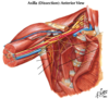
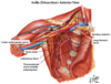
Axillary Sheath
Continuation of the fascia associated with the anterior and middle scalene muscle that continues laterally as a tubular sheath surrounding the nerves of the brachial plexus and the axillary artery.
The axillary vein is not surrounded by the sheath.

Apex - directed upward and medialward, ending in the cervicoaxillary canal that leads to the posterior triangle of the neck
Base - formed by the axillary fascia and skin (axilla)
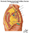
Walls (4)
Anterior - pectoralis major and minor, clavipectoral fascia
Posterior - subscapularis, teres major, latissimus dorsi
Medial wall - first 4 ribs and intercostal muscles, upper part of serratus anterior
Lateral wall - humerus, coracobrachialis, biceps brachii
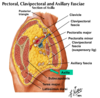

Apex - directed upward and medialward, ending in the cervicoaxillary canal that leads to the posterior triangle of the neck

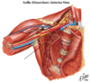


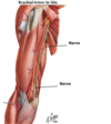
The axillary artery begins at the lateral border of the first rib. The 1st part of the axillary artery has one artery, the superior thoracic artery. The second part has two branches, the thoracoacromial trunk and the lateral thoracic. The lateral thoracic is significant because it supplies the lateral side of the breast. The third part has 3 arteries. The subscapular is the largest. It descends and divides into the circumflex scapular artery and the thoracodorsal artery. The thoracodorsal artery accompanies the thoracodorsal nerve to the latissimus muscle, while the circumflex scapular enters the infraspinous fossa and forms an anastomosis with the suprascapular artery (generally are larger – branching to the circumflex scapular artery (which forms anastamoses with superior scapular artery) and the thoracodorsal artery).

The posterior circumflex humeral artery accompanies the axillary nerve through the quadrilateral space and encircles the humerus to anastomose with the anterior circumflex humeral.
What is the posterior triangle of the neck?

Which spinal nerves make up the 3 anterior and posterior divisions?
Anterior division of superior trunk
C5 - C6
Anterior division of middle trunk
C7
Anterior division of inferior trunk
C8 - T1
Posterior division of superior trunk
C5 - C6
Posterior division of middle trunk
C7
Posterior division of inferior trunk
C8 - T1


Which spinal nerves make up with cords?
Lateral cord
C5, C6, C7
Medial cord
C8, T1
Posterior cord
C5, C6, C7, C8, T1
Upper brachial plexus injury
Injury to C5 – C6 spinal roots
Occurs with an increase in angle between the neck and the shoulder during a fall or during delivery of the baby
Clinical features: adducted arm, medial rotation of the arm (hand faces backward), extended elbow
Since the C5 and C6 ventral rami supply the lateral rotators (infraspinatus and teres minor) and the abductors of the arm (supraspinatus and deltoid), the arm will be medially rotated and adducted.
Lower brachial plexus injury
Injury to C8 – T1 spinal roots
Occurs when the upper limb is suddenly pulled superiorly or a baby’s limb is pulled excessively during delivery
Clinical features: atrophy of the intrinsic (short) muscles of the hand.
Since the ulnar is a major nerve derived from C8 and T1, and supplies only one and one-half muscles in the forearm and all but 5 muscles in the hand, weakness and atrophy of the small muscles of the hand will occur. C8 and T1 also supply the median nerve, and since the median nerve supplies the other 5 muscles of the hand, these muscles also atrophy.
Claw hand - Can’t flex 4th and 5th digit of hand
The medial cutaneous nerve of the arm and the medial cutaneous nerve of the forearm are sensory nerves derived from the medial cord of the plexus and provide sensory innervation to the medial areas of the upper extremity.


Origin: subscapular fossa
Insertion: lesser tubercle of humerus
Blood Supply: transverse cervical artery, subscapular artery
Innervation: upper subscapular nerve, lower subscapular nerve (C5, C6)
Actions: internally (medially) rotates humerus; stabilizes shoulder


The latissimus dorsi is supplied by the thoracodorsal nerve (middle subscapular nerve) derived from the posterior cord of the brachial plexus.
Remember that the posterior cord mostly supplies extensors.
The teres major attaches to the humerus medial to the insertion of the latissimus dorsi. It is innervated by the lower subscapular nerve.
The long thoracic nerve runs on the outer surface of the serratus anterior muscle which it innervates.

Acromioclavicular joint – synovial joint between the acromion and the clavicle. An incomplete articular disc is usually present in the joint.
Acromioclavicular ligament – joins the acromion and the clavicle
Coracoclavicular ligament – a strong ligament composed of two parts (trapezoid and conoid) that anchors the distal clavicle to the coracoid process
Coracoacromial ligament – a ligament extending from the acromion to the coracoid process that provides a protective arch above the head of the humerus
Fractures occur medialyll to the coracoclaviuclar ligament

