Backup Flashcards
Simple harmonic motion
Displacement =
Frequency = =
Period =
x = A cos (ωt + δ)
displacement = max displacement cos( angular velocity x time + phase angle)
f = 1/T = ω/2π
T = 2π/ω
Atomic radius by element - graph

Electron affinity - define and graph
Electron affinity is defined as the change in energy (in kJ/mole) of a neutral atom (in the gaseous phase) when an electron is added to the atom to form a negative ion. In other words, the neutral atom’s likelihood of gaining an electron.
X + e− → X− + energy
Half filled subshells (eg N P) are more difficult to add to as the new electron has to go close to an existing one.

Electronegativity - define and graph
First 20 elements of periodic table
with electronegativity of relevant Organic chemistry elements
Ionisation energy - define and graph
Energy required to REMOVE the most loosely held electron of 1 mol of gaseous atoms
Thermodynamics
zeroth law
The zeroth law of thermodynamics states that
if two thermodynamic systems are each in thermal equilibrium with a third, then they are in thermal equilibrium with each other.
The physical meaning of the law was expressed by Maxwell in the words: “All heat is of the same kind”
Two systems are said to be in the relation of thermal equilibrium if they are linked by a wall permeable only to heat, and do not change over time
Periodic table families - show
- Alkali metals
- Alkali earth metals
- Halogens
- Noble gases
d-block Transition elements
4f Lanthanide
5f Actinide
Ionic naming
Cations
The preferred method is to use the metal name followed in parentheses by the ionic charge written as a Roman numeral: Iron(III).
But an older naming method, which is still in use, is to use -ous and -ic endings. The ion with the lower oxidation state (lower numerical charge, ignoring the + or -) is given an -ous ending, and the ion with the higher oxidation state (higher numerical charge) is given an -ic ending.
Element Cation Preferred Name Other Name
copper Cu+ copper(I) cuprous
Cu2+ copper(II) cupric
iron Fe2+ iron(II) ferrous
Fe3+ iron(III) ferric
lead Pb2+ lead(II) plumbous
Pb4+ lead(IV) plumbic
mercury Hg22+ mercury(I) mercurous
Hg2+ mercury(II) mercuric
tin Sn2+ tin(II) stannous
Sn4+ tin(IV) stannic
1+ …ous
2+ …ic
Non-metal …ium eg ammonium
Ionic naming
Anions
1- …ide
With OXYGEN
Cl O- ….hypo chlor ite
Cl O2- ….chlor ite
Cl O3- …chlor ate
Cl O4- …per chlor ate
With HYDROGEN
H CO3-hydrogen carbonate
Acids
Naming convention
…ide –> hydro…ic H2S hydro sulf ic
with OXYGEN
H2 SO3 Sulfur ous acid SO32- Sulf ite ion
H2 SO4 Sulfur ic acid SO42- Sulf ate ion
Binary molecules
Naming convention
Lowest left in periodic table FIRST
eg N2 O4
di nitrogen tetr ox ide
10 O & OH
+ 4 C only
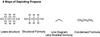
Functional groups
(no S N or Halogen)
Draw and name
Black folder
Organic 1 pdf
Four equations of kinematics
one dimension
uniform acceleration
s = v0t + 1/2 at2
s = 1/2(v + v0)t
v = v0 + at
v2 = v02 + 2as
acceleration in uniform circular motion
formula
name
direction
a = v2 / r
Centripital acceleration
to centre
Newton’s laws of motion
Number
title
formula
1st Law - The law of Inertia
If net force = 0 then acceleration = 0
2nd Law - Net force causes acceleration
F = ma
3rd Law - Action and reaction
F12 = F21
Friction forces
Types
Formulae
Static friction
Fsmaximum = usN (coefficient of static friction mus, Normal force)
Kinetic friction
Fk = ukN (coefficient of kinetic friction muk, Normal force)
Classical fundamental forces
How many
Names
Formulae
Gravitational force F = G m1 m2 / r2
Electrostatic force F = k q1 q2 / r2
Magnetic force F = q B v sin theta
Work
define in words
formula
Work = force x component of displacement in the direction of the force
W = (F cos theta) s
Kinetic energy
formula
K = 1/2 m v2
Potential energy
describe in words
Gravity - formula general
Gravity - formula Earths gravity
Two charges interacting by Electric force - formula
In a stretched spring - formula
The energy of position
Ug = -G m1 m2 /r
U = mgh
Ue = k q1 q2 /r
Uspr = 1/2 k x2
Conservation of energy
formula
Utotal = initial (K + U) = final (K + U)
Power
Definition
formula
Power in terms of force - formula
Rate of energy expenditure OR work done
P = W/t
P = F v


Osteogenic cells differentiate and develop into osteoblasts which, in turn, are responsible for forming new bone.
Osteoblasts synthesize and secrete a collagen matrix and calcium salts.
When the area surrounding an osteoblast calcifies, the osteoblast becomes trapped and transforms into an osteocyte, the most common and mature type of bone cell.
Osteoclasts, the cells that break down and reabsorb bone, stem from monocytes and macrophages rather than osteogenic cells..
There is a continual balance between osteoblasts generating new bone and osteoclasts breaking down bone.


2) Acetic acid (formula = HC2H3O2) is the most common weak acid example used by instructors.
3) Another way to write acetic acid's formula is CH3COOH.
4) A common abbreviation for acetic acid is HAc, where Ac¯ refers to the acetate polyatomic ion. The following equation describes the reaction between acetic acid and water: HAc + H2O H3O+ + Ac¯ Note that it is an equilibrium condition. The equilibrium constant for this reaction is written as follows: Kc = ( [H3O+] [Ac¯] ) / ( [HAc] [H2O] ) However, in pure liquid water, [H2O] is a constant value. To demonstrate this, consider 1000 mL of water with a density of 1.00 g/mL. This 1.00 liter (1000 mL) would weigh 1000 grams. This mass divided by the molecular weight of water (18.0152 g/mol) gives 55.5 moles. The "molarity" of this water would then be 55.5 mol / 1.00 liter or 55.5 M. The solutions studied in introductory chemistry are so dilute that the "concentration" of water is unaffected. So 55.5 molar can be considered to be a constant if the solution is dilute enough. Moving [H2O] to the other side gives: Kc [H2O] = ( [H3O+] [Ac¯] ) / [HAc] Since the term Kc [H2O] is a constant, let it be symbolized by Ka, giving: **Ka = ( [H3O+] [Ac¯] ) / [HAc]** This constant, Ka, is called the **acid ionization constant**.
2) Ammonia (formula = NH3) is the most common weak base example used by instructors. The following equation describes the reaction between ammonia and water: NH3 + H2O NH4+ + OH¯ Note that it is an equilibrium condition. The equilibrium constant for this reaction is written as follows: Kc = ( [NH4+] [OH¯] ) / ( [NH3] [H2O] ) However, in pure liquid water, [H2O] is a constant value. To demonstrate this, consider 1000 mL of water with a density of 1.00 g/mL. This 1.00 liter (1000 mL) would weigh 1000 grams. This mass divided by the molecular weight of water (18.0152 g/mol) gives 55.5 moles. The "molarity" of this water would then be 55.5 mol / 1.00 liter or 55.5 M. The solutions studied in introductory chemistry are so dilute that the "concentration" of water is unaffected. So 55.5 molar can be considered to be a constant if the solution is dilute enough. Moving [H2O] to the other side gives: Kc [H2O] = ( [NH4+] [OH¯] ) / [NH3] Since the term Kc [H2O] is a constant, let it be symbolized by Kb, giving: **Kb = ( [NH4+] [OH¯] ) / [NH3]** This constant, Kb, is called the **base ionization constant**.
The half-equivalence point is can be used to determine the acid dissociation and pKa of the acid used in titration. In acid-base titration the ratio between the acid and corresponding base is exactly 1:1 at the half-equivalence point. [Video webpage image LINK](http://chemistry.tutorvista.com/analytical-chemistry/equivalence-point.html)


A substitution implies that one group replaces another. Nucleophilic substitution reactions occur when an **electron rich species, the nucleophile**, reacts at an **electrophilic saturated C atom** attached to an **electronegative** group (important), the **leaving** group The electrophilic C can be recognised by looking for the polar σ bond due to the presence of an electronegative substituent (esp. C-Cl, C-Br, C-I and C-O) Nucleophilic substitution reactions are an important class of reactions that allow the interconversion of functional groups. Of particular importance are the reactions of alkyl halides (R-X) and alcohols (R-OH) For alcohols, the range of substitution reactions possible can be increased by utilising the tosylates (R-OTs), an alternative method of converting the -OH to a better leaving group.

A substitution implies that one group replaces another. Nucleophilic substitution reactions occur when an electron rich species, the nucleophile, reacts at an electrophilic saturated C atom attached to an electronegative group (important), the leaving group The electrophilic C can be recognised by looking for the **polar σ bond** due to the presence of an **electronegative** substituent (esp. C-Cl, C-Br, C-I and C-O) Nucleophilic substitution reactions are an important class of reactions that allow the interconversion of functional groups. Of particular importance are the reactions of alkyl halides (R-X) and alcohols (R-OH) For alcohols, the range of substitution reactions possible can be increased by utilising the tosylates (R-OTs), an alternative method of converting the -OH to a better leaving group.


