Biochemistry of Vision - SRS Flashcards
(49 cards)
The retina is made up of the Ora Serrata, nonsensory retinal pigment epithelium, and sensory retina. What are the three parts of the sensory retina?
–Macula lutea
–Fovea centralis
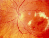
–Optic disk
What are the cell types of the retina?
Neurons
Retinal Pigment cells (RPE)
Neuron support cells
What types of neurons are found in the retina?
Photoreceptor cells (rods and cones)
Retinal ganglion cells
Interneurons (integrating neurons)
What are the types of interneurons found in the retina?
Bipolar cells
Horizontal cells
Amacrine cells
Where are RPE’s found?
Retinal pigment epithelial cells are found in the outermost layer seperating the retina from the choroid.
What are the neuron support cells of the retina known as?
Mueller cells
In order of signal transduction, name the ten layers of the retina! Go!
- Pigment epithelial cells (RPE)
- Photoreceptor cells
- Outer limiting membrane
- Outer nuclear layer
- Outer plexiform layer
- Inner nuclear layer
- Inner plexiform layer
- Ganglion cell layer
- Optic nerve fibers
- Inner limiting membrane
Rods pick up light of differing intensities and are peripheral, what photopigment do they posess?
Rhodopsin
Cones are located in the fovea and detect blue, red and green. What photopigment do they contain?
Iodopsin
What connects the photoreceptors and the RPE?
Interphotoreceptor matrix
The interphotoreceptor matrix is important in recycling and uses the interstitial retinoid binding protein (IRBP) to do what?
What else does this matrix do?
Transports retinol to the RPE and Retinal to the photoreceptor
Also, shedding of older disks
RPE contains melanin grandules and phagocytose shed disks. These disks are degraded in lysosomes and released into choriocapillaries. What enzye does the RPE express and what function does it serve?
Retinol re-isomerization enzyme
•Enzymatic conversion of 11-cis retinal to retinol
Rods end in rod sperule, what components of cells are involved in this?
Dendrites of bipolar cells
Neurites of horizontal cells
Cones end in cone pedicles, what cell processes are involved here?
Dendrites of bipolar cells
Neurites of horizontal cells
What is iodopsin composed of?
Opsins and chromophore (11-cis retinal)
When blue, green and red are stimulated together, what light is seen?
White
In a process called bleaching, a photopigment absorbs a photon of light and changes conformation of 11-cis-retinal. What does the photopigment then act as?
A GPCR -
- Induces GMP from cGMP
- cGMP dependent Na+ channels close
Leber congenital amaurosis type II involves the RPE65 gene and isomerohydrolase conversion of all-trans retinol to 11-cis-retinal. What is the inheritance pattern? What is the delivery vector?
Autosomal recessive
Adenoviral delivery vector
Bipolar cells recieve impulses from photoreceptor cells . What are the types that recieve input from multiple photoreceptors?
Diffuse cone bipolar cells
Rod bipolar cells
Midget cone receptors recieve input from one photoreceptor and communicate with?
One ganglion cell
Ganglion cells have dendrites in the inner plexiform layer and come in two varieties. What are they?
Diffuse ganglion cells - contact with several bipolar cells
Midget ganglion cells - contact with a single bipolar cell
Association neurons integrate signaling. What are the two types of these?
Amacrine cells
Horizontal cells
Where do amacrine neurites end?
Axon terminals of bipolar cells and the ganglion cell dendrites and bodies
Where do the neurites of horizontal cells end?
On cone pedicals and rod spherules