{ "@context": "https://schema.org", "@type": "Organization", "name": "Brainscape", "url": "https://www.brainscape.com/", "logo": "https://www.brainscape.com/pks/images/cms/public-views/shared/Brainscape-logo-c4e172b280b4616f7fda.svg", "sameAs": [ "https://www.facebook.com/Brainscape", "https://x.com/brainscape", "https://www.linkedin.com/company/brainscape", "https://www.instagram.com/brainscape/", "https://www.tiktok.com/@brainscapeu", "https://www.pinterest.com/brainscape/", "https://www.youtube.com/@BrainscapeNY" ], "contactPoint": { "@type": "ContactPoint", "telephone": "(929) 334-4005", "contactType": "customer service", "availableLanguage": ["English"] }, "founder": { "@type": "Person", "name": "Andrew Cohen" }, "description": "Brainscape’s spaced repetition system is proven to DOUBLE learning results! Find, make, and study flashcards online or in our mobile app. Serious learners only.", "address": { "@type": "PostalAddress", "streetAddress": "159 W 25th St, Ste 517", "addressLocality": "New York", "addressRegion": "NY", "postalCode": "10001", "addressCountry": "USA" } }

Block 3 - CNS Flashcards

(113 cards)

1
Q

___ aggregates are seen in Alzheimer’s, Pick disease, and progressive supranuclear palsy

Microtubule associated protein

Soluble in monomeric form

Insoluble fibrillary aggregates escape degradation and form neurofibrillary tangles

In some patients, the enzyme that cleaves the protein can be altered ( familial AD and PD)

A

tau

2
Q

___ aggregates seen in Parkinson’s, dementia with lewy bodies, and multiple system atrophy

A

synuclein

3
Q

____ aggregates seen in Alzheimer’s and cerebral amyloid angiopathy

A

beta-amyloid

4
Q

___ protein aggregates seen in CJD, vCJD, FFI, and animal diseases

A

prion

5
Q

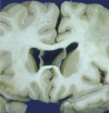
Type of degeneration seen in alzheimer’s

A

diffuse cortical

6
Q

type of degeneration seen in diffuse lewy body disease

A

diffuse cortical

7
Q

degeneration seen in frontotemperol dementias

A

diffuse cortical

8
Q

type of degeneration seen in parkinson disease

A

midbrain/brainstem

9
Q

type of degeneration seen in progressive supranuclear palsy

A

midbrain/brainstem

10
Q

type of degeneration seen in huntington disease

A

caudate nucleus

11
Q

type of degeneration seen in amyotrophic lateral sclerosis

A

motor neurons

12
Q

chromosome associated with beta-amyloid precursor protein in Alzheimer’s disease

A

21

(alzheimer’s increased in Down syndrome)

13
Q

gene associated with presenilin 1, associated with Alzheimer’s

A

14q24.3

14
Q

gene associated with presenilin 2, associated with Alzheimer’s

A

1q31-q42

15
Q

gene associated with tau, associated with Alzheimer’s

A

17q21.1

16
Q

____ plaques as seen in alzheimer’s

A

amyloid

17
Q

____ ____ of Tau protein as seen in alzheimer’s disease

A

neurofibrillary tangles

18
Q

Superficial cortical dark spots denote ____ deposition in Alzheimer’s

A

hemosiderin

19
Q

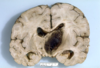
Parkinson’s disease involves the deposition of ____ inclusions (Lewy bodies) with progressive loss of neurons, visible grossly in the substantia nigra.

A

synuclein

20
Q

Parkinson’s disease involves the deposition of synuclein inclusions, called ______ _____, with progressive loss of neurons, visible grossly in the substantia nigra.

A

Lewy bodies

21
Q

Parkinson’s disease involves the deposition of synuclein inclusions (Lewy bodies) with progressive loss of neurons, visible grossly in the ____ ____.

A

substantia nigra

22
Q

Huntington chorea involves chromosome ___, which codifies Huntingtin, containing a polyglutamine sequence due to CAG repeats. Repeats increase with each subsequent generation, increasing the severity of the disease.

A

4p

23
Q

Huntington chorea involves chromosome 4p, which codifies Huntingtin, containing a polyglutamine sequence due to CAG repeats. Repeats increase with each subsequent generation, increasing the severity of the disease. This phenomenon is called ___.

A

anticipation

24
Q

Huntington’s disease shows ___ nucleus atrophy and neuronal loss. Causes significant dilation of the lateral ventricles.

A

caudate

25
amyotrophic lateral sclerosis involves the loss of ____ neurons
motor (upper and lower)
26
amyotrophic lateral sclerosis involves the loss of motor neurons, which appears as degeneration of the ____ tracts.
corticospinal
27
Anencephaly is one of the more common neural tube defects due to closure failure of the ___ \_\_\_, resulting of the cerebrum and calvarium.
frontal neuropore
28
Anencephaly is associated with ___ deficiency
folic acid
29
\_\_\_\_ is closure failure of the frontal to caudal neuropores, exposing the spinal cord and adjacent nerve roots.
Craniorachischisis
30
Failure of closure of part of the spinal cord. Spinal cord/roots can protrude from the defect.
meningomyelocele
31
Meningomyelocele is associated with ___ deficiency.
folic acid
32
Meninges and/or brain herniate through a mesodermal defect. Usually occipital, secondly frontoethmoidal.
encephalocele
33
fluid accumulation in the central canal of spinal cord.
hydromyelia
34
downward herniation of cerebellar tonsils through the foramen magnum. Asymptomatic or neck pain, lower CN palsies, sleep apnea, and sudden death. Cerebellar atxia and long tract signs. Syringomyelia is common.
chiari malformation type 1
35
downward herniation of tonsils and brainstem. Highly associated with lumbosacral myelomeningocele and hydrocephalus.
chiari malformation type 2
36
failure to divide the brain into hemispheres. Associated with mutations: SHH, SIC2, SIX3, TGIF1
holoprocencephaly
37
holoprocencephaly is associated with abnormalities of which chromosome?
trisomy 13
38
agenesis of the ___ \_\_\_ is total or partial disruption of cerebral interhemispheric axonal migration across the midline during development. Asymptomatic or seizures and cognitive impairment.
corpus callosum
39
Agenesis of the corpus callosum shows remnants of ____ matter bundles called bundles of Probst.
white
40
Common incidental finding on imaging or autopsy that involves the failure of the two halves of the septum pellusidum go fuse at the midline. Typically asymptomatic.
cavum septum pellusidum
41
cerebral cortical defects are due to impairment of ____ migration and cortical differentiation.
neuroblast
42
\_\_\_\_ is the absence of normal convolutions on the brain, "smooth brain." Seen in neuronal migration disorders.
lissencephaly
43
autosomal dominant syndrome of defective neuronal migration that results in lissencephaly
miller-dieker syndrome
44
chromosomal abnormality for miller-dieker syndrome
17p13.3
45
x linked syndrome associated with defective neuronal migration, resulting in lissencephaly
filamin A and doublecortin (DCX)
46
autosomal recessive syndrome associated with defective neuronal migration, resulting in lissencephaly
Walker-warburg syndrome
47
2 genes associated with Walker-warburg syndrome
POMT1 and POMT2
48
group of conditions of various etiologies that show nodules of abnormally placed gray matter. Arrest of neurons during migration, which stay around ventricles and white matter. Asymptomatic or seizures.
heterotopias
49
disruption of normal gyration (too many, too small). Diffuse, focal, bilateral, or unilateral. Seizures and severe psychomotor retardation. Results from fetal infection, prenatal hypoxia, metabolic disease, or genetics.
polymicrogyria
50
The least severe degree of CNS destruction from stroke during gestation. any fluid-filled cavity in the fetal or neonatal brain. A thin membrane may separate the cavity from the lateral ventricle or the subarachnoid space.
porencephaly
51
Moderately severe degree of CNS destruction from stroke during gestation. ongenital clefts in the cerebral mantle.
schizencephaly
52
the most severe degree of CNS degradation from stroke during gestation. erebral hemispheres are replaced by a thin-walled, fluid-filled cyst. The aqueduct is usually atretic, and increased fluid pressure causes the cyst (and the head) to enlarge.
hydranencephaly
53
Mycobacterial infection commonly seen in the brains of AIDS patients.
TB
54
Fungal infection that begins as a pulmonary infection and spreads hematogenous to the brain in immunosuppressed patients. Angioinvasion leads to infarcts. Fibrin and hyphae are seen in the artery wall.
Aspergillosis
55
Infection that spreads hematogenously from the lung in immunosuppressed patients. Abscesses, thick & slimy meninges, and parenchymal cysts. Yeast with thick capsules invade macrophages.
cryptococcosis
56
RNA virus that replicates in the muscle and migrates to the CNS via centripetal axonal transport --\> salivary and lacrimal glands. Neurons with cytoplasmic Negri bodies.
Rabies
57
Herpes virus causes necrosis of the ____ lobes, called necrotizing panencephalitis. Causes severe memory disturbances and other deficits
temporal
58
Diffuse white matter damage caused by JC virus. Shows small, round, confluent white matter lesions (demyelination).
Progressive multifocal leukoencephalopathy (PML)
59
JC virus goes latent in which two organs?
kidneys and tonsils (B cells)
60
Infection in immunosuppressed patients, especially pregnant women transplacentally to fetus, that encysts and remains dormant in the CNS. Reactivation due to immunosuppression. Shows multifocal areas of necrosis and inflammation, bradyzoite cysts and tachyzoites.
toxoplasmosis
61
Cystic infection that reactivates in immunosuppressed patients.
Toxoplasmosis
62
Most common CNS parasitic infection and a leading cause of epilepsy. Tapeworms develop cysts in any organ. Inflammation follows the parasite's death and eventual calcification
Neurocysticercosis
63
\_\_\_ hematoma occurs between the dura and the skull. Associated with skull fracture. Progressive mass effect, so patient may have a lucid intervan with subsequent mental decline. Biconcave (lens) shaped and does not cross suture lines.
Epidural (extradural)
64
\_\_\_\_ hematoma occurs between the dura and the brain. Associated with trauma and injury to bridging veins, which may be more common in elderly patients due to fragility of the veins. Increased risk with anticoagulation and alcoholism. Progress quickly. May become chronic Banana shaped and may cross suture lines.
65
\_\_\_ hemorrhage between the arachnoid membrane and CNS parenchyma. Commonly caused by ruptured aneurysms, AVM, or cortical contusion. "Worse headache of my life." High fatality due to hemorrhage and vasospasm. Blood can be seen in the CSF on LP or anywhere that CSF is present.
Subarachnoid
66
Aneurysms are commonly seen at the ____ of an artery
bifurcation
67
\_\_\_\_\_ hemorrhage is located within the cortex. Related to HTN (basal ganglia or thalamus), metastases, or vascular lesions Hemorrhage after CNS infarct. Presents as sudden headache, loss of function, weakness, and hemiplegia
Parenchymal
68
Traumatic brain injury can lead to progressive brain atrophy and cortical ___ deposition in deep sulci
Tau
69
Cerebral contusions usually occur in the ___ gyri and ___ lobe. Can cause diffuse axonal and vascular rinjury.
supraorbital temporal
70
Cerebral contusions can present with ____ lesions, where the brain hits the skull on the opposite side from the trauma.
countercoup
71
diffuse axonal and vascular injury, typically caused by a ____ contusion.
cerebral
72
\_\_\_\_ herniation involved the movement of part of the temporal lobe under the free edge of the tentorium, compressing the midbrain and the oculomotor nerve (ipsilateral palsy). This is usually caused by a rapidly expanding middle cranial fossa epidural, subdural, or infratemporal lobe hematoma.
uncal
73
In ____ herniation, the part of both the cerebral hemispheres are squeezed through a notch in the tentorium cerebelli.
Diencephalic/central
74
\_\_\_\_\_ herniation is the most common form of intracranial herniation and occurs when brain tissue is displaced under the falx cerebri. The cingulate gyrus is herniated under the falx, and if progression occurs, other areas of the frontal lobe are involved.
subfalcine
75
\_\_\_\_ ____ can be drilled in the skull to relieve intracranial pressure
Burr holes
76
A ____ herniation is characterized by the descent of the cerebellar tonsils through the foramen magnum, which compresses the medulla against the clivus/odontoid process. It is described as "coning" as the brain tissue is squeezed down through the foramen like being squeezed into a cone.
tonsillar
77
\_\_\_\_ ___ phenomenon occurs with uncal herniation, which causes contralateral pupillary dilation and ipsilateral oculomotor nerve weakness.
Kernohan's notch
78
Secondary ___ hemorrhages in the brainstem occur after uncal herniation, due to overstretching of the vessels.
Duret
79
Brief episodes of CNS symptoms lasting \<1 hr, due to ischemia and thromboembolism.
TIA (transient ischemic attack)
80
Abrupt onset of CNA symptoms lasting \>24 hours, due to tissue infarct and necrosis from thromboembolism.
Stroke
81
\_\_\_\_ injury occurs when an infarcted area of the cortex bleeds after treatment to reestablish blood flow.
Reperfusion
82
Dural based tumors in adults. Slow growing with good prognosis. Women \> men.Increased in neurofibromatosis and post-radiation. May grow through bone without being histologically malignant. Well-circumscribed. Uniform tumor cells grow in whorls with no evidence of malignancy.
Meningioma
83
Ill-defined, infiltrating tumor. Occur anywhere in the CNS but more commonly supratentorial. Cause seizures, headaches, and focal signs. The tendency to become malignant. MRI shows ill-defined hyperdensity (hypercellular) with higher water content than normal white matter.
astrocytoma
84
Most frequent glial tumor in adults. \>supratentorial. Highly malignant. Present as seizures, headaches, focal deficits. Hypercellular, especially at rim. Cells have marked atypia and mitosis.
Glioblastoma
85
benign tumor in adults. More common in peripheral nerves, but within the CNS, more common in vestibular branch of CN 8 (cerebellopontine angle). sporadic = solitary Neurofibromatosis 2 = bilateral Well circumscribed
schwannoma
86
Well-circumscribed tumor, typically intraventricular or in the spinal cord, centrally placed. Any age. Causes hydrocephalus, increased ICP, and atxia.
ependymoma
87
\_\_\_\_\_ frequently form perivascular "pseudo rosettes." Centered by blood vessels and tumor cells arrange around them.
ependymoma
88
Most frequent glial tumor in children. Occur mostly in the cerebellum, but also in the optic nerve, thalamus, hypothalamus, and supratentorially. Well circumscribed, solid and cystic. Present with increased ICP and cerebellar signs (ataxia). Spindle cells with dense, fibrillary cytoplasm. Low cellularity and characteristic Rosenthal fibers.
pilocytic astrocytoma
89
Common malignant tumor in children, \<15yo. Typically in the posterior fossa, vermis. Present with ataxia and signs of increased ICP. Tendency to disseminate along the subarachnoid space (drop metastases). Mt: SHH, WSH Solid, typically in the midline. Markedly cellular, frequently with "rosettes." Some neuroblasts within these tumors form nodules of more differentiated cells, called neurocites.
medulloblastoma
90
Steroid myopathy involves (proximal/distal) muscle weakness
proximal
91
Steroid myopathy preferentially affects (type 1/type 2) muscle fibers
type 2
92
Dermatomyositis preferentially affects the (proximal/distal) muscles autoimmune, women Elevated CK Underlying malignancy Heliotrope erythema, Gottron sign, mechanics hands
proximal
93
autoantibodies associated with dermatomyositis 1 = gottron papules and heliotrope rash 2 = arthritis and skin rash, interstitial lung disease 3 = paraneoplastic/juvenile
Anti-Mi2 (against helicase) Anti-Jo1 (against histidyl t-RNA synthetase) Anti-P155/P140
94
Lymphocyte-mediated muscle injury leading to muscle pain and weakness
polymyositis
95
polymyositis preferentially affects (proximal/distal) muscles, symmetrically
Proximal
96
Durg-induced myopathy high CK
Toxic/vacuolar
97
Inherited myopathy (AD or AR) that can be due to defects in several cellular enzymes Scattered fibers show more subsarcolemmal mitochondria on NADH staining, indicating increased proliferation Typical "ragged-red fibers"
Mitochondrial myopathy
98
X-linked (male) progressive muscle weakness Floppy baby or asymptomatic at birth High CK
Duchenne musculodystrophy
99
Duchene MD favors (proximal/distal) muscles
proximal
100
Mild dystrophy that also presents with heart disease, typically around 12yo.
Becker Muscular dystrophy
101
Sarcoglycanopathy favors (proximal/distal) muscles Intolerance to exercise/cramps
proximal (limb girdle)
102
Spongiform encephalopathies are a group of progressive, lethal diseases that involve the accumulation of abnormal ___ protein in the CNS and the formation of ___ plaques.
prion amyloid
103
Prion PrPc protein is associated with Chromosome \_\_\_. Expressed in all cells, particularly neurons. Decrease in size of a-helices, and increase in size of b-sheets causes the protein to become resistant to digestion and liable to form aggregates.
20
104
Rapidly progressive dementia presentiing in the 7th decade of life. Also shows myoclonus. Progressive and fatal in months. Increased signal in the basal ganglis and cortex. Vauolization of the gray matter neutrophil (sponge-like) Progressive neuronal loss and reactive gliosis PrP deposition as amyloid plaques
sporadic Creutxfeldt-Jakob disease (CJD)
105
sporadic Creutxfeldt-Jakob disease (CJD) shows vacuolization of (gray/white) matter
gray
106
sporadic Creutxfeldt-Jakob disease (CJD) involves ___ protein deposition in the form of amyloid plaques
PrP
107
PrP protein disease that appears in younger patients in the UK. Initially as sensory and psychiatric disturbances and unsteadiness. Slowly progressive dementia to akinetic mutism. Death around 14 months. Plaques and vacuolization Spongiform degeneration in the basal ganglia and thalamus PrPsc also found in follicular dendritic cells of tonsil, LN, spleen, thymus and GALT (all immune tissues)
variant Creutxfeldt-Jakob disease (CJD)
108
Prion protein diseases are transmissible but are not \_\_\_\_.
contagious
109
•Destruction of myelin in CNS or peripheral nerves, with relative preservation of axons Multifocal with lesions of different ages Autoimmune ; women \> men Increased incidence with increased latitude * Variable presentation with weakness, paresthesia, sensory loss of one or more limbs, optic neuritis, diplopia, incoordination, vertigo * Others with loss of vision, dysarthria, disturbances of micturition, painful muscle spasms, seizures * Usually associated with oligoclonal bands of immunoglobulins on electrophoresis of CSF
Multiple sclerosis
110
Classify the MS: ## Footnote –multiple acute attacks, each followed by clinical improvement
relapsing remitting
111
Classify the MS: ## Footnote –after years of RRMS, the patient enters a stage with no recovery between attacks
secondary progressive
112
Classify the MS: progressive disease without episodes of recovery
primary progressive
113
Classify the MS: repeated acute attacks superimposed on progressive disease without episodes of recovery
relapsing progresssive