Cardiovascular System II Flashcards
What are the most common aorticopulmonary septum defects?

- Persistent Truncus Arteriosus -> conotruncal ridges do not fuse, shared primitive outflow tract (truncus arteriosus)
- Transposition of the Great Arteries (TGA) -> septum fails to follow normal spiral course and runs straight down between vessels
- Unequal division -> tetralogy of Fallot (TOF), Pulmonary stenosis
Persistent Truncus Arteriosus
-1-2% of congenital heart abnormalities, shared primitive outflow tract
Symptoms (infants first couple weeks): cyanosis, tachypnea, poor feeding, diaphoresis
-Usually patients have a large ventricular septal defect as well, surgical fixation involves complete primary repair, and closure of the ventricular septal defect

Transposition of the Great Arteries (TGA)
How is it surgically repaired?
Aorticopulmonary septum fails to rotate, and right ventricle joins the aorta while the left ventricle joins the pulmonary trunk
Symptoms: “Blue babies”
Arterial Switch Procedure

Arterial Switch Procedure
- Cut PA and aorta, remove L. and R. pulmonary arteries from aorta
- PA moved in front of aorta, aorta reconnected to left ventricle and L. and R. pulmonary arteries reconnected to aorta
- Pulmonary artery reconnected to right ventricle

Tetralogy of Fallot (TOF)
- Narrowing of pulmonary valve
- Thickening of right ventricle
- Aorta displaced over ventricular septal defect
- Ventricular septal defect
Results in insufficiently oxygenated blood pumped to the body, can be surgically repaired with long life span (Shawn White)

Pulmonary Stenosis
Thickening of pulmonary valve, fusion of leaflets and small valve opening
- Sometimes the valve is absent and the right ventricle is underdeveloped
- Usually results in the right side of the heart having to work harder = hypertrophy

What is the ductus arteriosus? What does it eventually turn into?

Vascular structure that connects main pulmonary artery near left branch to the proximal descending aorta
- Eventually it closes and turns into the ligamentum arteriosum
- Aggrevation of the L. recurrent laryngeal nerve located near the ligamentum arteriosum is possible with people with lung cancer

What do the coronary arteries supply?
Myocardium and epicardium (visceral layer of the serous pericardium)

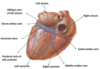
Describe the circulation and distribution of the right coronary artery (RCA)
Anterior aortic sinus -> through the right atrioventricular sulcus (coronary sulcus) -> branches to SA node, right atrium -> inferior right marginal branch feeds right ventricle
Posterior interventricular artery wraps around heart feeding posterior AV node and L. and R. ventricles running through the interventricular sulcus

Describe the distribution of the RCA on the posterior surface
RCA continues in the atrioventricular sulcus until junction with the posterior interventricular sulcus -> Here it descends and turns into the posterior descending artery where it supplies the AV node, adjacent sides of the right and left ventricles, and part of the interventricular septum

RCA supplies what heart structures, and what are the main 4 branches?

Describe the circulation and distribution of the left coronary artery (LCA)
Left posterior aortic sinus -> behind left pulmonary trunk -> divides into left anterior descending (LAD) and the circumflex coronary arteries
LAD -> passes inferiorly through the anterior interventricular sulcus, supplying L. and R. ventricles, and the major part of the interventricular septum

Describe the branches of the LAD
Branches to left ventricle = diagonal branches
LAD turns around inferior margin of heart and heads up the posterior interventricular sulcus a short distance to anastomose with the posterior interventricular (descending) artery

Where are some common coronary artery blockages?

What does the circumflex artery supply?
Winds around left margin of the heart to posterior surface where it supplies the left atrium and left ventricle
Obtuse marginal branches -> large branches to left ventricle
Before descending down the posterior interventricular sulcus, it decreases in size and anastomoses with a branch of the RCA
AV nodal branch 10-20%

How do you determine a right dominant or left dominant heart?
Dominance -> is from the artery that gives rise to the posterior interventricular artery
Right ~67% RCA
Left ~15% LCA
Codominance -> RCA and LCA have branches that course in or near the posterior interventricular groove
~18%
And a few people only have one coronary artery (Pistol-Pete)

Coronary artery variations

Arteriosclerosis
Hardening of the arteries
Athersclerosis
Fatty deposits and occlusion (plaque)
Risk factors: Genetics, hypercholesterolemia, male, age, smoking, hypertension
Treatment: angioplasty, surgery

What is the treatment for coronary artery disease (CAD)?
Coronary artery bypass graft (CABG) or ballon angioplasty, as well as lifestyle changes, medications

Thrombus
Damaged epithelial lining due to disease, trauma, vascular lesion can occur such as a thrombus -> clot that forms inside of a vessel wall
-Can dislodge and travel (emboli) to other vessels and cause blockage of normal blood flow

Bacteremia
Presence of viable bacteria in the blood that travels through bloodstream can turn into sever, lifethreatening bacteremia called sepsis

Myocardial infarction (MI)
Occlusion of majory artery usually from embolus where region affected eventually becomes necrotic
-Most commonly caused from coronary artery insufficiency from atherosclerosis

Explain venous drainage from the heart
Mostly* all cardiac veins empty into the coronary sinus -> runs toward right side of heart and drains into right atrium
-Great cardiac vein -> located anterior interventricular sulcus
-Middle cardiac vein -> located posterior interventricular sulcus
Small cardiac vein -> located posterior atrioventricular sulcus near marginal artery
*Exception is Anterior cardiac veins -> drain into right atrium