Cartilage and Bone Flashcards
(38 cards)
Functions of Cartilage
- Support of soft tissues
- Forms articular surface of long bones
- Growth in length of long bones
Cartilage ECM
Type II collagen maintains shape and provides tensile strength
Proteoglycan aggregates (between layers of collagen) provide resilience
Composition permits cartilage to bear mechanical stress without permanent distortion
GAGs: chondroitin sulfate 4, chondroitin sulfate 6, keratin sulfate
How to make a proteoglycan aggregate
- Begin with core proteins
- Add GAGs (creates proteoglycans)
- Bind proteoglycans to core of hyaluronic acid with link proteins
Morphological features of Chondrocytes
The only cells found in healthy cartilage
Round diffuse nucleus with prominent nucleolus
Cytoplasm rich in RER, well-developed Golgi and mitochondria (basophlic)

Identify

Hyaline cartilage (most common form) surrounded by perichondrium
Located on joint surfaces, tracheal rings, ventral end of ribs, nose, larynx, trachea, bronchi, epiphyseal plate
ECM: Type II collagen, basophilic
Chondrocytes live within lacunae - spaces in matrix.
Rich in sulfated GAGs in cytoplasm - makes ECM basophilic (blue)
Consequences of cartilage lacking blood vessels (avascular)
- Size limitation
- Low metabolic rate
- Poor potential for repair (except in young children)
- Systemic drug treatment is difficult (medically relevant)
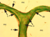
Identify

Elastic cartilage (arrow to elastic fibers): located where flexible support is needed - external ear, epiglottis, eustachian tube, larynx
ECM: more flexible than hyaline, less homogenous, numberous elastic fibers (stain with orcein dyes)
Chondrocytes look identical to those in hyaline
Less susceptible to deneration/age change sthan hyaline
Identify

Fibrocartilage: Intervertebral discs (annulus fibrosus), pubic symphasis, menisci, some tendons
ECM : eosiniphilic (pink) ground substance reduced, collagen increased (type I predominates)
Chondrocytes look the same as those in elastic and hyaline
No perichondrium
Distinguish from dense regular CT: has irregular fiber distribution, fewer cells per unit area and rounder chondrocytes
Medical Conditions Assoicated with Cartilage
- Calcification of the matrix: hyaline cartilage is most susceptible, occurs with aging
- Osteoarthritis: loss/change in physical properties of articular cartilage, occurs with aging
- Chondroma: benign tumors
- Chondrosarcoma: slow growing malignant tumors
Functions of bone
- Supports fleshy structures
- Protects vital organs
- Harbors bone marrow
- Reservoir of calcium, phosphate, etc.
- Involved in body movement
Similarities between cartilage and bone
- Both supportive CT
- Bone consists mostly of ECM
- Osteocytes reside within lacunae
- Bone is surrounded by periosteum (specialized CT)
- Bone can grow by means of appositional growth
Differences between cartilage and bone
- Interstitial growth does not occur in bone
- Bone has a more regular arrangment of cells and fibers
- Bone is vascularized and has nerves
- Calcification of ECM is a normal process in bone
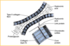
General structure of long bone
Diaphysis: cylindrical part, thick outer layer of compact (aka cortical) bone with thin marrow cavity containing spongy bone (aka cancellous or trabecular)
Epiphysis: bulbous ends, spongy bone covered by thin layer of compact bone

Canaliculi
In compact bone, small channels that radiate in all directions thru the ECM from each lacuna.
Connect (gap junctions) with canaliculi of adjacent lacunae for communication
Nutrients from intersitital fluid (in contact with capillaries)
Red squiggles in picture
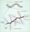
Lamellae
Concentric layers of compact bone in an osteon that surround a central canal (Haversian Canal)
Osteocytes
Reside in lacunae, have processes that extend into canaliculi.
Adjacent osteocytes are in contact with one another via gap junctions - allows exchange of ions and small molecules (can provide nourishment for a chain of about 10 cells)
Types of Lamellar Organization (compact bone)
- Haversian Canals: parallel to long axis of bone, have small blood vessels, loose CT, and nerves
Haversian system = osteon
- Volksman Canals: oblique angles to long axis of bone, connect Haversian canals to each other and to the free surface, not completely surrounded by concentric lamellae
- Interstitial lamellae: wedge-shaped regions between osteons, fragments of previous Haversian systems (continually made and destroyed)
- Inner circumferential lamellae: beneath endosteum
- Outer circumferential lamellae: beneath periosteum
(4 and 5 observed at external and internal surfaces)

ECM of Bone: Organic Matrix (osteoid)
Responsible for toughness and resilience
- Type 1 collagen
- Non-collagenous proteins
- Ground substance (GAGs and proteoglycans)
ECM of Bone: Inorganic Matrix
Responsible for hardness
- Calcium phosphate (hydroxyapatite): thin plates or crystals, associated with collagen fibers
Identify

Osteoblasts
Identify

Osteoprogenitor cells
Present in adults, have a low profile
Not actively making bone, but can be reactivated
Compact chromatin
Less basophilic cytoplasm
Identify

- Haversian canal (lined by endosteum)
- Volksman canal
- Bone marrow
Periosteum on outside
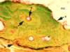
Identify

- Spongy bone
- Marrow cavity
- Compact bone
Functional Adaptation (REMODELING)
- Both spongy and compact bone are destroyed and replaced in a constant process throughout life
- Involves relative activity of osteoblasts (make new bone) and osteoclasts (destroy bone). Need balanced activity to maintain skeletal integrity
- Bone adapts to mechanical load placed on it - osteocytes are mechanotransducers of local strain
Spongy bone more responsivle to changes in load than compact bone


