Cartilage and Bone Flashcards

(54 cards)

1
Q

Types of cartilage (specialized CT)

A

Hyaline

Elastic

Fibrocartilage

2
Q

Cartilage

A
  • contains collagen II
  • contains sulfated proteoglygans (e.g. aggrecan) plus other ECM proteins dependent on cartilage type
  • net negative charge (basophilic)
  • avascular & aneural
  • chondrocytes in isogenous groups
3
Q

Types of bone tissue (specialized CT)

A
  • Woven
  • Lamellar
  • Compact (includes Haversian bone)
  • Spongy (cancellous, trabecular)

all contain calcium hydroxyapatite

4
Q

Hyaline Cartilage

A

translucent (think: Glass)

very hydrated; resists compression

smooth

located in all articular joint surfaces

  • isogenous groups (back-to-back D’s: D)
  • territorial matrix - TM (very basophilic)
  • homogeneous interterritorial matrix - ITM (less basophilic)
  • abundant sulfated PGs (e.g. aggrecan, thus very hydrated)
  • cells retracted from lacunae (L. “lake” or “lagoon”)
  • perichondrium at edges (but NOT at articular surfaces)
  • grows by both interstitial & appositional deposition
5
Q

Elastic Cartilage

A

similar to hyaline but also contains elastic fibers

found in external ear, eustachian tube, epiglottis

  • isogenous groups (back-to-back D’s:D)
  • territorial matrix
  • elastic fibers present surrounding cells and in interterritorial matrix
  • perichondrium
  • interstitial/appositional growth
6
Q

Fibrocartilage

A

hydrated (resists compression) but also contains collagen I so also high in tensile strength

found in intervertebral discs, temporomandibular joints, public symphysis

  • isogenous groups (back-to-back D’s:D)
  • territorial matrix not obvious
  • wavy collagen fibers (type I) present, thus eosinophilic
  • NO perichondrium
  • has fibroblasts
  • interstitial growth
7
Q

Interstitial growth

A

pushes to side

8
Q

Appositional growth

A

on top of eachother

9
Q

Location of hyaline cartilage

A

articular surfaces

10
Q

Elastic fibers

A

look like wires

11
Q

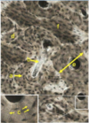
ID the type of tissue and the type of growth

A

Hyaline Cartilage

Appositional growth (left) and Interstitial growth (right)

12
Q

Chondrocytes

A

Cartilage cells

13
Q

Osteoarthritis

A

progressive, wear & tear joint disease of unknown cause

14
Q

Rheumatoid arthritis

A

Autoimmune, destruction of cartilage

15
Q

Woven Bone

A
  • immature bone
  • disorderly arrangement of collagen
16
Q

Lamellar Bone

A

Compact

  • Haversian systems
  • concentric lamellae
  • osteocytes interconnected by processes within canaliculi
  • central Haversian canal
  • interstitial lamellae
  • periosteum & endosteum

Spongy (aka cancellous, trabecular)

  • lamellae
  • osteocytes interconnected by processes within canaliculi
  • endosteum
17
Q

Histological preparation of bone tissues

A
18
Q

ID Type

A

Woven bone (immature bone)

19
Q

ID Type of Bone and Labeled sections

A

Compact Bone

Volkman’s Canal (A) and Interstitial Lamallae (IL)

20
Q

Interstitial Lamallae

A

space between osteons, remnants of osteons that were partially resorbed during the process of bone remodeling

21
Q

Canaliculi

A

microscopic canals between the lacunae of ossified bone

Interconnect osteocytes in life

22
Q

Lacunae

A

where osteocytes lived

a small space containing an osteocyte in bone or chondrocyte in cartilage

23
Q

Periosteum and Endosteum

A

endosteum (plural endostea) is a thin vascular membrane of connective tissue that lines the inner surface of the bony tissue that forms the medullary cavity of long bones

periosteum is the membrane that covers the outer surface of all bones,[1]except at the joints of long bones

24
Q

Spongy (cancellous) bone

A

contains bone lamallae but not haversian systems

25
Cells in bone tissue
**osteogenic cells** * mesenchymal derivative * osteoblasts (Ob) * osteocytes (Oc) **osteoclasts** * MPS-derivative * multinucleated * acidophilic (many mitochondria & enzymes)
26
Continual balance of of “blast” and “clast” activities for
- bone remodeling to accommodate stress changes - maintenance of Ca2+ levels
27
Osteoporosis
- bone degradation \> bone formation - estrogen reduction causes increased resorption (physiologic estrogen levels inhibit bone resorption) - aging and hormone related (physiologic estrogen levels inhibit bone resoption) - female incidence \> male
28
Osteogenesis Imperfecta (OI)
- aka brittle bone disease - most result from mutations in type I collagen (frequently affecting glycine) - collagen fibrils (fibers) are defective and production is reduced - many collagen-rich tissues are affected (teeth, sclera, etc.)
29
Identify: * woven bone * osteoid * osteoblasts * osteoclasts * osteocytes * calcified cartilage
30
ID this tissue What do you call the region labeled with the asterisks? Is the tissue growing or quiescent?
Hyaline cartilage Interterritorial Matrix (ITM) Growing because cells are dividing (double Ds and groups of 4)
31
ID Tissue type and the labels
Hyaline Cartilage ## Footnote territorial matrix - TM (very basophilic) homogeneous interterritorial matrix - ITM (less basophilic) cells retracted from lacunae (L. "lake" or "lagoon") perichondrium at edges (but NOT at articular surfaces) Capsula is very basophilic (dark outline of cell), surrounding territorial matrix, during growth -- not so basophilic when fully matured
32
ID Tissue type
Elastic Cartilage ## Footnote - perichondrium w/ chrondrogenic cells - hyaline cartilage-like w/ branching elastic fibers (special stains) - avascular ECM - territorial & interterritorial matrices; basophilia mainly due to chondroitin sulfates - appositional and interstitial growth
33
ID Tissue type
Fibrocartilage ## Footnote * Hybrid of hyaline + dense CT * No perichondrium * Collagen I + collagen II * Chondrocytes + fibroblasts * Interstitial growth
34
ID Tissue Type
Fibrocartilage ## Footnote * Hybrid of hyaline + dense CT * No perichondrium * Collagen I + collagen II * Chondrocytes + fibroblasts * Interstitial growth
35
ID Tissue Type and Type of staining
Fibrocartilage H&E (left) and Trichrome (right)
36
Arthritis
[Gr arthron =joint + -itis =inflammation] is a condition characterized by inflammation of joints due to degeneration of articular cartilage resulting in pain, stiffness, swelling & restriction of joint mobility **Osteoarthritis**: typically a disease of aging; caused by “wear & tear” of articular cartilages; also may be due to injury or infection **Rheumatoid arthritis**: autoimmune disease involving cartilage, bone & synovial membranes
37
Territorial matrix is highly
Basophilic. ITM is less basophilic Capsula is *most* basophilic
38
Volkmann's Canal
only found in compact bone any of the small channels in the bone that transmit blood vessels from the periosteum into the bone and that communicate with the haversian canals
39
Haversian System
Only found in compact bone osteon or haversian system is the fundamental functional unit of much compact bone
40
ID Tissue and Stain
Compact Bone Ground section ? or Decalcified (fine detail less visible)
41
ID Labels
a. lacunae b. Volkmann Canal c. Canuliculi d. Interstitial Lamellae e. Osteon f. Lamellar bone
42
Diagram of Bone tissue (No Question)
43
ID Type of Tissue and Labels
Compact Bone H = Haversian System O = Osteocytes L = Lamellae P = Periosteum C = circumferential lamellae
44
ID Labels
a. Osteocyte b. Osteoclast c. Osteoid d. Decalcified Bone e. Osteoblast
45
Osteoblasts
Bone-makers Osteoprogenitor cells, osteoblasts and osteocytes are all derived from mesenchymal precursors form a single layer on the surfaces of developing bone and lay down osteoid, which mineralizes to become bone tissue. They are intensely basophilic, owing to the presence of abundant rER for active protein synthesis. Osteoblasts are derived from flattened, fibroblast-like osteogenic cells that migrate from the stem cell niches of the periosteum or endosteum. Osteoblasts become trapped within lacunae and differentiate into osteocytes (O)
46
ID Cell Type
Osteoblasts, bone-makers
47
ID Cell Types
48
Osteoclasts
Bone-breakers mononuclear phagocyte system (MPS) lineage: derived from monocytes break down bone and are multinucleate cells with highly eosinophilic cytoplasm
49
Sharpey Fibers
Bundles of penetrating collagen fibers that anchor tendons and ligaments to the underlying bone tissue
50
Osteon
AKA Haversian System Cylindrical, structural units that are formed by lamellar bone arranged concentrically around longitudinally oriented vascular channels
51
ID Selection
Woven bone (immature bone)
52
ID labels
a. osteocyte b. osteoblast c. osteoid d. woven bone e. osteoclast
53
Cartilage Types Summary (No question)
54
Bone Types Summary (No question)